

Review
doi: 10.1097/JES.0b013e3181b7b7c5.
Exercise and insulin: Convergence or divergence at AS160 and TBC1D1?
Affiliations
- PMID: 19955868
- PMCID: PMC2789346
- DOI: 10.1097/JES.0b013e3181b7b7c5
Item in Clipboard
Review
Exercise and insulin: Convergence or divergence at AS160 and TBC1D1?
Exerc Sport Sci Rev.
2009 Oct.
Abstract
Akt substrate of 160 kDa (called AS160 or TBC1D4) and TBC1D1, Rab GTPase-activating proteins that regulate glucose transport, become phosphorylated with exercise or insulin stimulation. Evidence suggests that this convergence may prove to be imperfect, and each stimulus will produce a unique phosphosignature, providing a plausible mechanism for their apparently unique and overlapping roles in exercise- and insulin-stimulated glucose transport.
Figures
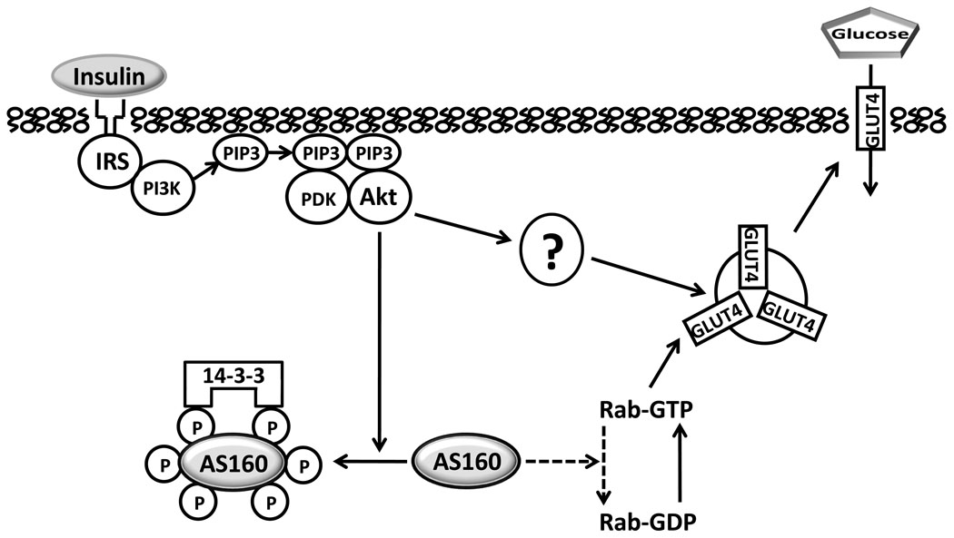
Under basal conditions, the Rab GTPase activating domain of non-phosphorylated Akt Substrate of 160 kDa (AS160) catalyzes hydrolysis of Rab-bound guanosine triphosphate (GTP), producing Rab-bound guanosine diphosphate (GDP), which inhibits exocytosis of insulin responsive glucose transporter protein (GLUT4) vesicles. Insulin-stimulated, Akt-induced phosphorylation of AS160 releases this inhibition and results in more GTP-bound Rab, favoring GLUT4 translocation toward the cell surface. Proximal insulin signaling includes, insulin binding to its receptor, leading to tyrosine phosphorylation of insulin receptor substrates (IRS) which bind phosphatidylinositol-3 kinase (PI3K) and catalyze production of phosphatidylinositol 3,4,5-phosphate (PIP3) which binds phosphoinsitide-dependent kinase (PDK) and Akt, causing phosphorylation of Akt, which in turn, phosphorylates Akt Substrate of 160 kDa (AS160) on multiple Akt-phosphomotifs. Members of the 14-3-3 family of proteins bind to specific phosphomotifs of AS160, and this appears to promote AS160 release from membranes and/or to inhibit Rab GAP activity. There also appear to be unidentified Akt-dependent, but AS160 dependent mechanisms (indicated by “?”). Arrows with solid lines indicate a mechanism favoring increased GLUT4 translocation and glucose transport. Dashed lines indicate a mechanism favoring retention of intracellular GLUT4 and lower glucose transport.
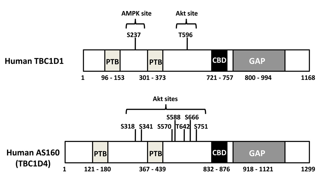
Schematic diagrams depict some key elements of two human Rab GTPase activating proteins (GAP): Akt substrate of 160 kDa (AS160 or TBC1D4) and TBC1D1 (, , –25, 30). Each protein includes two phosphotyrosine binding (PTB) domains, a calmodulin binding domain (CBD) and a GAP domain. AS160 includes more Akt phosphomotifs than TBC1D1. TBC1D1 includes an adenosine monophosphate (AMP)-activated protein kinase (AMPK) phosphomotif between the PTB domains that is absent from AS160. For clarity, the list of phosphomotifs identified in the figure is incomplete. TBC1D indicates tre-2/USP6, BUB2, cdc16 domain.
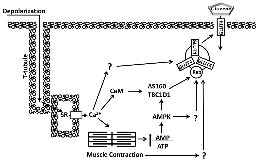
Contraction stimulation of skeletal muscle glucose transport involves multiple inputs that lead to increased cell surface insulin responsive glucose transporter protein (GLUT4). T-tubule depolarization causes calcium (Ca2+) release from the sarcoplasmic reticulum (SR) which triggers actin and myosin interaction. The energy demand of contraction increases the ratio of adenosine monophosphate (AMP)/ adenosine triphosphate (ATP) which stimulates AMP-associated protein kinase (AMPK). AMPK can phosphorylate both AS160 and TBC1D1, as well as other unknown substrates, which potentially contribute to increased glucose transport. Both Akt substrate of 160 kDa (AS160) and TBC1D1 have a calmodulin (CaM) binding domain (CBD). The CBD of AS160 has been implicated in contraction-stimulated glucose transport, but the role of TBC1D1’s CBD is unknown. Increased glucose transport with contraction may also involve other Ca2+-dependent and Ca2+-independent processes (indicated by “?”).TBC1D indicates tre-2/USP6, BUB2, cdc16 domain.
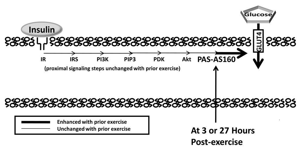
Increased insulin-stimulated glucose transport, as the result of greater insulin responsive glucose transporter protein (GLUT4) translocation, can occur a few or many hours after acute exercise. A number of proximal insulin signaling steps (insulin receptor, IR, binding; tyrosine phosphorylation of IR and insulin receptor substrates, IRS; IRS-associated phosphatidylinositol-3 kinase, PI3K, activity; and Akt serine phosphorylation) are not enhanced a few hours after acute exercise. At either 3 or 27 hours after acute exercise, Akt Substrate of 160 kDa (AS160) phosphorylation on sites recognized by anti-PAS (PAS-AS160) was found to be increased in rat skeletal muscle in the absence of insulin (4, 12). We hypothesize that the persistent increase in AS160 phosphorylation plays a role in the increased insulin-stimulated glucose transport after acute exercise. PIP3 indicates phosphatidylinositol 3,4,5-phosphate; PDK, phosphoinsitide-dependent kinase; PAS, phospho-(Ser/Thr) Akt substrate.
Comment in
-
Exercise and insulin - understanding the molecular interactions.Exerc Sport Sci Rev. 2009 Oct;37(4):156. doi: 10.1097/JES.0b013e3181b95462. Exerc Sport Sci Rev. 2009. PMID: 19955863 No abstract available.
References
-
- Arias EB, Kim J, Cartee GD. Prolonged incubation in PUGNAc results in increased protein O-Linked glycosylation and insulin resistance in rat skeletal muscle. Diabetes. 2004;53:921–930. - PubMed
-
- Arias EB, Kim J, Funai K, Cartee GD. Prior exercise increases phosphorylation of Akt substrate of 160 kDa (AS160) in rat skeletal muscle. Am J Physiol Endocrinol Metab. 2007;292:E1191–E1200. - PubMed
-
- Bruss MD, Arias EB, Lienhard GE, Cartee GD. Increased phosphorylation of Akt substrate of 160 kDa (AS160) in rat skeletal muscle in response to insulin or contractile activity. Diabetes. 2005;54:41–50. - PubMed
-
- Cartee GD, Wojtaszewski JF. Role of Akt substrate of 160 kDa in insulin-stimulated and contraction-stimulated glucose transport. Appl Physiol Nutr Metab. 2007;32:557–566. - PubMed
Publication types
MeSH terms
Substances
Grants and funding
LinkOut - more resources
Full Text Sources
Medical
