. 2009 Nov 24:15:2448-63.

The membrane proteome of the mouse lens fiber cell

Affiliations

The membrane proteome of the mouse lens fiber cell

Steven Bassnett et al. Mol Vis. .

Abstract

Purpose: Fiber cells of the ocular lens are bounded by a highly specialized plasma membrane. Despite the pivotal role that membrane proteins play in the physiology and pathophysiology of the lens, our knowledge of the structure and composition of the fiber cell plasma membrane remains fragmentary. In the current study, we utilized mass spectrometry-based shotgun proteomics to provide a comprehensive survey of the mouse lens fiber cell membrane proteome.

Methods: Membranes were purified from young mouse lenses and subjected to MudPIT (Multidimensional protein identification technology) analysis. The resulting proteomic data were analyzed further by reference to publically available microarray databases.

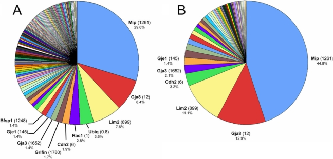
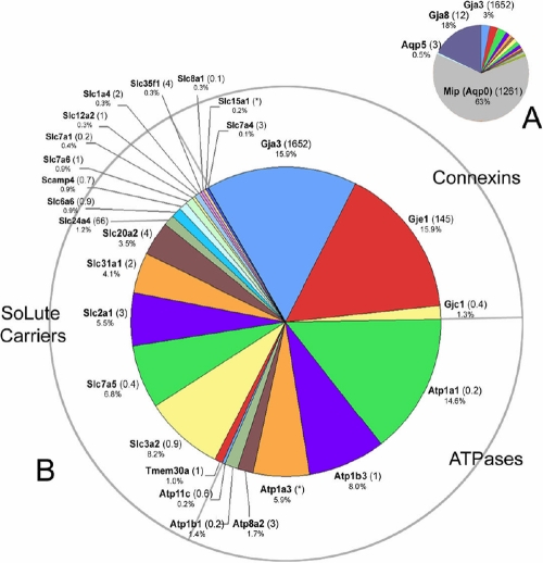
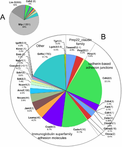
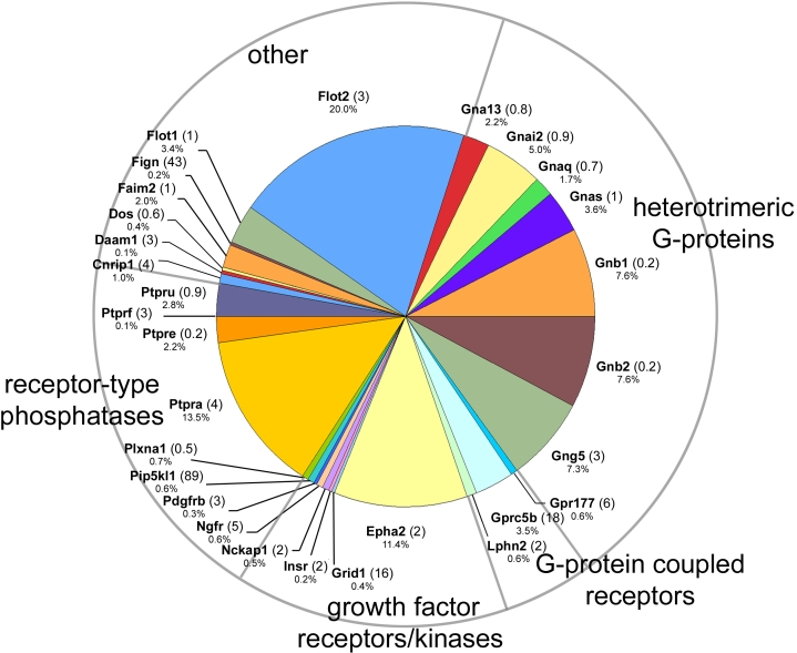
Results: More than 200 membrane proteins were identified by MudPIT, including Type I, Type II, Type III (multi-pass), lipid-anchored, and GPI-anchored membrane proteins, in addition to membrane-associated cytoskeletal elements and extracellular matrix components. The membrane proteins of highest apparent abundance included Mip, Lim2, and the lens-specific connexin proteins Gja3, Gja8, and Gje1. Significantly, many proteins previously unsuspected in the lens were also detected, including proteins with roles in cell adhesion, solute transport, and cell signaling.

Conclusions: The MudPIT technique constitutes a powerful technique for the analysis of the lens membrane proteome and provides valuable insights into the composition of the lens fiber cell unit membrane.


Figures

Figure 1
Figure 1
MudPIT analysis of the lens fiber cell membrane proteome. A: More than 200 membrane proteins and membrane-associated proteins were identified. The ten proteins of highest apparent abundance are indicated. For each protein, the number shown in parentheses is the lens preferred expression index (LPEI), a measure of the relative level of expression of the RNA for that protein in the lens compared to a panel of 96 other tissues and cell types (see text for details). The percentage value for each protein relates to its representation on that particular pie chart. B: Eighty-seven integral membrane proteins were detected. Six proteins (including three connexin proteins; Gja3, Gja8, Gje1) account for >75% of the integral plasma membrane proteins detected in MudPIT analysis of lens fiber membranes.
Figure 2
Figure 2
Transport proteins detected in the lens membrane proteome. A: Mip, a lens-specific aquaporin protein, and Gja8, a connexin protein, together dominate this category of proteins. B: To better visualize the results, Mip and Gja8 have been removed from the data set. Three classes of membrane transport protein were detected: connexins, ATPases and Slc transporters. The numbers in parentheses indicate the lens preferred expression index (LPEI, see text for details). Occasionally, it was not possible to calculate the LPEI because of inadequate hybridization signals, these instances are indicated by an asterisk.
Figure 3
Figure 3
Cell adhesion proteins. A: Mip, Lim2, and Cdh2 were the adhesion proteins with highest apparent abundance (together constituting ≈ 85% of identified spectra). In B, Mip and Lim2 have been removed to help visualize the identities of adhesion proteins of lower apparent abundance. The numbers in parentheses indicate the lens preferred expression index (LPEI, see text for details).
Figure 4
Figure 4
Components of the membrane cytoskeleton identified in the fiber cell membrane proteome. Actin-binding proteins, intermediate filament proteins and elements of the spectrin cytoskeleton were detected. The numbers in parentheses indicate the lens preferred expression index (LPEI, see text for details). Occasionally, it was not possible to calculate the LPEI because of inadequate hybridization signals, these instances are indicated by an asterisk.
Figure 5
Figure 5
Cell signaling components identified in the fiber cell membrane proteome.
Figure 6
Figure 6
Members of the Ras superfamily of small GTPases identified in the fiber cell membrane proteome.
Figure 7
Figure 7
Extracellular matrix and matrix-interacting proteins in the fiber cell membrane proteome.

References

    1. Delamere NA, Tamiya S. Expression, regulation and function of Na,K-ATPase in the lens. Prog Retin Eye Res. 2004;23:593–615. - PubMed
    1. Shiels A, Hejtmancik JF. Genetic origins of cataract. Arch Ophthalmol. 2007;125:165–73. - PubMed
    1. Shiels A, Bennett TM, Knopf HL, Maraini G, Li A, Jiao X, Hejtmancik JF. The EPHA2 gene is associated with cataracts linked to chromosome 1p. Mol Vis. 2008;14:2042–55. - PMC - PubMed
    1. Shiels A, King JM, Mackay DS, Bassnett S. Refractive defects and cataracts in mice lacking lens intrinsic membrane protein-2. Invest Ophthalmol Vis Sci. 2007;48:500–8. - PubMed
    1. Wu CC, MacCoss MJ, Howell KE, Yates JR., 3rd A method for the comprehensive proteomic analysis of membrane proteins. Nat Biotechnol. 2003;21:532–8. - PubMed

Publication types

LinkOut - more resources