. 2009 Mar 5;1(3):267-82.

Triptolide induces anti-inflammatory cellular responses

Triptolide induces anti-inflammatory cellular responses

Ranyia Matta et al. Am J Transl Res. .

Abstract

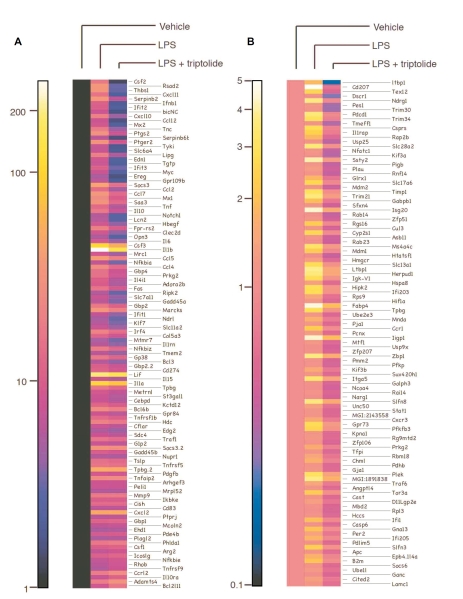
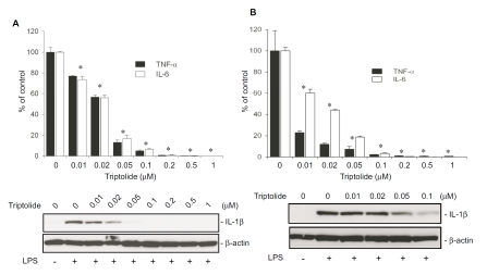
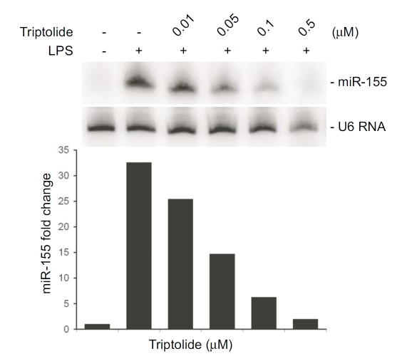
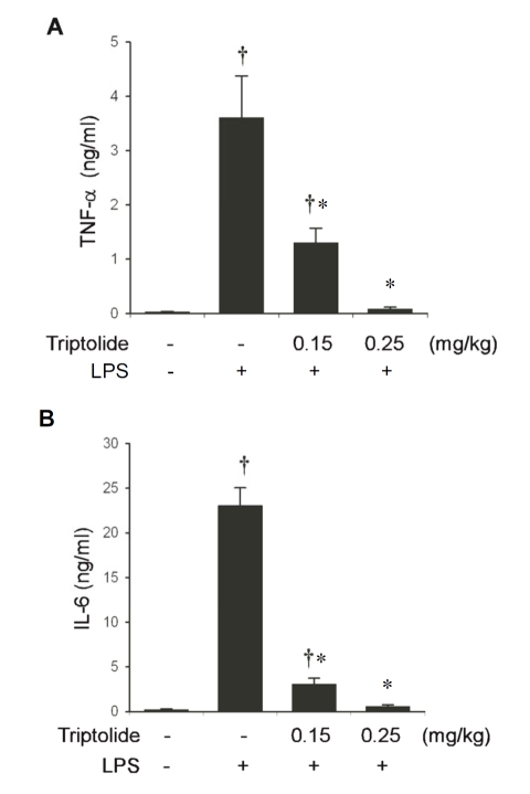
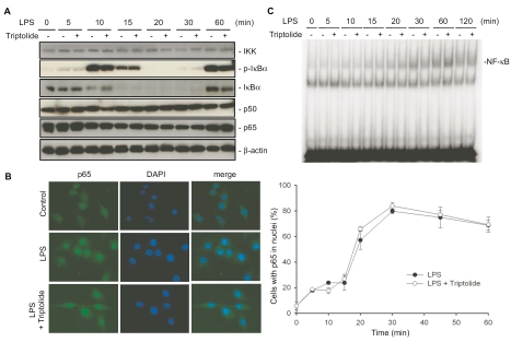
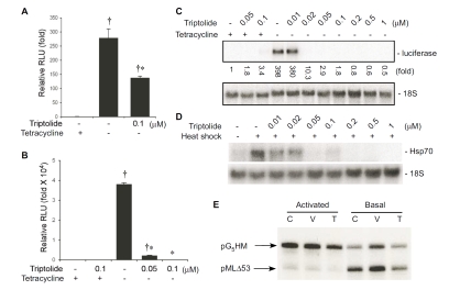
Tripterygium wilfordii Hook F. has been used for centuries in traditional Chinese medicine to treat rheumatoid arthritis, an autoimmune disease associated with increased production of the pro-inflammatory cytokine, tumor necrosis factor (TNF)-alpha. Triptolide is a compound originally purified from T. wilfordii Hook F. and has potent anti-inflammatory and immunosuppressant activities. In this study, we investigated the effect of triptolide on the global gene expression patterns of macrophages treated with lipopolysaccharide (LPS). We found that LPS stimulation resulted in >5-fold increase in expression of 117 genes, and triptolide caused a >50% inhibition in 47 of the LPS-inducible 117 genes. A large portion of the genes that were strongly induced by LPS and significantly inhibited by triptolide were pro-inflammatory cytokine and chemokine genes, including TNF-alpha, IL-1beta, and IL-6. Interestingly, LPS also induced the expression of micro-RNA-155 (miR-155) precursor, BIC, which was inhibited by triptolide. Confirming the cDNA array results, we demonstrated that triptolide blocked the induction of these pro-inflammatory cytokines as well as miR-155 in a dose-dependent manner. Profound inhibition of pro-inflammatory cytokine expression was observed at concentrations as low as 10-50 nM. However, triptolide neither inhibited the phosphorylation or degradation of IkappaBalpha after LPS stimulation, nor affected the DNA-binding activity of NF-kappaB. Surprisingly, we found that triptolide not only inhibited NF-kappaB-regulated reporter transcription, but also dramatically blocked the activity of other transcription factors. Our study offers a plausible explanation of the therapeutic mechanism of T. wilfordii Hook F.

Keywords: Chinese medicine; Inflammation; Tripterygium wilfordii; cytokines; rheumatoid arthritis; transcription.


Figures

Figure 1
Figure 1
Triptolide selectively inhibits the expression of a subset of genes involved in the immune response. RAW264.7 cells were either treated with vehicle or LPS in the presence or absence of triptolide, and microarray analyses were performed. Heat maps were generated representing cluster analyses of genes exhibiting 5-fold or greater induction by LPS (A) or 1–5-fold induction by LPS but inhibited by triptolide at least 50% (B).
Figure 2
Figure 2
Triptolide potently inhibits the expression of genes that were potently induced by LPS. The graph depicts the effect of triptolide on the genes highly induced (>5-fold) by LPS. Dots above the line represent genes whose expression is enhanced by triptolide. Dots below the line represent genes whose expression is inhibited by triptolide.
Figure 3
Figure 3
Triptolide blocks the induction of pro-inflammatory cytokine mRNA in LPS-stimulated macrophages. RAW264.7 cells (A) and primary peritoneal macrophages (B) were pretreated with indicated doses of triptolide for 30 min, and then stimulated with LPS (100 ng/ml) for 4 h. Total RNA was harvested and analyzed by Northern blotting. The IL-1β and TNF-α mRNA signals were normalized to 18S rRNA signals, and expressed as fold increase relative to control. Numbers below the blots indicate fold of changes in gene expression relative to untreated controls.
Figure 4
Figure 4
Triptolide potently inhibits the production of pro-inflammatory cytokine proteins in macrophages treated with LPS. RAW264.7 cells (A) and primary peritoneal macrophages (B) were pretreated with indicated doses of triptolide for 30 min followed by stimulation with 100 ng/ml LPS for 6 h. The negative control was only treated with equal volume of vehicle (DMSO). Positive controls were those first treated with vehicle (DMSO) then stimulated with LPS. Upper panels represent the effect of triptolide on TNF-α and IL-6 secretion, which were assayed by ELISA. Results in the graph represent % relative to the mean value of the positive controls. Data were expressed as means ± S.E. from 3 independent experiments. *, p<0.05, compared to the group stimulated with LPS after pretreatment with vehicle. Lower panels demonstrate the effect of triptolide on IL-1βproduction detected by Western blot analysis. Presented are the representative results of at least three experiments.
Figure 5
Figure 5
Triptolide inhibits the induction of miR-155 in LPS-stimulated macrophages. RAW264.7 cells were pretreated with the indicated doses of triptolide followed by stimulation with LPS for 4 h. Total RNA was harvested and Northern blot analysis was performed using a urea-PAGE denaturing gel. U6 was used as a loading control. Graph depicts the fold of changes in miR-155 expression in a representative experiment.
Figure 6
Figure 6
Triptolide attenuates LPS-induced cytokine production in vivo in a dose-dependent manner. C57BL6 mice (5–6 weeks old) were given the indicated doses of triptolide, or equal volumes of vehicle i.p, and then challenged with LPS (5 mg/kg body weight). Cytokine concentrations in the serum were determined by ELISA. (A) Effects of triptolide on TNF-α biosynthesis in vivo. (B) Effects of triptolide on IL-6 production in vivo. Values represent the mean ± S.E. from 8–12 animals. †, p < 0.05, compared to control group (not stimulated with LPS). *, p<0.05, compared to the group received vehicle and then stimulated with LPS.
Figure 7
Figure 7
Triptolide has no effect on IκBα degradation and p65 nuclear translocation, and does not affect NF-κB DNA-binding activity in LPS-stimulated macrophages. (A) Effects of triptolide on the components of the NF-κB pathway in LPS-stimulated macrophages. RAW264.7 cells were pretreated with 0.1 μM triptolide for 30 minutes, then stimulated with 100 ng/ml LPS for the indicated periods. Cells lysates were harvested and Western blotting was performed. Data shown were from a representative experiment. (B) Effect of triptolide on nuclear translocation of NF-κB p65 subunit. RAW264.7 cells were treated as in A, and immunofluorescence was performed with a p65 antibody (green), and then stained with DAPI (blue). The percentage of cells where p65 was localized in the nuclei was scored from at least 10 fields chosen randomly. Values in the graph on the right side represent mean ± S.E. from 3 independent experiments. (C) Effect of triptolide on NF-κB DNA-binding activity. RAW264.7 cells were treated as in A, and nuclear proteins were extracted. EMSA was performed using end-labeled double-stranded oligonucleotides containing a consensus NF-κB-binding element. Results were from a representative experiment.
Figure 8
Figure 8
Triptolide blocks gene transcriptional induction regulated by a variety of transcriptional factors. Effect of triptolide on reporter expression mediated by the transactivation domains of p65 NF-κB (A) and VP16 (B). RAW264.7 (A) and CHO-AA8-Luc (B) cells were treated as described. Cells were then lysed and luciferase activity in the lysates was measured. Relative luciferase units were normalized to protein concentrations. Luciferase activities in both A and B were expressed in the graphs as fold change relative to cells kept in tetracycline-containing medium (controls). Values represent mean ± SE from 3 independent experiments. † different from control group (with tetracycline), p<0.05. *, different from the group without tetracycline but treated with vehicle (DMSO), p<0.05. (C) Effect of triptolide on luciferase reporter mRNA expression mediated by the VP16 transactivation domain. CHO-AA8-Luc cells were treated as in B. Cells were harvested to isolate total RNA, and Northern blot analysis was performed. Luciferase expression was normalized to 18S rRNA, and expressed as fold increase relative to control (signal of cells grown in medium containing only tetracycline). (D) Triptolide inhibits the induction of Hsp70 by heat shock. RAW264.7 cells were subjected to heat shock in the presence of indicated doses of triptolide, and Northern blot analysis was performed. (E) Triptolide does not inhibit gene transcription by RNA polymerase II in vitro. RAW264.7 cells were either left untreated (C) or treated with vehicle (V) or triptolide (T), and in vitro transcription assays were performed using the indicated templates.

Similar articles

Cited by

References

    1. Chen BJ. Triptolide, a novel immunosuppressive and anti-inflammatory agent purified from a Chinese herb Tripterygium wilfordii Hook F. Leuk Lymphoma. 2001;42:253–265. - PubMed
    1. Tao X, Cush JJ, Garret M, Lipsky PE. A phase I study of ethyl acetate extract of the chinese antirheumatic herb Tripterygium wilfordii hook F in rheumatoid arthritis. J Rheumatol. 2001;28:2160–2167. - PubMed
    1. Tao X, Younger J, Fan FZ, Wang B, Lipsky PE. Benefit of an extract of Tripterygium Wilfordii Hook F in patients with rheumatoid arthritis: a double-blind, placebo-controlled study. Arthritis Rheum. 2002;46:1735–1743. - PubMed
    1. Cibere J, Deng Z, Lin Y, Ou R, He Y, Wang Z, Thorne A, Lehman AJ, Tsang IK, Esdaile JM. A randomized double blind, placebo controlled trial of topical Tripterygium wilfordii in rheumatoid arthritis: reanalysis using logistic regression analysis. J Rheumatol. 2003;30:465–467. - PubMed
    1. Qiu D, Kao PN. Immunosuppressive and anti-inflammatory mechanisms of triptolide, the principal active diterpenoid from the Chinese medicinal herb Tripterygium wilfordii Hook. f. Drugs R D. 2003;4:1–18. - PubMed

LinkOut - more resources