

Posterior lumbar interbody fusion using a unilateral single cage and a local morselized bone graft in the degenerative lumbar spine
- PMID: 19956479
- PMCID: PMC2784962
- DOI: 10.4055/cios.2009.1.4.214
Posterior lumbar interbody fusion using a unilateral single cage and a local morselized bone graft in the degenerative lumbar spine
Abstract
Background: We retrospectively evaluated the clinical and radiological outcomes of posterior lumbar interbody fusion (PLIF) with using a unilateral single cage and a local morselized bone graft.
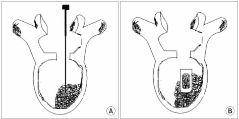
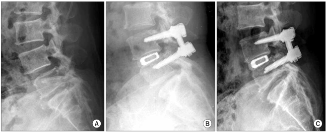
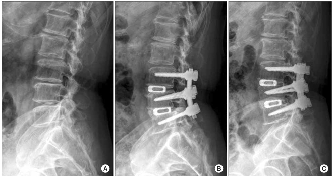
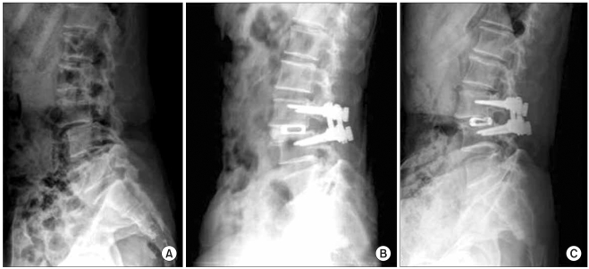
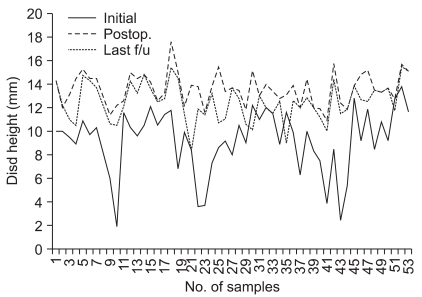
Methods: Fifty three patients who underwent PLIF with a unilateral single cage filled with local morselized bone graft were enrolled in this study. The average follow-up duration was 31.1 months. The clinical outcomes were evaluated with using the visual analogue scale (VAS) at the pre-operative period, at 1 year post-operation and at the last follow-up, the Oswestry Disability Index, the Prolo scale and the Kim & Kim criteria at the last follow-up; the radiological outcomes were evaluated according to the change of bone bridging, the radiolucency, the instability and the disc height.
Results: For the clinical evaluation, the VAS pain index, the Oswestry Disability Index, the Prolo scale and the Kim & Kim criteria showed excellent outcomes. For the the radiological evaluation, 52 cases showed complete bone union at the last follow-up. Regarding the complications, only 1 patient had cage breakage during follow-up.
Conclusions: PLIF using a unilateral single cage filled with a local morselized bone graft has the advantages of a shorter operation time, less blood loss and a shorter hospital stay, as compared with the PLIF using bilateral cages, for treating degenerative lumbar spine disease. This technique also provides excellent outcomes according to the clinical and radiological evaluation.
Keywords: Local morselized graft; Posterior lumbar interbody fusion; Spinal fusion; Unilateral single cage.
Figures
Similar articles
-
[Adjacent segment degeneration after lumbosacral fusion in spondylolisthesis: a retrospective radiological and clinical analysis].Acta Chir Orthop Traumatol Cech. 2010 Apr;77(2):124-30. Acta Chir Orthop Traumatol Cech. 2010. PMID: 20447355 Czech.
-
Clinical results of single-level posterior lumbar interbody fusion using the Brantigan I/F carbon cage filled with a mixture of local morselized bone and bioactive ceramic granules.Spine (Phila Pa 1976). 2002 Feb 1;27(3):258-62. doi: 10.1097/00007632-200202010-00011. Spine (Phila Pa 1976). 2002. PMID: 11805688
-
Minimally Invasive Transforaminal Lumbar Interbody Fusion and Unilateral Fixation for Degenerative Lumbar Disease.Orthop Surg. 2017 Aug;9(3):277-283. doi: 10.1111/os.12345. Orthop Surg. 2017. PMID: 28960820 Free PMC article.
-
Double-level lumbar spondylolysis and spondylolisthesis: A retrospective study.J Orthop Surg Res. 2018 Mar 16;13(1):55. doi: 10.1186/s13018-018-0723-3. J Orthop Surg Res. 2018. PMID: 29548343 Free PMC article. Review.
-
Unilateral transforaminal lumbar interbody fusion: a review of the technique, indications and graft materials.J Int Med Res. 2009 May-Jun;37(3):908-17. doi: 10.1177/147323000903700337. J Int Med Res. 2009. PMID: 19589277 Review.
Cited by
-
Dislodgment Effects of Different Cage Arrangements in Posterior Lumbar Interbody Fusion: A Finite Element Study.Bioengineering (Basel). 2024 May 31;11(6):558. doi: 10.3390/bioengineering11060558. Bioengineering (Basel). 2024. PMID: 38927794 Free PMC article.
-
Chitosan-Hydroxyapatite Scaffold for Tissue Engineering in Experimental Lumbar Laminectomy and Posterolateral Spinal Fusion in Wistar Rats.Asian Spine J. 2020 Apr;14(2):139-147. doi: 10.31616/asj.2019.0091. Epub 2019 Nov 5. Asian Spine J. 2020. PMID: 31679322 Free PMC article.
-
Minimally invasive versus open posterior lumbar interbody fusion: a systematic review.Clin Orthop Relat Res. 2014 Jun;472(6):1792-9. doi: 10.1007/s11999-014-3619-5. Clin Orthop Relat Res. 2014. PMID: 24748069 Free PMC article.
-
The Prolo Scale: history, evolution and psychometric properties.J Orthop Traumatol. 2013 Dec;14(4):235-45. doi: 10.1007/s10195-013-0243-1. Epub 2013 May 10. J Orthop Traumatol. 2013. PMID: 23660865 Free PMC article.
-
Comparison of Fusion Rates among Various Demineralized Bone Matrices in Posterior Lumbar Interbody Fusion.Medicina (Kaunas). 2024 Feb 2;60(2):265. doi: 10.3390/medicina60020265. Medicina (Kaunas). 2024. PMID: 38399552 Free PMC article.
References
-
- Chiang MF, Zhong ZC, Chen CS, Cheng CK, Shih SL. Biomechanical comparison of instrumented posterior lumbar interbody fusion with one or two cages by finite element analysis. Spine (Phila Pa 1976) 2006;31(19):E682–E689. - PubMed
-
- Huang KF, Chen TY. Clinical results of a single central interbody fusion cage and transpedicle screws fixation for recurrent herniated lumbar disc and low-grade spondylolisthesis. Chang Gung Med J. 2003;26(3):170–177. - PubMed
-
- Bagby GW. Arthrodesis by the distraction-compression method using a stainless steel implant. Orthopedics. 1988;11(6):931–934. - PubMed
-
- Axelsson P, Johnsson R, Stromqvist B, Arvidsson M, Herrlin K. Posterolateral lumbar fusion: outcome of 71 consecutive operations after 4 (2-7) years. Acta Orthop Scand. 1994;65(3):309–314. - PubMed
-
- Diedrich O, Luring C, Pennekamp PH, Perlick L, Wallny T, Kraft CN. Effect of posterior lumbar interbody fusion on the lumbar sagittal spinal profile. Z Orthop Ihre Grenzgeb. 2003;141(4):425–432. - PubMed
MeSH terms
LinkOut - more resources
Full Text Sources
Other Literature Sources
Medical
