. 2009 Nov 30;4(11):e8027.
doi: 10.1371/journal.pone.0008027.

Rearrangements of the actin cytoskeleton and E-cadherin-based adherens junctions caused by neoplasic transformation change cell-cell interactions

Affiliations

Rearrangements of the actin cytoskeleton and E-cadherin-based adherens junctions caused by neoplasic transformation change cell-cell interactions

Dmitry V Ayollo et al. PLoS One. .

Abstract

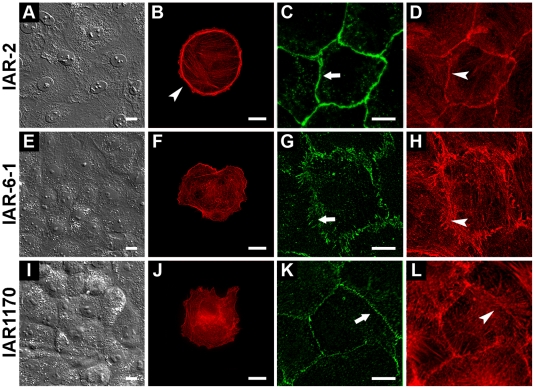
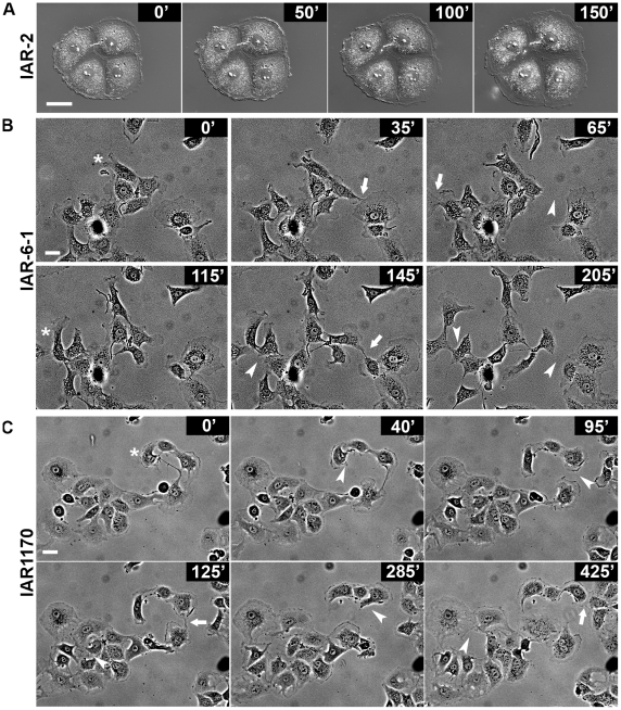
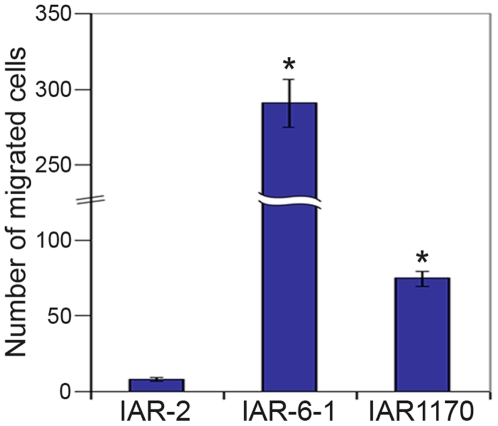
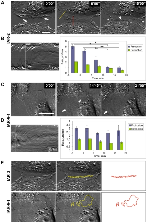
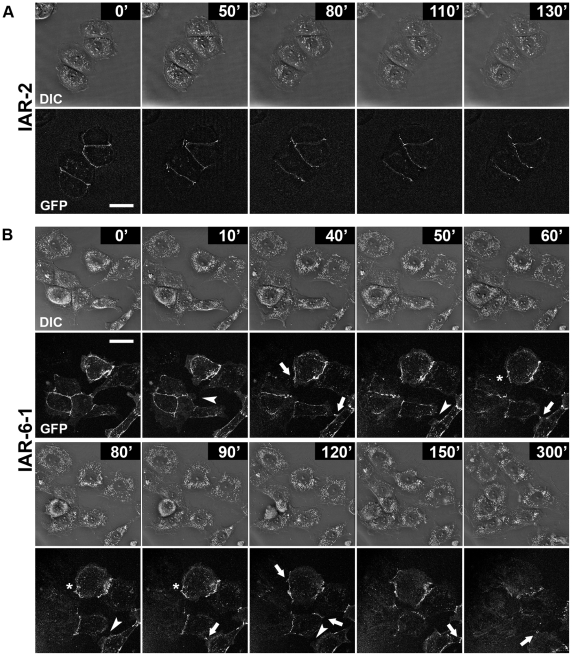
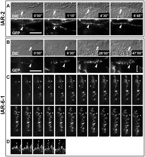
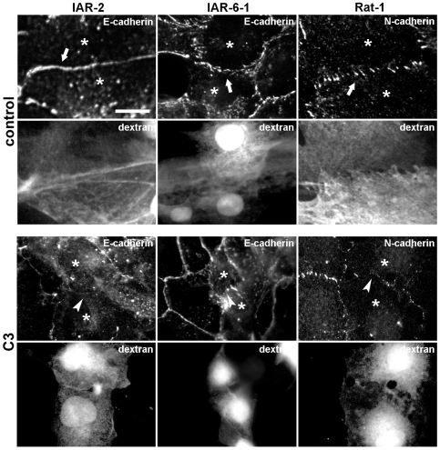
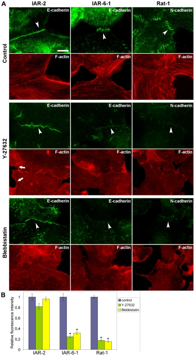
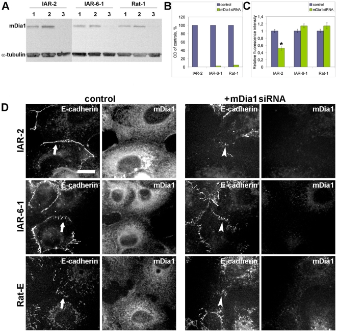
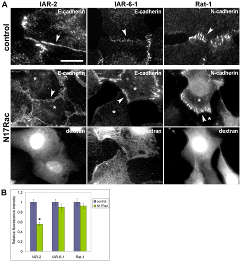
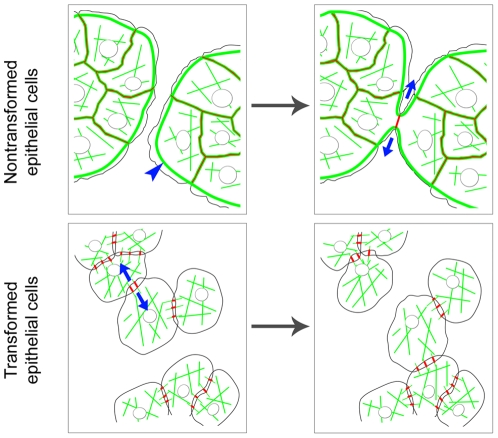
E-cadherin-mediated cell-cell adhesion, which is essential for the maintenance of the architecture and integrity of epithelial tissues, is often lost during carcinoma progression. To better understand the nature of alterations of cell-cell interactions at the early stages of neoplastic evolution of epithelial cells, we examined the line of nontransformed IAR-2 epithelial cells and their descendants, lines of IAR-6-1 epithelial cells transformed with dimethylnitrosamine and IAR1170 cells transformed with N-RasG12D. IAR-6-1 and IAR1170 cells retained E-cadherin, displayed discoid or polygonal morphology, and formed monolayers similar to IAR-2 monolayer. Fluorescence staining, however, showed that in IAR1170 and IAR-6-1 cells the marginal actin bundle, which is typical of nontransformed IAR-2 cells, disappeared, and the continuous adhesion belt (tangential adherens junctions (AJs)) was replaced by radially oriented E-cadherin-based AJs. Time-lapse imaging of IAR-6-1 cells stably transfected with GFP-E-cadherin revealed that AJs in transformed cells are very dynamic and unstable. The regulation of AJ assembly by Rho family small GTPases was different in nontransformed and in transformed IAR epithelial cells. As our experiments with the ROCK inhibitor Y-27632 and the myosin II inhibitor blebbistatin have shown, the formation and maintenance of radial AJs critically depend on myosin II-mediated contractility. Using the RNAi technique for the depletion of mDia1 and loading cells with N17Rac, we established that mDia1 and Rac are involved in the assembly of tangential AJs in nontransformed epithelial cells but not in radial AJs in transformed cells. Neoplastic transformation changed cell-cell interactions, preventing contact paralysis after the establishment of cell-cell contact and promoting dynamic cell-cell adhesion and motile behavior of cells. It is suggested that the disappearance of the marginal actin bundle and rearrangements of AJs may change the adhesive function of E-cadherin and play an active role in migratory activity of carcinoma cells.


Conflict of interest statement

Competing Interests: The authors have declared that no competing interests exist.

Figures

Figure 1
Figure 1. Nontransformed and transformed IAR epithelial cell lines.
(A) In dense culture, nontransformed IAR-2 epithelial cells form a monolayer. (B) A characteristic feature of the actin cytoskeleton of an IAR-2 epithelial cell is the marginal actin bundle (arrowhead). (C, D) E-cadherin-based AJs (tangential AJs) (arrow) in an IAR-2 monolayer organized into adhesion belts along the cell-cell boundaries and colocalized with perijunctional actin bundles (arrowhead). (E–H) An IAR-6-1 line transformed with dimethylnitrosamine. (E) In dense culture, transformed IAR-6-1 epithelial cells form a monolayer. (F) IAR-6-1 cells have a slightly changed morphology. They are polygonal. Note the disappearance of the marginal actin bundle at the cell periphery. (G, H) Double staining for E-cadherin and F-actin shows that AJs in IAR-6-1 cells appear as radial strands (arrow) that are colocalized with straight actin bundles (arrowhead). (I–L) Ras-transformed IAR1170 epithelial cells (clone H5). (I) In dense culture, IAR1170 cells form a monolayer. (J) The shapes of IAR1170 cells in sparse cultures are polygonal. The actin cytoskeleton is present in the form of randomly oriented actin bundles; the marginal actin bundle disappears. (K–L) Punctate clasters of E-cadherin at the cell-cell boundaries in an IAR1170 monolayer (arrow). Circumferential actin bundles are disorganized. Straight actin bundles oriented perpendicularly to the cell-cell boundaries (arrowhead) are seen. Bar, 10 µm.
Figure 2
Figure 2. The alterations of motile behavior of transformed IAR-6-1 and IAR1170 cells in comparison with those of nontransformed IAR-2 cells.
(A–C) Selected frames from the time-lapse sequences of sparse cultures showing cell behavior and cell-cell interactions. (A) Nontransformed IAR-2 cells form an island. Cell-cell contacts are stable during the entire period of observation (see also Video S1). (B) Transformed IAR-6-1 cells; (C) transformed IAR1170 cells (clone H5). In cultures of transformed IAR-6-1 and IAR1170 cells, significant alterations in cell-cell interactions are seen. Cells extend lamellipodia at different sites of the free edges and establish contacts with different neighboring cells (arrows). These contacts are unstable and often break (arrowheads). Cells could move in different directions (asterisks) (see also Video S2 and Video S3). Bar, 20 µm.
Figure 3
Figure 3. Transformed IAR-6-1 and IAR1170 cells display enhanced motility in a migration assay.
IAR-2, IAR-6-1, and IAR-1170 (clone H5) cells were plated on membranes with 8-µm pores in Bio-Coat migration chambers. After 20 h of incubation, the cells on the lower surface of membranes were fixed and stained. The number of migrated cells was determined from an average of the number of stained cells on membranes in 15 randomly selected fields. The data are presented as means±SEM of triplicate assays for each cell line in three independent experiments. Asterisks indicate the values that differ significantly from control (p<0.001, t-test).
Figure 4
Figure 4. Dynamics of cell-cell interactions in a narrow wound.
Time-lapse images of colliding cells. (A, B) Nontransformed IAR-2 cells extend lamellipodia (arrows) and establish a cell-cell contact that expands laterally. By 15–20 min, protrusion of lamellipodia at the site of the contact is inhibited (contact paralysis) (arrowhead) (see also Video S4). Protrusive activity at the free edges of contacting cells also decreases (asterisks). Kymographs were generated along straight lines in different regions. (B) Left: kymograph generated at the site of the cell-cell contact (red line in A) shows contact paralysis of protrusive activity by 15–20 min after the establishment of the contact. White line on kymograph indicates the time of the establishment of the contact. Right: quantification of the rates of lamellipodial protrusion and retraction at the sites of cell-cell contacts. Data are presented as means±SEM. Brackets indicate statistically significant differences between the mean rates of lamellipodial protrusion (*, p<0.001; **, p<0.005, n = 25 cells, t-test). (C, D) Transformed IAR-6-1 cells. During formation of the cell-cell contact, protrusive activity persists both at the site of the contact (arrowhead) and at the free edges, where new lamellipodia are formed (arrows) (see also Video S5). (D) Left: kymograph generated at the site of the contact (left) shows the disappearance of contact paralysis. White line on kymograph indicates the time of the establishment of the contact. Right: the rates of lamellipodial protrusion and retraction at the sites of cell-cell contacts did not change within the time of observation. Data are presented as means±SEM. (E). Measurement of overlapping areas of contacting cells (15 min after formation of cell-cell contact). In cultures of transformed IAR-6-1 cells, there is prominent overlapping of lamellae of contacting cells. Bar, 10 µm.
Figure 5
Figure 5. E-cadherin dynamics at the sites of cell–cell contacts.
Nontransformed IAR-2 cells and transformed IAR-6-1 cells were stably transfected with GFP-E-cadherin. (A) In IAR-2 cells, GFP-E-cadherin accumulates in the zones of cell-cell interactions, forming stable tangential AJs (see also Video S6). (B) IAR-6-1 cells form lamellipodia along their entire edges. When a protrusion contacts another cell, E-cadherin begins to aggregate in punctate AJs in the zone of contact (arrows). AJs can grow, rearrange, and relocate (asterisks). The disruption of AJs results in detachment of one cell from another (arrowheads) (see also Video S7). Bar, 10 µm.
Figure 6
Figure 6. E-cadherin dynamics during cell–cell collision in a narrow wound.
Nontransformed IAR-2 cells and transformed IAR-6-1 cells were stably transfected with GFP-E-cadherin. (A) IAR-2 cells. DIC image shows lateral expansion of the cell-cell contact (arrows). During cell-cell contact formation, GFP-E-cadherin accumulates in a tangential line (arrowheads) at the boundary between two cells (see also Video S8). (B, C, D; see also Video S9) IAR-6-1 cells. (B) During cell-cell contact formation, there is overlapping of lamellae in the area of the contact (arrows). Radial AJs (arrowheads) assemble in the zone of overlapping. (C) Time-lapse images of the same cell-cell contact acquired at 1-min intervals. GFP-E-cadherin initially aggregates into dot-like clusters. A majority of GFP-E-cadherin dots grow and elongate (asterisks). Other AJs can disappear (arrowheads). (D) Because of cell movement, pre-existing AJs (bracket in B) elongate and break (arrowheads). Interval time, 2 min. Bar, 10 µm.
Figure 7
Figure 7. Rho activity is required for the assembly of tangential and radial AJs.
Cell monolayers were wounded in the presence of C3 transferase and TRITC-labeled dextran (as a marker). Cells were fixed and stained for E-cadherin or N-cadherin. In control cultures, AJs are formed at the sites of cell-cell contacts (arrows). The IAR-2, IAR-6-1, and IAR1170 cells loaded with C3 are unable to assemble new AJs (arrowheads). Bar, 10 µm.
Figure 8
Figure 8. Effect of myosin II inactivation on AJ formation.
Cell monolayers were wounded and transferred to the medium with the Rho-kinase inhibitor Y-27632 or with the myosin II ATPase inhibitor blebbistatin for 2 h. Cells were fixed and stained for F-actin, E-cadherin, or N-cadherin. (A) Incubation with Y-27632 (30 µM) leads to total disappearance of actin bundles. In IAR-2 cells, both marginal (arrows) and straight actin bundles disappear. IAR-2 cells can form wave-like tangential AJs (arrowhead) in the presence of 30 µM Y-27632. IAR-6-1 cells treated with 30 µM Y-27632 do not assemble radial AJs. E-cadherin-based junctions are seen as dots at the cell-cell boundary (arrowhead). Y-27632 (30 µM) blocks the formation of radial AJs in Rat-1 fibroblasts (arrowhead). Blebbistatin abolishes actin bundles in all types of cells. IAR-2 cells in the presence of 50 µM blebbistatin can accumulate E-cadherin in wave-like tangential AJs (arrowhead). Myosin II activity is necessary for the assembly of radial AJs. IAR-6-1 cells treated with 50 µM blebbistatin accumulate E-cadherin in dot-like clusters (arrowhead) and do not form radial AJs. In Rat-1 fibroblasts, blebbistatin (15 µM) inhibits the formation of radial AJs (arrowhead). (B) Accumulation of E-cadherin and N-cadherin at cell-cell contacts was quantified by measuring fluorescence intensity on immunofluorescence images. Fluorescence intensity at cell-cell contacts of drug-treated cells relative to fluorescence intensity at cell-cell contacts of control cells is shown. Vertical lines indicate SEM. Asterisks indicate the values that differ significantly from corresponding controls (t-test, p<0.001, n = 25–32 contacts). Bar, 10 µm.
Figure 9
Figure 9. Effect of mDia1 depletion on AJ formation.
Subconfluent cultures were transfected with mDia1 siRNA or control GFP siRNA and incubated for 48 h. (A, B) Immunoblot analysis of cell lysates. Total lysate protein (20 µg) was separated with SDS-PAGE and immunoblotted for mDia1 (α-tubulin was used as a loading control). Immunoblotting (A) shows the extent of mDia1 suppression by RNAi in different cell lines. Samples 1, 2, and 3 represent GFP siRNA-transfected cells, control cells, and mDia1 siRNA-transfected cells, respectively. Densitometry (B) shows the significant reduction of mDia1 after RNAi application (OD, optical density). (C) Effect of mDia1 suppression on cadherin accumulation at cell-cell contacts. Fluorescence intensity at cell-cell contacts of mDia1 siRNA-transfected cells relative to fluorescence intensity at cell-cell contacts of GFP siRNA-transfected cells on immunofluorescence images is shown. Asterisk indicates the value that differs significantly from corresponding control (t-test, p<0.001, n = 25 contacts). (D) Effect of mDia1 suppression on the formation of AJs in a narrow wound. Cell monolayers were wounded, fixed 4 h later, and stained for E-cadherin and mDia1 simultaneously. Arrows show the newly formed AJs in control cells. The staining for mDia1 shows a significantly lower level of mDia1 expression in the cells transfected with mDia1 siRNA than in control cells. IAR-2 cells transfected with mDia1 siRNA fail to form tangential AJs (arrowhead). mDia1 is not required for the formation of radial AJs either in transformed IAR-6-1 cells or in Rat-E fibroblasts (Rat-1 fibroblasts stably expressing E-cadherin). Radial AJs (arrowheads) are formed at the sites of contacts between mDia1 siRNA-transfected cells. Bar, 10 µm.
Figure 10
Figure 10. Effect of N17Rac on AJ formation in different cell types.
Cell monolayers were wounded in the presence of N17Rac and TRITC-labeled dextran as a marker. (A) IAR-2 cells loaded with N17Rac fail to form continuous tangential AJs (arrowhead). Inhibition of Rac activity does not affect the formation of radial AJs. IAR-6-1 cells loaded with N17Rac assemble radial AJs (arrowhead). Rat-1 fibroblasts loaded with N17Rac also form radial AJs (arrowhead). (B) Effect of N17Rac loading on cadherin accumulation at cell-cell contacts. Fluorescence intensity of E-cadherin or N-cadherin at cell-cell contacts of N17Rac-loaded cells relative to fluorescence intensity at cell-cell contacts of control cells is shown. Asterisk indicates the value that differs significantly from corresponding control (t-test, p<0.001, n = 27 contacts). Bar, 10 µm.
Figure 11
Figure 11. Hypothetical scheme for alterations of cell–cell interactions between epithelial cells at the early stages of morphologic transformation.
The actin cytoskeleton (green) and AJs (red) in nontransformed and transformed epithelial cells. The characteristic feature of the actin cytoskeleton in nontransformed epithelial cells is marginal actin bundles at the free edges (blue arrowhead). In nontransformed cells, the formation of a tangential E-cadherin-based cell-cell contact (red line) leads to the disruption of marginal bundles at the site of the contact. The remaining segments of marginal bundles of adjacent cells form two arc-like bundles. The tangential tension of arcs (blue arrows) decreases protrusive activity both at the site of the cell-cell contact (contact paralysis) and at the free edges of contacting cells. In transformed cells, the crucial alteration of the actin cytoskeleton is the disappearance of marginal actin bundles. Upon the formation of the cell-cell contact, the deficiency of tangential tension as a result of the disappearance of marginal bundles prevents contact paralysis and leads to overlapping of lamellae of contacting cells. Radial AJs assemble in the area of overlapping. These AJs are associated with straight actin bundles. The enlargement of radial AJs depends on centripetal myosin II-mediated tension (blue arrows). AJs in transformed cells are very dynamic and unstable. Reorganization of the actin cytoskeleton and remodeling of AJs caused by neoplastic transformation results in a decrease in cell-cell adhesion and alters motile behavior of epithelial cells.

References

    1. Abercrombie M. Contact inhibition in tissue culture. In Vitro. 1970;6:128–142. - PubMed
    1. Carmona-Fontaine C, Matthews HK, Kuriyama S, Moreno M, Dunn GA, et al. Contact Inhibition of Locomotion in vivo controls neural crest directional migration. Nature. 2008;456:957–961. - PMC - PubMed
    1. Nelson WJ. Regulation of cell-cell adhesion by the cadherin-catenin complex. Biochem Soc Trans. 2008;36:149–155. - PMC - PubMed
    1. Takeichi M. Cadherins in cancer: implications for invasion and metastasis. Curr Opin Cell Biol. 1993;5:806–811. - PubMed
    1. Thiery JP. Epithelial-mesenchymal transitions in tumour progression. Nat Rev Cancer. 2002;2:442–454. - PubMed

Publication types