. 2009 Nov 30;4(11):e8017.
doi: 10.1371/journal.pone.0008017.

The mycobacterial MsDps2 protein is a nucleoid-forming DNA binding protein regulated by sigma factors sigma and sigma

Affiliations

The mycobacterial MsDps2 protein is a nucleoid-forming DNA binding protein regulated by sigma factors sigma and sigma

Ramachandran Saraswathi et al. PLoS One. .

Abstract

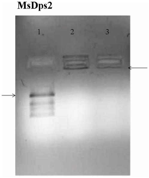
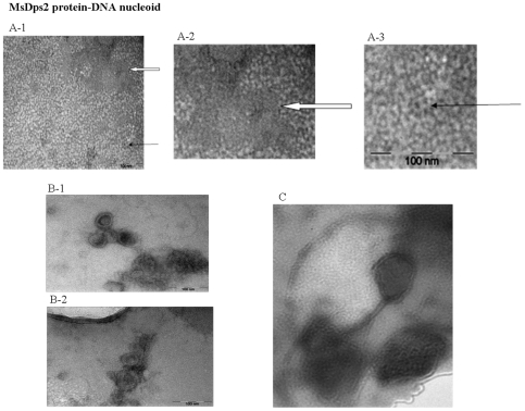
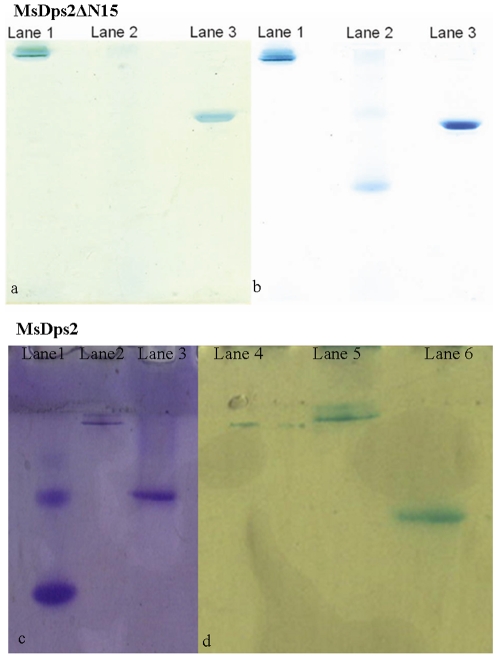
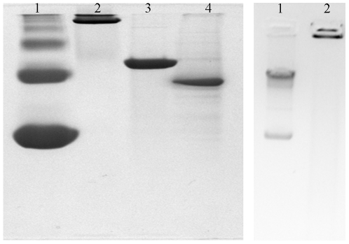
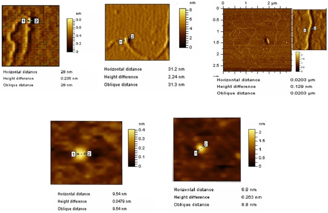
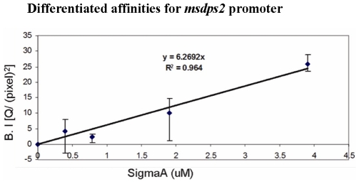
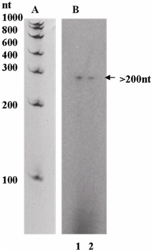
The Dps (DNA-binding protein from starved cells) proteins from Mycobacterium smegmatis MsDps1 and MsDps2 are both DNA-binding proteins with some differences. While MsDps1 has two oligomeric states, with one of them responsible for DNA binding, MsDps2 has only one DNA-binding oligomeric state. Both the proteins however, show iron-binding activity. The MsDps1 protein has been shown previously to be induced under conditions of starvation and osmotic stress and is regulated by the extra cellular sigma factors sigma(H) and sigma(F). We show here, that the second Dps homologue in M. smegmatis, namely MsDps2, is purified in a DNA-bound form and exhibits nucleoid-like structures under the atomic force microscope. It appears that the N-terminal sequence of Dps2 plays a role in nucleoid formation. MsDps2, unlike MsDps1, does not show elevated expression in nutritionally starved or stationary phase conditions; rather its promoter is recognized by RNA polymerase containing sigma(A) or sigma(B), under in vitro conditions. We propose that due to the nucleoid-condensing ability, the expression of MsDps2 is tightly regulated inside the cells.


Conflict of interest statement

Competing Interests: The authors have declared that no competing interests exist.

Figures

Figure 1
Figure 1. MsDps2 is purified as a DNA- bound complex.
MsDps2 protein in 20 mM Tris-HCl (pH 7.9), 200 mM NaCl, purified by DE52 ion-exchange chromatography was checked on a 0.8% agarose gel for bound DNA. Lane 1: free plasmid DNA pGEM; lane 2: DNA+ MsDps2 (1∶104 molar ratio); lane 3: purified MsDps2. The arrows indicate the position of the DNA entered in the agarose gel. Quantification of intensities of the DNA in the wells in Gel Retardation assay was done using Multi-Gauge V 2.3 (Fujifilm) software. The relative intensity/ (pixel) 2 for DNA in samples of MsDps2 with respect to the blank (lane 1) were 12.14 for MsDps2 with DNA (lane 2) versus 8.28 (lane 3) for purified MsDps2 protein alone.
Figure 2
Figure 2. MsDps2 forms protein-DNA nucleoid-like structures under transmission electron microscope. A (1–3) MsDps2 forms ring-like structures and arrays.
Purified MsDps2 protein shows ring-like doughnut-shaped dodecamers under the transmission electron microscope. A single particle is shown with the thin arrow. The protein was also seen to form arrays as shown in the region covered with the solid arrow. B (1–2) MsDps2 protein forms nucleoid-like structures in vitro . Transmission electron microscopic analysis of the MsDps2 protein bound to DNA showed nucleoid-like structures similar to those observed for the E.coli suggesting a role in the condensation and organization of the mycobacterial nucleoid for the MsDps2 protein. C) MsDps2 protein shows tightly compacted stationary phase nucleoid-like structure.
Figure 3
Figure 3. MsDps2ΔN15 has an intact oligomeric structure.
Iron-binding assay with the MsDps2ΔN15 shows that the protein accumulates iron like the full-length MsDps2. a) shows the staining for iron by the Prussian blue method. Lane 1: Ferritin; Lane 2: BSA; Lane 3: MsDps2ΔN15. b) shows the same gel stained with Commassie blue. c) MsDps2 protein is seen to accommodate externally added iron in Fe3+ state as analysed with potassium ferricynaide method. Here spleen ferritin and BSA were used as positive and negative controls, respectively, on a 10% Native PAGE. d) Subsequent to staining for iron the gel was stained with Coomassie blue (left panel). Lane 1: BSA, Lane 2: Ferritin and Lane 3: MsDps2 protein.
Figure 4
Figure 4. Oligomeric status of MsDps2ΔN15 and its DNA bound complex.
A) The N-terminal deleted protein MsDps2ΔN15 is a dodecamer like the full-length protein. Lane 1 and 2 are the markers, BSA and Ferritin respectively, on a 10% native gel; Lane 3 is MsDps2; while lane 4 shows MsDps2ΔN15. B) MsDps2ΔN15 protein in 20 mM Tris-HCl (pH 7.9), 200 mM NaCl, purified by DE52 ion-exchange chromatography was checked on a 0.8% agarose gel for bound DNA.Lane 1: Free plasmid DNA; Lane 2: DNA+ MsDps2NΔ15 (1∶104 molar ratio).
Figure 5
Figure 5. Comparison between the interaction of MsDps2 and MsDps2ΔN15 with DNA by AFM analysis.
A) MsDps2ΔN15 forming a complex with DNA. B) MsDps2 forming a complex with DNA. C) Plasmid DNA. D) MsDps2 (without DNA). E) MsDps2ΔN15 (without DNA).
Figure 6
Figure 6. Standard calibration curve using known molar concentrations of pure σA protein from M. tuberculosis.
The band intensity (B.I) in Y axis represents the relative intensities of the western blots obtained with different concentrations of M. tuberculosis σA. In silico analysis was carried out with the Fujifilm Multi-Gauge software. All experiments were performed in triplicates and the average value was taken.
Figure 7
Figure 7. Single-round heparin-resistant run-off transcription at the msdps2 promoter carried out with M. smegmatis reconstituted holo-RNA polymerases.
A) A 10% polyacrylamide gel containing 6M urea shows an RNA ladder run separately and matched as described before (Chowdhury et al., 2007). B) M. tuberculosis σA and σB were reconstituted with M. smegmatis core RNA polymerase. The intensity of each transcript band as obtained from phosphorimager analysis showed the mRNA transcripts for EσA (lane 1) and EσB (lane 2).
Figure 8
Figure 8. Phylogenetic analysis of mycobacterial MsDps2.
Phylogenetic tree of the Dps proteins reveals that MsDps1 and MsDps2 represent two distinct groups among mycobacterial Dps proteins. M. avium paratuberculosis has one only Dps which falls into the MsDps1 category.

Similar articles

Cited by

References

    1. Matin A, Auger EA, Blum PH, Schultz JE. Genetic basis of starvation survival in non differentiating bacteria. Annu Rev Microbiol. 1989;43:293–316. - PubMed
    1. Frenkiel-Krispin D, Minsky A. Nucleoid organization and the maintenance of DNA integrity in E. coli, B. subtilis and D. radiodurans. J Struct Biol. 2006;156:311–319. - PubMed
    1. Almirón M, Link AJ, Furlong D, Kolter R. A novel DNA-binding protein with regulatory and protective roles in starved Escherichia coli. Genes Dev. 1992;6:2646–2654. - PubMed
    1. Kim J, Yoshimura SH, Hizume K, Ohniwa RL, Ishihama A, et al. Fundamental structural units of the Escherichia coli nucleoid revealed by atomic force microscopy. Nucleic Acids Res. 2004;32:1982–1992. - PMC - PubMed
    1. Gupta S, Pandit SB, Srinivasan N, Chatterji D. Proteomics analysis of carbon-starved Mycobacterium smegmatis: induction of Dps-like protein. Protein Eng. 2002;15:503–512. - PubMed

Publication types