

Immature blood vessels in rheumatoid synovium are selectively depleted in response to anti-TNF therapy
- PMID: 19956574
- PMCID: PMC2779850
- DOI: 10.1371/journal.pone.0008131
Immature blood vessels in rheumatoid synovium are selectively depleted in response to anti-TNF therapy
Abstract
Background: Angiogenesis is considered an important factor in the pathogenesis of Rheumatoid Arthritis (RA) where it has been proposed as a therapeutic target. In other settings, active angiogenesis is characterized by pathologic, immature vessels that lack periendothelial cells. We searched for the presence of immature vessels in RA synovium and analyzed the dynamics of synovial vasculature along the course of the disease, particularly after therapeutic response to TNF antagonists.
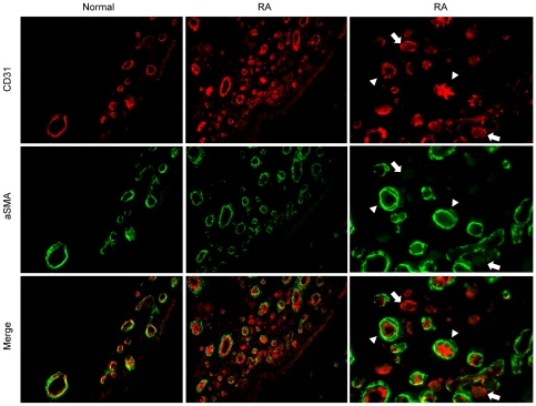
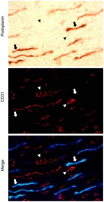
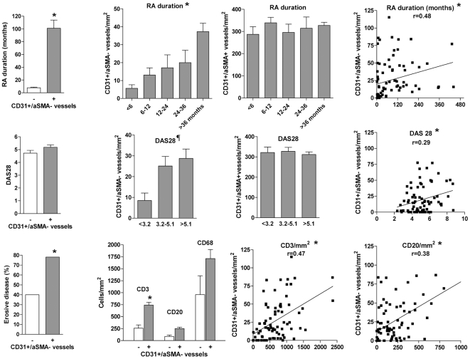
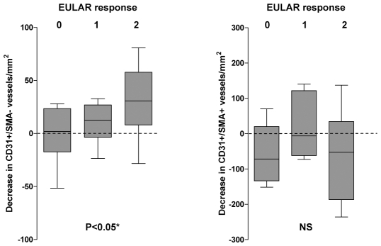
Methodology/principal findings: Synovial arthroscopic biopsies from RA, osteoarthritis (OA) and normal controls were analyzed by double labeling of endothelium and pericytes/smooth muscle mural cells to identify and quantify mature/immature blood vessels. To analyze clinicopathological correlations, a cross-sectional study on 82 synovial biopsies from RA patients with variable disease duration and severity was performed. A longitudinal analysis was performed in 25 patients with active disease rebiopsied after anti-TNF-alpha therapy. We found that most RA synovial tissues contained a significant fraction of immature blood vessels lacking periendothelial coverage, whereas they were rare in OA, and inexistent in normal synovial tissues. Immature vessels were observed from the earliest phases of the disease but their presence or density was significantly increased in patients with longer disease duration, higher activity and severity, and stronger inflammatory cell infiltration. In patients that responded to anti-TNF-alpha therapy, immature vessels were selectively depleted. The mature vasculature was similarly expanded in early or late disease and unchanged by therapy.
Conclusion/significance: RA synovium contains a significant fraction of neoangiogenic, immature blood vessels. Progression of the disease increases the presence and density of immature but not mature vessels and only immature vessels are depleted in response to anti-TNFalpha therapy. The different dynamics of the mature and immature vascular fractions has important implications for the development of anti-angiogenic interventions in RA.
Conflict of interest statement
Figures
Similar articles
-
Clinicopathological correlations of podoplanin (gp38) expression in rheumatoid synovium and its potential contribution to fibroblast platelet crosstalk.PLoS One. 2014 Jun 16;9(6):e99607. doi: 10.1371/journal.pone.0099607. eCollection 2014. PLoS One. 2014. PMID: 24932813 Free PMC article.
-
Clinical significance of synovial lymphoid neogenesis and its reversal after anti-tumour necrosis factor alpha therapy in rheumatoid arthritis.Ann Rheum Dis. 2009 May;68(5):751-6. doi: 10.1136/ard.2008.089284. Epub 2008 May 21. Ann Rheum Dis. 2009. PMID: 18495732
-
The relationship between synovial lymphocyte aggregates and the clinical response to infliximab in rheumatoid arthritis: a prospective study.Arthritis Rheum. 2009 Nov;60(11):3217-24. doi: 10.1002/art.24913. Arthritis Rheum. 2009. PMID: 19877042
-
Targeting synovial neoangiogenesis in rheumatoid arthritis.Autoimmun Rev. 2017 Jun;16(6):594-601. doi: 10.1016/j.autrev.2017.04.005. Epub 2017 Apr 14. Autoimmun Rev. 2017. PMID: 28414154 Review.
-
The vasculature in rheumatoid arthritis: cause or consequence?Int J Exp Pathol. 2009 Jun;90(3):249-61. doi: 10.1111/j.1365-2613.2009.00640.x. Int J Exp Pathol. 2009. PMID: 19563609 Free PMC article. Review.
Cited by
-
Patients with rheumatoid arthritis in clinical remission and ultrasound-defined active synovitis exhibit higher disease activity and increased serum levels of angiogenic biomarkers.Arthritis Res Ther. 2014 Jan 8;16(1):R5. doi: 10.1186/ar4431. Arthritis Res Ther. 2014. PMID: 24398122 Free PMC article.
-
Engagement of toll-like receptor 3 induces vascular endothelial growth factor and interleukin-8 in human rheumatoid synovial fibroblasts.Korean J Intern Med. 2010 Dec;25(4):429-35. doi: 10.3904/kjim.2010.25.4.429. Epub 2010 Nov 27. Korean J Intern Med. 2010. PMID: 21179282 Free PMC article.
-
Nailfold Videocapillaroscopy in Patients with Rheumatoid Arthritis and Psoriatic Arthropathy on ANTI-TNF-ALPHA Therapy.Diagnostics (Basel). 2023 Jun 15;13(12):2079. doi: 10.3390/diagnostics13122079. Diagnostics (Basel). 2023. PMID: 37370974 Free PMC article.
-
Anti-IL-17A treatment reduces serum inflammatory, angiogenic and tissue remodeling biomarkers accompanied by less synovial high endothelial venules in peripheral spondyloarthritis.Sci Rep. 2020 Dec 3;10(1):21094. doi: 10.1038/s41598-020-78204-6. Sci Rep. 2020. PMID: 33273664 Free PMC article.
-
Towards endometriosis diagnosis by gadofosveset-trisodium enhanced magnetic resonance imaging.PLoS One. 2012;7(3):e33241. doi: 10.1371/journal.pone.0033241. Epub 2012 Mar 22. PLoS One. 2012. PMID: 22457748 Free PMC article.
References
-
- Pessler F, Dai L, Diaz-Torne C, Gomez-Vaquero C, Paessler ME, et al. The synovitis of “non-inflammatory” orthopaedic arthropathies: a quantitative histological and immunohistochemical analysis. Ann Rheum Dis. 2008;67:1184–7. - PubMed
-
- Giatromanolaki A, Sivridis E, Athanassou N, Zois E, Thorpe PE, et al. The angiogenic pathway “vascular endothelial growth factor/flk-1(KDR)-receptor” in rheumatoid arthritis and osteoarthritis. J Pathol. 2001;194:101–8. - PubMed
-
- Koch AE, Harlow LA, Haines GK, Amento EP, Unemori EN, et al. Vascular endothelial growth factor: a cytokine modulating endothelial function in rheumatoid arthritis. J Immunol. 1994;152:4149–56. - PubMed
