. 2009 Dec 2;4(12):e8122.
doi: 10.1371/journal.pone.0008122.

Adult mouse subventricular zone stem and progenitor cells are sessile and epidermal growth factor receptor negatively regulates neuroblast migration

Affiliations

Adult mouse subventricular zone stem and progenitor cells are sessile and epidermal growth factor receptor negatively regulates neuroblast migration

Yongsoo Kim et al. PLoS One. .

Abstract

Background: The adult subventricular zone (SVZ) contains stem and progenitor cells that generate neuroblasts throughout life. Although it is well accepted that SVZ neuroblasts are migratory, recent evidence suggests their progenitor cells may also exhibit motility. Since stem and progenitor cells are proliferative and multipotential, if they were also able to move would have important implications for SVZ neurogenesis and its potential for repair.

Methodology/principal findings: We studied whether SVZ stem and/or progenitor cells are motile in transgenic GFP+ slices with two photon time lapse microscopy and post hoc immunohistochemistry. We found that stem and progenitor cells; mGFAP-GFP+ cells, bright nestin-GFP+ cells and Mash1+ cells were stationary in the SVZ and rostral migratory stream (RMS). In our search for motile progenitor cells, we uncovered a population of motile betaIII-tubulin+ neuroblasts that expressed low levels of epidermal growth factor receptor (EGFr). This was intriguing since EGFr drives proliferation in the SVZ and affects migration in other systems. Thus we examined the potential role of EGFr in modulating SVZ migration. Interestingly, EGFr(low) neuroblasts moved slower and in more tortuous patterns than EGFr-negative neuroblasts. We next questioned whether EGFr stimulation affects SVZ cell migration by imaging Gad65-GFP+ neuroblasts in the presence of transforming growth factor alpha (TGF-alpha), an EGFr-selective agonist. Indeed, acute exposure to TGF-alpha decreased the percentage of motile cells by approximately 40%.

Conclusions/significance: In summary, the present study directly shows that SVZ stem and progenitor cells are static, that EGFr is retained on some neuroblasts, and that EGFr stimulation negatively regulates migration. This result suggests an additional role for EGFr signaling in the SVZ.


Conflict of interest statement

Competing Interests: The authors have declared that no competing interests exist.

Figures

Figure 1
Figure 1. Subventricular zone cell types selectively labeled with GFP.
A: Stem cells in the SVZ express GFAP and give rise to EGFr+ and Mash1+ transit-amplifying progenitors. These, in turn generate migratory neuroblasts that express βIII-tubulin, Dcx, and PSA-NCAM. Note that the expression and loss of some markers, such as EGFr is gradual. B: Model of a typical SVZ neuroblast chain (red) with cluster of transit-amplifying progenitors (purple) and GFAP+ astrocytes (blue) surrounding it. For the sake of clarity only a few progenitors (top of chain) and GFAP+ cells (bottom of chain) are shown. mGFAP-GFP mice only label GFAP+ cells. Nestin-GFP labels a subset of all three cell types and Dcx-GFP labels all and only neuroblasts. The CSH-nestin-GFP line labels stem cells and transit-amplifying progenitors GFPbright and neuroblasts GFPdim. Unexpectedly, the Mash1-GFP mouse labels not only the transit-amplifying progenitors but also neuroblasts. The Gad65-GFP mouse labels a subset of neuroblasts. Adapted from .
Figure 2
Figure 2. Stem and progenitor cells are stationary in the SVZ.
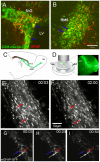
A–B: CSH-nestin-GFP showed bright GFP+ cells colocalized with GFAP immunohistochemistry (blue arrows) in the SVZ (A) and the RMS (B), (coronal sections). Scale bar = 50 µm. C: Location of two photon imaging in the SVZ and RMS of sagittal slices. LV = lateral ventricle, OB = olfactory bulb. D: Schematic of two photon imaging and a 5X image of the RMS in a CSH-nestin-GFP mouse. E-F: Bright CSH-nestin-GFP+ cells (ex. red arrows) showed no local movement in the RMS in two photon time lapse imaging. Time stamp is in hr:min in all figures and movies. G–I: All mGFAP-GFP+ cells in the movie were stationary during imaging. Examples of individual cells are indicated with arrows. Scale bar = 30 µm.
Figure 3
Figure 3. Mash1+ progenitor cells are not motile in the RMS.
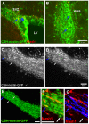
A–B: Many bright cells from CSH-nestin-GFP slices were colocalized with Mash1 immunohistochemistry (blue arrows) in the SVZ (A) and the RMS (B). Scale bar = 50 µm. C–D: Motility of CSH-nestin-GFP positive cells was followed with two photon imaging. Most bright cells were stationary (ex. white arrows). Blue arrow: example of a cell that was followed with post hoc immunohistochemistry (E–G). Scale bar = 50 µm. E–G: the two photon imaged area was found with confocal microscopy. Arrows indicate cells matched with the last frame of two photon imaging. Mash1+ (F) and Dcx-negative (G) cell shown with blue arrow. Scale bars = 50 µm.
Figure 4
Figure 4. A subset of neuroblasts express EGFr.
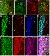
A: βIII-tubulin and EGFr double immunohistochemistry in a coronal section through the RMS. Many EGFrhigh cells were βIII-tubulin-negative (white arrows). A1–A3 shows high magnification of inset in A. Note EGFrhigh cell that is βIII-tubulin-negative (white arrow). Yellow arrow shows a cell that expressed both EGFr and βIII-tubulin. Cells that expressed the highest levels of βIII-tubulin+ (white arrowhead) had EGFr immunofluorescence similar to background levels. Scale bars = 10 µm. (Please see Movie S3). B–E: Dcx (B), EGFr (C), and PSA-NCAM (D) triple immunohistochemistry in the RMS. Simlar colocalization of EGFr with neuroblasts is seen as in A. White arrows point to EGFrhigh cells that are negative for Dcx and PSA-NCAM. Yellow arrows point to cells that express immunodetectable levels of all three markers. White arrowheads point to neuroblasts that had EGFr immunofluorescence similar to background levels. F–I: Confocal microscopy shows near perfect overlap between βIII-tubulin, Dcx, and PSA-NCAM in RMS neuroblasts (coronal section).
Figure 5
Figure 5. EGFr expression is correlated with differences in motility.
A–C: Two photon time lapse imaging of nestin-GFP cells in the RMS. Blue and red arrows indicate migratory and exploratory cells, respectively. Scale bar = 50 µm. (Please see corresponding Movie S4.). D: Confocal image corresponding to two photon image shown in C. Scale bar = 50 µm. E: After fixation and EGFr immunohistochemistry, confocal microscopy was used to find individual cells imaged with two photon microscopy (same cell as in A–C shown with red arrow) Scale bar = 50 µm. E1: high magnification 3-D confocal microscopy showing EGFrlow expression (red) on cell exhibiting exploratory motility. Scale bar = 20 µm. F: 3D view of motile cell trajectory. The blue and red trajectories indicate the migratory EGFr negative and exploratory EGFrlow cells shown in A–E. 1 unit = 42.9 µm. (Please see corresponding Movie S5.). G: Cell movement distances between frames (3 min apart) of the EGFr-negative (blue) and EGFrlow (red) nestin-GFP+ cells shown in A–F. EGFr negative cells were significantly faster than EGFrlow exploratory cells.
Figure 6
Figure 6. TGF-α decreased the percentage of motile cells.
A–D: Gad65-GFP labels a subset of βIII-tubulin+ neuroblasts (arrowheads), but not EGFrhigh progenitor cells (arrows). E: Low magnification view of Gad65-GFP sagittal section showing area analyzed. F: First frame of two photon time lapse imaging. Each distinguishable cell was labeled (yellow numbers) and analyzed to determine cell motility. Corresponds to first frame of Movie S6 (pretreatment). G: TGF-α schedule and analyzed segments. H: Percentage of motile cells before and after TGF-α treatment. TGF-α caused significant decreases compared to pretreatment and aCSF. **P<0.01. I: Percentage decrease after TGF-α compared to pre-treatment. *P<0.05.

Similar articles

Cited by

References

    1. Alvarez-Buylla A, Garcia-Verdugo JM. Neurogenesis in adult subventricular zone. J Neurosci. 2002;22:629–634. - PMC - PubMed
    1. Doetsch F, Garcia-Verdugo JM, Alvarez-Buylla A. Cellular composition and three-dimensional organization of the subventricular germinal zone in the adult mammalian brain. J Neurosci. 1997;17:5046–5061. - PMC - PubMed
    1. Pastrana E, Cheng LC, Doetsch F. Simultaneous prospective purification of adult subventricular zone neural stem cells and their progeny. Proc Natl Acad Sci U S A 2009 - PMC - PubMed
    1. Doetsch F, Caille I, Lim DA, Garcia-Verdugo JM, Alvarez-Buylla A. Subventricular zone astrocytes are neural stem cells in the adult mammalian brain. Cell. 1999;97:703–716. - PubMed
    1. Garcia AD, Doan NB, Imura T, Bush TG, Sofroniew MV. GFAP-expressing progenitors are the principal source of constitutive neurogenesis in adult mouse forebrain. Nat Neurosci. 2004;7:1233–1241. - PubMed

Publication types

Substances