. 2009 Nov 30;4(11):e8058.
doi: 10.1371/journal.pone.0008058.

Fetuin-A/albumin-mineral complexes resembling serum calcium granules and putative nanobacteria: demonstration of a dual inhibition-seeding concept

Affiliations

Fetuin-A/albumin-mineral complexes resembling serum calcium granules and putative nanobacteria: demonstration of a dual inhibition-seeding concept

Cheng-Yeu Wu et al. PLoS One. .

Abstract

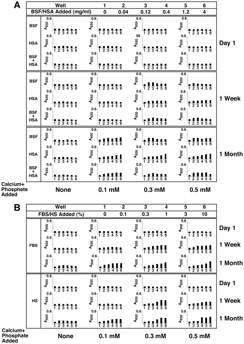
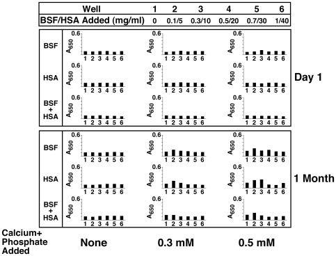
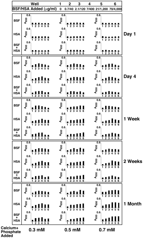
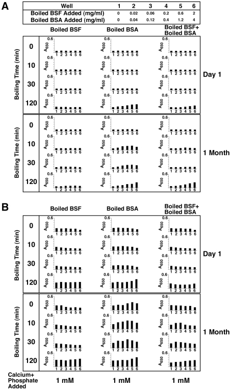
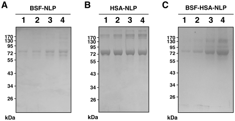
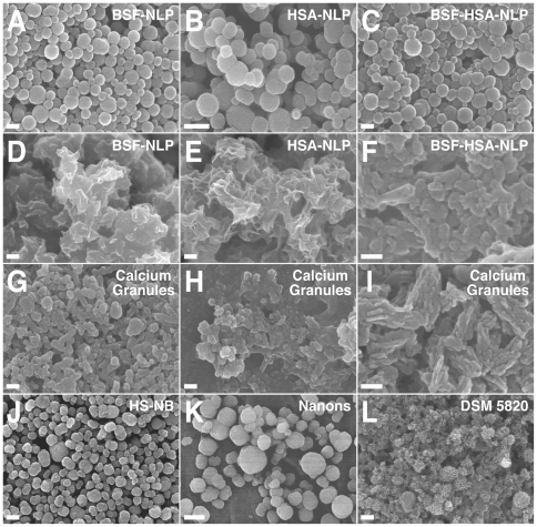
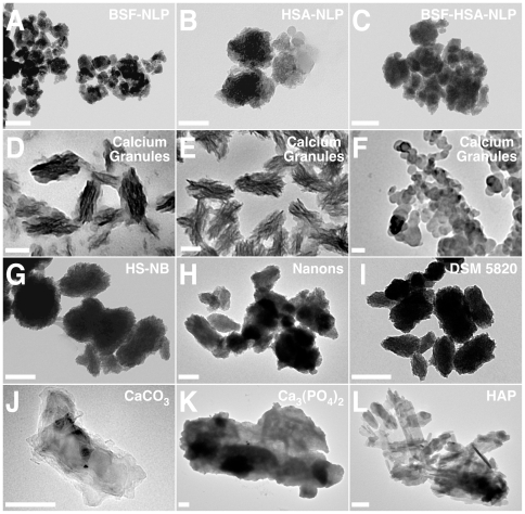
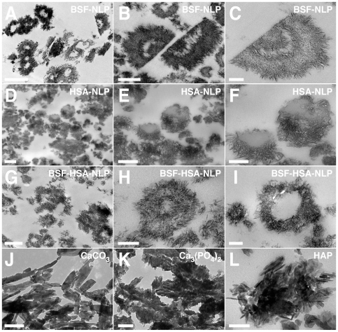
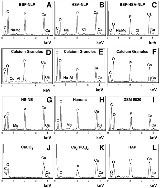
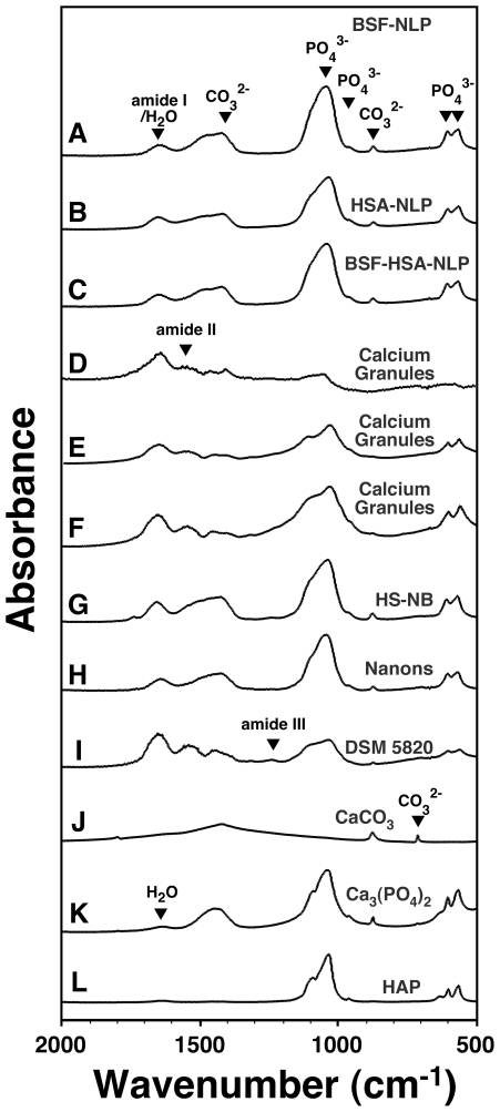
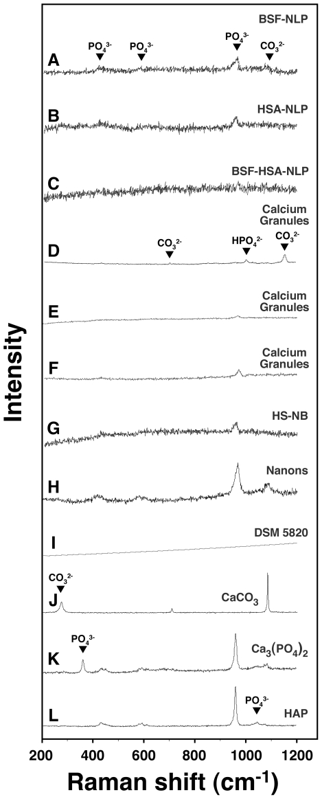
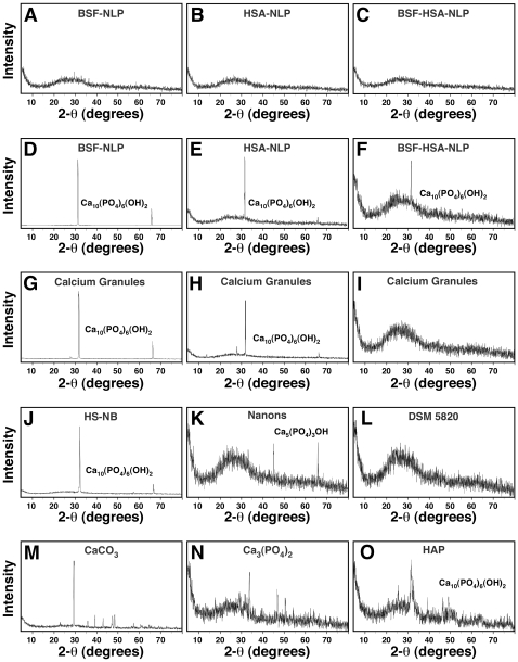
Serum-derived granulations and purported nanobacteria (NB) are pleomorphic apatite structures shown to resemble calcium granules widely distributed in nature. They appear to be assembled through a dual inhibitory-seeding mechanism involving proteinaceous factors, as determined by protease (trypsin and chymotrypsin) and heat inactivation studies. When inoculated into cell culture medium, the purified proteins fetuin-A and albumin fail to induce mineralization, but they will readily combine with exogenously added calcium and phosphate, even in submillimolar amounts, to form complexes that will undergo morphological transitions from nanoparticles to spindles, films, and aggregates. As a mineralization inhibitor, fetuin-A is much more potent than albumin, and it will only seed particles at higher mineral-to-protein concentrations. Both proteins display a bell-shaped, dose-dependent relationship, indicative of the same dual inhibitory-seeding mechanism seen with whole serum. As ascertained by both seeding experiments and gel electrophoresis, fetuin-A is not only more dominant but it appears to compete avidly for nanoparticle binding at the expense of albumin. The nanoparticles formed in the presence of fetuin-A are smaller than their albumin counterparts, and they have a greater tendency to display a multi-layered ring morphology. In comparison, the particles seeded by albumin appear mostly incomplete, with single walls. Chemically, spectroscopically, and morphologically, the protein-mineral particles resemble closely serum granules and NB. These particles are thus seen to undergo an amorphous to crystalline transformation, the kinetics and completeness of which depend on the protein-to-mineral ratios, with low ratios favoring faster conversion to crystals. Our results point to a dual inhibitory-seeding, de-repression model for the assembly of particles in supersaturated solutions like serum. The presence of proteins and other inhibitory factors tend to block apatite nuclei formation or to stabilize the nascent nuclei as amorphous or semi-crystalline spherical nanoparticles, until the same inhibitory influences are overwhelmed or de-repressed, whereby the apatite nuclei grow in size to coalesce into crystalline spindles and films-a mechanism that may explain not only the formation of calcium granules in nature but also normal or ectopic calcification in the body.


Conflict of interest statement

Competing Interests: The authors have declared that no competing interests exist.

Figures

Figure 1
Figure 1. Slow formation of NB-like particles by serum inoculated into DMEM.
(A) Formation of NB-like particles by FBS. FBS was inoculated into DMEM to the indicated % by volume. Visual inspection and A650 turbidity reading (insets) were performed within 1 hour following inoculation (top panel, “Day 1”) as well as after incubation at 37°C in cell culture conditions for “1 Month” (middle panel) or “2 Months” (bottom panel). Notice the gradual increase in turbidity observed with time as well as the bell-shaped increase in turbidity as a function of the amount of serum inoculated, with maximum turbidity reached at 0.3% FBS, while higher concentrations of FBS resulted in increasingly lower turbidities. (B) Formation of NB-like particles by HS, performed as described in (A), except that higher serum concentrations varying from 1 to 10% were used. Notice the bell-shaped increase in turbidity, with peak reached at 1-to-3% HS at “1 Month” and 3% at “2 Months.”
Figure 2
Figure 2. Formation of NB-like particles from protease-treated serum.
(A) NB-like particles cultured from protease-treated FBS. Stock solutions of 5% (w/v) trypsin or chymotrypsin were diluted into FBS at the final concentration of 0.5% (v/v), followed by incubation at 37°C for 2 hours. After incubation, the two protease-treated FBS solutions were inoculated into DMEM (columns labeled as “Trypsin” or “Chymotrypsin”) to the concentrations indicated on the top heading, starting from 0% (e.g. untreated, well 1) up to 10% (well 6). FBS was also treated with trypsin or chymotrypsin that had been inactivated by heat at 95°C for 1 hour (“Boiled Trypsin” or “Boiled Chymotrypsin”). For each treatment, A650 turbidity readings were performed within 1 hour following inoculation (“Day 1”) and after incubation in cell culture conditions for “1 Week” or “1 Month”, as indicated on the right. Untreated FBS (“None” column) or FBS that was treated with either boiled trypsin or boiled chymotrypsin produced a low bell-shaped curve of turbidity increase after 1 month of incubation. In contrast, inoculation of FBS that was treated with trypsin or chymotrypsin resulted in a more prominent straight dose-dependent turbidity increase after 1 month of incubation. (B) NB-like particles cultured from protease-treated HS were obtained as in (A). Notice the straight dose-dependent increase in turbidity, noticeable at “1 Month,” while both untreated HS and pre-boiled HS produced a low bell-shaped turbidity response. (C) Control experiment, performed with both untreated and boiled proteases inoculated directly into DMEM at the indicated % (w/v). No increase in turbidity was noticed for these control treatments following incubation.
Figure 3
Figure 3. Formation of NB-like particles from boiled serum in metastable versus supersaturated medium.
(A) Metastable medium: FBS and 25% HS were boiled at 95°C for the time indicated on the left panel (0, 10, 30, and 120 min). “0 min” refers to control, untreated serum. The boiled sera were then inoculated into DMEM to the concentrations shown on the top heading. (B) Supersaturated medium: both FBS and HS were treated as described in (A), except that 1 mM each of CaCl2 and NaH2PO4 (labeled as “Calcium+Phosphate Added”) was added following the inoculation of the boiled serum into DMEM. Note the marked differences in turbidity changes between the various panels shown in (A) versus (B). See the text for explanation and interpretation.
Figure 4
Figure 4. Seeding of NB-like particles by fetuin-A, albumin, and serum in metastable versus supersaturated medium.
(A) Fetuin-A and albumin in both metastable and supersaturated media: bovine serum fetuin-A (BSF) and human serum albumin (HSA) were diluted into DMEM, separately or together, to the final concentrations indicated on the top. Where indicated, CaCl2 and NaH2PO4 were added to the final concentrations shown at the bottom (labeled as “Calcium+Phosphate Added”), followed by incubation in cell culture conditions for “1 Week” or “1 Month”, as indicated on the right of each panel. “None” refers to the absence of any exogenously added calcium and phosphate, that is, to metastable medium DMEM. Seeding of the proteins alone without additional ion input did not produce any turbidity change even following incubation for 1 month (“None” column). Low amounts of precipitation were noticed when precipitating ions were added (supersaturated solutions) that increased gradually over time through either bell-shaped (BSF, BSF and HSA) or straight, dose-dependent (HSA) relationships. (B) Serum in supersaturated medium: FBS (top panel) or HS (lower panel) were added to the final concentrations indicated on the top, followed by addition of precipitating reagents to the concentrations shown at the bottom. Addition of precipitating reagents at either 0.1, 0.3, or 0.5 mM into DMEM containing FBS or HS produced turbidities which increased steadily as seen after “1 Week” either through bell-shaped (0.1 mM calcium and phosphate) or straight, dose-dependent (0.3 and 0.5 mM calcium and phosphate) relationships. After further incubation, all treatments showed straight dose-dependent turbidities that continued to increase in a slow and gradual manner (“1 Month”).
Figure 5
Figure 5. Effect of high concentrations of fetuin-A and albumin on the formation of NB-like particles in metastable versus supersaturated medium.
Stock solutions of BSF and/or HSA were diluted into DMEM to the concentrations indicated on the top, so as to mimick the concentration range of these proteins seen in HS. When indicated, the precipitating reagents CaCl2 and NaH2PO4 were added to the concentrations shown at the bottom (labeled as “Calcium+Phosphate Added”). A650 turbidity readings were performed following inoculation (“Day 1”) and after incubation in cell culture conditions for “1 Month”, as indicated on the right. Seeding of the proteins alone without additional precipitating reagents did not produce any turbidity increase even following incubation for 1 month (“None” column). Notice the bell-shaped turbidity increases seen with 0.3 mM and 0.5 mM calcium and phosphate columns, observed at “1 Month,” but not “Day 1.”
Figure 6
Figure 6. Seeding of NB-like particles by fetuin-A and albumin at low concentrations in supersaturated ionic solutions.
Stock solutions of BSF and/or HSA were diluted into DMEM to the relatively low concentrations shown on the top. The precipitating reagents CaCl2 and NaH2PO4 were then added at either 0.3, 0.5, or 0.7 mM, as indicated at the bottom (labeled as “Calcium+Phosphate Added”). Turbidity was monitored by A650 turbidity reading following inoculation (“Day 1”) and after incubation in cell culture conditions for the time indicated on the right side of each panel. Bell-shaped curves of precipitation were noticed at “Day 4” for the three different combinations of proteins as well as the three different concentrations of added precipitating reagents. Precipitation in these conditions increased in a time-dependent manner with further incubation. When 0.3 mM of precipitating reagents was used, bell-shaped increases in turbidity were seen for the three protein combinations after 1 month of incubation. With calcium and phosphate added to 0.5 and 0.7 mM, the initial bell-shaped increase in turbidity observed at “Day 4” was seen to shift to the right at “1 Month” reading.
Figure 7
Figure 7. Seeding of NB-like particles by boiled fetuin-A and albumin in metastable versus supersaturated medium.
(A) Metastable medium: BSF and bovine serum albumin (BSA) were boiled for the time indicated on the left side. The boiled protein solutions were then diluted into DMEM, separately or together, to the final concentrations indicated on the top. A650 was then monitored following inoculation (“Day 1”) and after incubation in cell culture conditions for 1 month, as indicated on the right. Inoculation of either untreated BSF or BSF that had been boiled for either 10, 30, or 120 min did not result in any turbidity changes after 1 month of incubation. In contrast, pre-boiled BSA produced either bell-shaped or linear turbidity changes depending on the amount of boiling time. When added together, pre-boiled BSF and BSA produced additive effects. (B) Supersaturated medium: BSF and BSA were added to medium as in (A) that was also inoculated with 1 mM each of CaCl2 and NaH2PO4. Notice the various patterns of turbidity changes associated with the three protein combinations. See the text for further details.
Figure 8
Figure 8. Differences in the binding affinities between fetuin-A and albumin toward NB-like particles as revealed by SDS-PAGE.
NB-like particles were prepared from 3 mM each of CaCl2 and NaH2PO4 in 1 ml of DMEM containing either BSF, HSA, or both proteins at the following concentrations: 20 µg/ml, 40 µg/ml, 80 µg/ml, or 160 µg/ml of BSF, corresponding respectively to lanes 1–4 in (A); 0.2 mg/ml, 0.4 mg/ml, 0.8 mg/ml, and 1.6 mg/ml of HSA, respectively, lanes 1–4 in (B); or both proteins at these same concentrations for lanes 1–4 in (C), respectively. Following incubation in cell culture conditions for 1 month, the NB-like particles were pelleted by centrifugation, washed twice in HEPES buffer, resuspended in 50 µl of 50 mM EDTA, and processed for SDS-PAGE as described in the Materials and Methods . In the case of BSF-NLP shown in (A), fetuin-A appeared as a major band slightly above 72 kDa (lane 1), with additional bands of higher and lower molecular weights noticeable at higher protein concentrations (lanes 2 to 4). In HSA-NLP (B), albumin formed a major band of strong intensity at 72 kDa. Note a higher molecular band above the 170 kDa marker that appears to increase steadily from lanes 1 through 4, while the 72 kDa band in the 4 lanes appears to decrease slightly in intensity from left to right. In the case of BSF-HSA-NLP (C), note the increase in the intensity of bands at 72 kDa and at a higher molecular weight. The bands of higher molecular weights may be due to altered migration or aggregation of the purified proteins used.
Figure 9
Figure 9. Protein-mineral nanoparticles seeded by fetuin-A and albumin show morphological resemblance to NB and calcium granules when observed by SEM.
Protein-mineral nanoparticles were prepared by adding 0.3 mM of CaCl2 and NaH2PO4 to DMEM containing BSF at 2.1 µg/ml (A and D, labeled as “BSF-NLP”), HSA at 120 µg/ml (B and E, “HSA-NLP”), or both BSF and HSA at these same concentrations (C and F, “BSF-HSA-NLP”), followed by incubation for either 3 days (A–C) or 1 month (D–F) in cell culture conditions and preparation of the particles for TEM, as described in the Materials and Methods . Protein-mineral nanoparticles containing BSF and/or HSA showed a round morphology when observed after 3 days (A–C), but they tended to coalesce to form films or aggregates when incubated for a longer period of 1 month (D–F). Note the presence of structures resembling cells undergoing cell division in (B). Calcium granules were prepared from the addition of either CaCl2 (G), NaH2PO4 (H), or a combination of both (I) to FBS, followed by overnight incubation, centrifugation, and the washing steps described in the Materials and Methods . Calcium granules showed variable morphologies, consisting of either round particles (G) or film/aggregate-like structures (H and I). The morphologies of both the protein-mineral nanoparticles and the calcium granules were similar to NB obtained from 10% HS (J, “HS-NB”) or the NB strains “Nanons” (K) and “DSM 5820” (L) which were both maintained in 10% FBS. These NB samples revealed either round particles (J and K) or more crystalline structures harboring elongated crystal projections and aggregates (L). Scale bars: 100 nm (F, I); 200 nm (A, C–E, G, H, J, L); 400 nm (B); 1 µm (K).
Figure 10
Figure 10. Protein-mineral nanoparticles containing fetuin-A and albumin resemble NB and calcium granules as revealed by TEM.
Protein-mineral nanoparticles were prepared as described in Fig. 9, from solutions containing either BSF (A), HSA (B), or both proteins (C), followed by the addition of 0.3 mM each of CaCl2 and NaH2PO4, and incubation for 1 month in cell culture conditions. The samples were then prepared for TEM without fixation or staining. The various protein-seeded particles showed small, rounded formations that tended to aggregate (A–C). The samples of calcium granules prepared from addition of either CaCl2 (D), NaH2PO4 (E), or a combination of both (F) to FBS (see Materials and Methods ) showed stacks of crystalline spindles and stacks forming ellipsoid structures (D and E), while other granules appeared mostly as round aggregated particles (F). The morphologies of the protein-mineral nanoparticles and the calcium granules were similar to NB obtained from either 10% HS (G, “HS-NB”) or the two NB strains “Nanons” (H) and “DSM 5820” (I) which were both obtained from 10% FBS. In this case, the NB samples consisted of rounded particles with rough surfaces and variable sizes (G–I). Commercially available CaCO3 (J), Ca3(PO4)2 (K), and HAP (L) incubated in DMEM for 1 hour showed mainly large, crystalline, monolithic structures. Scale bars: 50 nm (A, F); 100 nm (B, D, E, G–J, L); 200 nm (C, K).
Figure 11
Figure 11. Thin-sections of protein-mineral nanoparticles seeded by fetuin-A or albumin show distinct morphologies.
Protein-mineral nanoparticles were prepared as described in Fig. 6, by diluting either BSF at 2.1 µg/ml (A–C), HSA at 120 µg/ml (D–F), or both proteins at these same concentrations (G–I) into DMEM and then adding 0.3 mM each of CaCl2 and NaH2PO4, followed by incubation in cell culture conditions for 1 month. Thin-sections were prepared without fixation or staining, as described in the Materials and Methods . BSF-NLP (A–C) resembled multi-layered laminations with alternate electron densities. HSA-NLP (D–F) appeared mostly as incompletely sealed, single-layered formations. The BSF-HSA-NLP particles had rough surfaces covered with elongated crystal projections (G), with some structures appearing either as multi-layered (H) or single-layered (I). Control, commercial grades of CaCO3 (J), Ca3(PO4)2 (K), and HAP (L) were incubated in DMEM as in Fig. 10. These controls showed mainly monolithic platelets or aggregates of crystalline formations that contrasted with the round nanoparticles shown above. Scale bars: 100 nm (H, I, L); 200 nm (C, K); 250 nm (F); 500 nm (B, D, E, G, J); 1 µm (A).
Figure 12
Figure 12. Energy-dispersive X-ray spectroscopy of protein-mineral nanoparticles shows elemental compositions indistinguishable from those of calcium granules and NB.
Protein-mineral nanoparticles were obtained as in Fig. 9, from solutions containing BSF (A), HSA (B), or both (C), to which 0.3 mM each of CaCl2 and NaH2PO4 was added, followed by incubation in cell culture conditions for 1 month. EDX spectra were also obtained for calcium granules prepared by adding either CaCl2 (D), NaH2PO4 (E), or a combination of both (F) to FBS, followed by overnight incubation, centrifugation, and washing, as described in the Materials and Methods . NB were cultured from 10% HS (G, “HS-NB”) or from 10% FBS (H and I, corresponding to “Nanons” and “DSM 5820”, respectively). In these specimens, major peaks of carbon, oxygen, calcium, and phosphorus were noted, concordant with the presence of a calcium phosphate mineral containing carbonate. The three controls CaCO3 (J), Ca3(PO4)2 (K), and HAP (L), diluted and washed in double-distilled water, were shown for comparison. The following Ca/P ratios were obtained: (A) 1.37; (B) 1.53; (C) 1.6; (D) 1.32; (E) 1.48; (F) 1.14; (G) 1.48; (H) 1.4; (I) 1.27; (K) 2.54; and (L) 1.48. Phosphorus was not detected in the CaCO3 samples shown in (J).
Figure 13
Figure 13. Fourier-transformed infrared spectroscopy of protein-mineral nanoparticles reveals the presence of both carbonate and phosphate.
Protein-mineral nanoparticles were obtained as described in Fig. 9, by diluting BSF (A), HSA (B), or both proteins (C) into DMEM, then adding 0.3 mM each of CaCl2 and NaH2PO4, and incubating the solutions in cell culture conditions for 1 month. Calcium granules were prepared by adding either CaCl2 (D) or NaH2PO4 (E) into FBS, or a combination of both CaCl2 and NaH2PO4 (F) into HS, as described in the Materials and Methods . NB were cultured from 10% HS (G, “HS-NB”) or 10% FBS (H and I, corresponding to “Nanons” and “DSM 5820”, respectively). The FTIR spectra of the protein-mineral nanoparticles revealed peaks similar to both calcium granules and NB as shown by the presence of phosphate peaks at 575 cm−1, 605 cm−1, 960 cm−1, and 1,000–1,150 cm−1 as well as carbonate peaks at 875 cm−1 and 1,400–1,430 cm−1. Some of the peaks corresponding to phosphate or carbonate were not detected in a few calcium granule samples such as the one shown in (D). In the various nanoparticle samples presented here, peaks corresponding to amide I, II, and III at 1,660 cm−1, 1,550 cm−1, and 1,250 cm−1, respectively, were observed and were attributed to the presence of serum proteins. Spectra for the controls CaCO3 (J), Ca3(PO4)2 (K), and HAP (L), diluted and washed in double-distilled water, were included as controls. Residual water (H2O) was observed at 1,650 cm−1 in some controls prepared in the absence of proteins (K and L); this peak could also have contributed to the intensity of the amide I peak seen at 1,660 cm−1 in the other samples shown.
Figure 14
Figure 14. Micro-Raman spectroscopy of protein-mineral nanoparticles shows chemical compositions similar to those of calcium granules and NB.
Protein-mineral nanoparticles were prepared as in Fig. 9, by adding 0.3 mM each of CaCl2 and NaH2PO4 to DMEM containing BSF (A), HSA (B), or both proteins (C), followed by incubation in cell culture conditions for 1 month and processing for micro-Raman spectroscopy. Calcium granules were obtained by adding CaCl2 (D), NaH2PO4 (E), or both (F) to FBS, followed by overnight incubation and preparation for micro-Raman spectroscopy as described in the Materials and Methods . Micro-Raman spectra were also acquired for NB that were initially cultured from 10% HS (G, “HS-NB”) or 10% FBS (H and I, “Nanons” and “DSM 5820”, respectively). These nanoparticle samples showed phosphate groups at 361 cm−1, 440 cm−1, 581 cm−1, 962 cm−1, 1,002 cm−1 (HPO4 2−), and 1,048 cm−1 and carbonate moieties at 280 cm−1, 712 cm−1, 1,080 cm−1, and 1,150 cm−1. The protein-mineral nanoparticles mainly showed peaks of phosphate and lower peaks of carbonate (A–C) while the calcium granules (D–F) and NB (G–I) samples showed carbonate and phosphate peaks of variable intensities. The three controls CaCO3 (J), Ca3(PO4)2 (K), and HAP (L), diluted and washed in double-distilled water, were included for comparison.
Figure 15
Figure 15. Powder X-ray diffraction spectra of protein-mineral nanoparticles reveal both amorphous and crystalline patterns.
Protein-mineral nanoparticles were obtained as described in Fig. 9, by diluting the proteins, separately (A and B) or together (C) into DMEM, followed by addition of the precipitating reagents CaCl2 and NaH2PO4 each to a final concentration of 0.3 mM and incubating the solutions in cell culture conditions for 1 month. The XRD spectra obtained for the protein-mineral nanoparticles obtained after 1 week of incubation represented mainly amorphous patterns as seen by the absence of diffraction peaks (A–C). Longer incubation of 1 month produced protein-mineral nanoparticles with crystalline peaks corresponding to HAP crystals (D–F, Ca10(PO4)6(OH)2). XRD spectra were also obtained for calcium granules that were prepared by adding either CaCl2 (G) or NaH2PO4 (H) into FBS or both CaCl2 and NaH2PO4 into HS (I), followed by sample preparation as described in the Materials and Methods . Peaks corresponding to Ca10(PO4)6(OH)2 were obtained for both calcium granules prepared in FBS (G and H) while the ones prepared in HS usually gave amorphous patterns (I). XRD spectra showing the presence of HAP crystals (J), a calcium phosphate compound (K, Ca5(PO4)3OH), or an amorphous pattern (L) were also acquired for NB cultured in 10% HS for 1 month (J, “HS-NB”) or in 10% FBS for 1 month (K, “Nanons”) or 1 week (L, “DSM 5820”). Commercial grades of CaCO3 (M), Ca3(PO4)2 (N), and HAP (O), used as controls, were diluted and washed in double-distilled water.

Similar articles

Cited by

References

    1. Martel J, Young JD. Purported nanobacteria in human blood as calcium carbonate nanoparticles. Proc Natl Acad Sci U S A. 2008;105:5549–5554. - PMC - PubMed
    1. Young JD, Martel J, Young L, Wu CY, Young A, et al. Putative nanobacteria represent physiological remnants and culture by-products of normal calcium homeostasis. PLoS One. 2009;4:e4417. - PMC - PubMed
    1. Young JD, Martel J, Young D, Young A, Hung CM, et al. Characterization of granulations of calcium and apatite in serum as pleomorphic mineralo-protein complexes and as precursors of putative nanobacteria. PLoS One. 2009;4:e5421. - PMC - PubMed
    1. Kajander EO, Kuronen I, Akerman K, Pelttari A, Ciftcioglu N. Nanobacteria from blood, the smallest culturable autonomously replicating agent on Earth. Proc Soc Photo Opt Instrum Eng. 1997;3111:420–428.
    1. Kajander EO, Ciftcioglu N. Nanobacteria: an alternative mechanism for pathogenic intra- and extracellular calcification and stone formation. Proc Natl Acad Sci U S A. 1998;95:8274–8279. - PMC - PubMed

Publication types