. 2009 Dec 1;4(12):e8104.
doi: 10.1371/journal.pone.0008104.

The E3 ubiquitin-ligase Bmi1/Ring1A controls the proteasomal degradation of Top2alpha cleavage complex - a potentially new drug target

Affiliations

The E3 ubiquitin-ligase Bmi1/Ring1A controls the proteasomal degradation of Top2alpha cleavage complex - a potentially new drug target

Iris Alchanati et al. PLoS One. .

Abstract

Background: The topoisomerases Top1, Top2alpha and Top2beta are important molecular targets for antitumor drugs, which specifically poison Top1 or Top2 isomers. While it was previously demonstrated that poisoned Top1 and Top2beta are subject to proteasomal degradation, this phenomena was not demonstrated for Top2alpha.

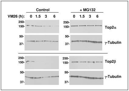
Methodology/principal findings: We show here that Top2alpha is subject to drug induced proteasomal degradation as well, although at a lower rate than Top2beta. Using an siRNA screen we identified Bmi1 and Ring1A as subunits of an E3 ubiquitin ligase involved in this process. We show that silencing of Bmi1 inhibits drug-induced Top2alpha degradation, increases the persistence of Top2alpha-DNA cleavage complex, and increases Top2 drug efficacy. The Bmi1/Ring1A ligase ubiquitinates Top2alpha in-vitro and cellular overexpression of Bmi1 increases drug induced Top2alpha ubiquitination. A small-molecular weight compound, identified in a screen for inhibitors of Bmi1/Ring1A ubiquitination activity, also prevents Top2alpha ubiquitination and drug-induced Top2alpha degradation. This ubiquitination inhibitor increases the efficacy of topoisomerase 2 poisons in a synergistic manner.

Conclusions/significance: The discovery that poisoned Top2alpha is undergoing proteasomal degradation combined with the involvement of Bmi1/Ring1A, allowed us to identify a small molecule that inhibits the degradation process. The Bmi1/Ring1A inhibitor sensitizes cells to Top2 drugs, suggesting that this type of drug combination will have a beneficial therapeutic outcome. As Bmi1 is also a known oncogene, elevated in numerous types of cancer, the identified Bmi1/Ring1A ubiquitin ligase inhibitors can also be potentially used to directly target the oncogenic properties of Bmi1.


Conflict of interest statement

Competing Interests: This work was carried out by employees of Proteologics LTD, for Proteologics LTD.

Figures

Figure 1
Figure 1. VM26 induces proteasomal degradation of Top2α.
HeLa cells were treated for the indicated time with 100 µM VM26 and with control solvent or 20 µM of proteasome inhibitor MG132. At the end of the incubation the medium was replaced with a fresh medium without drugs for 30 minutes to facilitate the recovery of ubiquitin and ubiquitin-like non-conjugated Top2 from the DNA. Top2α and β proteins were recovered from the DNA by alkaline lysis and S7 nuclease treatment and their level was determined by Western-blot with Top2α or β antibodies. The level of γ-Tubulin is shown as control.
Figure 2
Figure 2. Silencing of Bmi1 increases Top2-drug-induced toxicity, inhibits drug-induced Top2α degradation and stabilizes Top2α cleavage complex.
A. HeLa cells were transfected with three different siRNAs targeting Bmi1 (X63, X164, X165) and scramble siRNA (SC). Following transfection, the sensitivity to 1 µM VM26 was determined as described in the material and methods. Lower panel: Immuno-blot assay to determine the reduction of Bmi1 protein level. B. HeLa cells transfected with siRNA as described in panel A were treated 48 hours later with VM26 for the indicated time. Top2α protein was recovered from the DNA by alkaline lysis and S7 nuclease treatment and its level was determined by Western-blot with Top2α antibody. Levels of Top1 are shown as control. C. HeLa cells transfected with control or Bmi1 siRNA (X63) were treated 48 hours later with 100 µM VM26 and 25 µM MG132 for 3 hours as indicated. The level of Top2β was determined after alkaline lysis and S7 nuclease treatment by Western-blot with Top2β antibody. Levels of Top1 are shown as control. D. HeLa cells transfected with control (SC) or Bmi1 siRNA (X63) were treated 48 hours later with VM26 (100 µM) and MG132 (25 µM) as indicated. The level of Top2α cleavage complex in the cells was determined as described in the material and methods.
Figure 3
Figure 3. Increased toxicity of VM26 following siRNA mediated reduction of Bmi1.
HeLa, A549 and MDA-MB-231 cells were transfected with Control (SC) or Bmi1 (X63) siRNAs. Following transfection, the sensitivity of the cells to different concentrations of VM26 was determined as described in material and methods. The LD50 of VM26 was calculated using Prism software (see text). The reduction of Bmi1 protein level by the siRNA is presented in the insert.
Figure 4
Figure 4. Effect of Bmi1 silencing on Top2α protein levels in glucose deprived cells.
HeLa (A) and HT-29 cells (B) were transfected with either control (SC) or Bmi1 siRNA and then grown in normal medium (+Gluc) or in medium without glucose (w/o Gluc) for additional twenty-four hours. The levels of Top2α, Top1 and Bmi1 were determined by Western-blot analysis.
Figure 5
Figure 5. Ring1A but not Ring1B is required for VM26 induced degradation of Top2α HeLa cells transfected with siRNA targeting Bmi1, Ring1A, Ring1B or control siRNA (SC) were treated 48 hours later with 100 µM VM26 for the indicated time.
Top2α protein was recovered from the DNA by alkaline lysis and S7 nuclease treatment and its level was determined by Western-blot with Top2α antibody. Levels of Top1 are shown as control. The activity of Ring1A and Ring1B targeting siRNAs was verified by Western-blot analysis with the appropriate antibodies (lower panels).
Figure 6
Figure 6. Characterization of Bmi1 and Ring1A ubiquitination activity in cell-free systems.
Cell-free ubiquitination assays were carried with recombinant Bmi1 and Ring1A proteins and with different recombinant E2s. Levels of ubiquitination were determined by Western-blot analysis with indicated antibodies (A&B) or by HTRF® method (C & D). A. Ubiquitination reactions with either Ring1A (top) or Bmi1 (bottom) and with different E2 enzymes. B. Ubiquitination reactions containing both Bmi1 and Ring1A, with different E2 enzymes. Western-blot was carried out for tag present on either Ring1A (top) or Bmi1 (bottom). C. Ubiquitin-chain elongation assay carried out with recombinant Bmi1 and Ring1A proteins, either separately or in combination, using UbcH5a as an E2. D. Ubiquitin-chain elongation assay carried out with either a wild-type GST-Bmi1/Ring1A purified complex or purified complexes with different combinations of RING domain mutants. A reaction containing Bmi1 only serves as a background control. The E2 enzyme utilized is UbcH5a.
Figure 7
Figure 7. Bmi1 and Ring1A bind and ubiquitinate Top2α.
A. HeLa cells were transfected with Ring1A and V5-tagged Bmi1 as indicated, and treated with 50 µM MG132 for 30 minutes with or without 100 µM VM26. The cells were extracted by hot lysis followed by sonication. Proteins were separated by 6.5% SDS-PAGE and Top2α was detected by Western-blot. The levels of transfected Bmi1 and Ring1A in the total extracts were detected using the indicated antibodies. B. Recombinant GST-myc-Bmi1 and GST-HA-Ring1A were incubated with or without recombinant FLAG-Top2α. At the end of the incubation Top2α was immuno-precipitated with anti-Flag conjugated beads and the bound proteins were detected by Western-blot as indicated in the figure. C. Flag-tagged Top2α was transfected into HeLa cells. Cell extracts were immuno-precipitated with anti-Flag conjugated beads. The immuno-precipitate was used as a substrate in an ubiquitination reaction, using recombinant Bmi1/Ring1A, UbcH5a and biotin-tagged ubiquitin. At the end of the incubation the beads were washed, the bound proteins were separated on 6.5% SDS-PAGE and blotted with streptavidin-tagged HRP.
Figure 8
Figure 8. Compound PRT4165 inhibits both Bmi1/Ring1A self-ubiquitination and Top2α ubiquitination in-vitro.
A. Chemical structure of PRT4165. B. Inhibition of Bmi1/Ring1A self-ubiquitination as detected by HTRF® assay and determination of IC50 value. C. Inhibition of Bmi1-Ring1A self-ubiquitination by PRT4165 as detected by a Western-blot method. D. Inhibition of Bmi1/Ring1A-induced ubiquitination of immunopurified FLAG-Top2α. HeLa cells were transfected with FLAG-Top2α or empty vector. Twenty-four hours post transfection Top2α was immunopurified on anti-FLAG beads and used as a substrate for ubiquitination with recombinant Bmi1/Ring1A.
Figure 9
Figure 9. In-vivo activity of PRT4165.
A. Inhibition of Bmi1 ubiquitination. HeLa cells were transfected with Bmi1-FLAG, Ring1A and HA-ubiquitin. Twenty-four hours post transfection the cells were treated with either solvent (0.5% DMSO, 0.5% PEG400) or 50 µM PRT4165 for 5 hours. Bmi1-FLAG was immunoprecipitated from cell lysates, and conjugated ubiquitin was detected by Western-blot with anti-HA antibody. B. Prevention of Top2α drug induced degradation. HeLa cells were treated with 100 µM and with either 50 µM PRT4165 or solvent for the indicated time. Top2α protein was recovered from the DNA by alkaline lysis and S7 nuclease treatment and its level was determined by Western-blot with Top2α antibody. C. Disruption of Bmi1 nuclear localization. Cellular localization of Bmi1 was determined by immunoflouresence using Bmi1 directed antibodies in HeLa and A375 cells, treated for 3 hours with the indicated concentrations of PRT4165 or solvent.
Figure 10
Figure 10. The cytotoxicity of VM26 is increased by PRT4165 in a synergistic manner.
A. Dose dependent VM26 sensitivity of A549 lung carcinoma cells (top) and A375 malignant melanoma cells (bottom) in the presence of increasing amounts of PRT4165. The cells were treated with the drugs for twenty-four hours and then grown in medium without drugs for additional forty-eight hours before their viability was determined using WST1 reagent. B. Isobolgram analysis of combined drug sensitivity in A549 cells, using PRT4165 with either VM26 (top) or Taxol (bottom).

Similar articles

Cited by

References

    1. Holden JA. DNA topoisomerases as anticancer drug targets: from the laboratory to the clinic. Curr Med Chem Anticancer Agents. 2001;1:1–25. - PubMed
    1. Bandele OJ, Osheroff N. The efficacy of topoisomerase II-targeted anticancer agents reflects the persistence of drug-induced cleavage complexes in cells. Biochemistry. 2008;47:11900–11908. - PMC - PubMed
    1. Mao Y, Desai SD, Ting CY, Hwang J, Liu LF. 26 S proteasome-mediated degradation of topoisomerase II cleavable complexes. J Biol Chem. 2001;276:40652–40658. - PubMed
    1. Xiao H, Mao Y, Desai SD, Zhou N, Ting CY, et al. The topoisomerase IIbeta circular clamp arrests transcription and signals a 26S proteasome pathway. Proc Natl Acad Sci U S A. 2003;100:3239–3244. - PMC - PubMed
    1. Brown JM. The hypoxic cell: a target for selective cancer therapy–eighteenth Bruce F. Cain Memorial Award lecture. Cancer Res. 1999;59:5863–5870. - PubMed

Publication types

MeSH terms