. 2009 Nov 30;4(11):e8076.
doi: 10.1371/journal.pone.0008076.

Phosphatase-dependent regulation of epithelial mitogen-activated protein kinase responses to toxin-induced membrane pores

Affiliations

Phosphatase-dependent regulation of epithelial mitogen-activated protein kinase responses to toxin-induced membrane pores

Jorge L Aguilar et al. PLoS One. .

Abstract

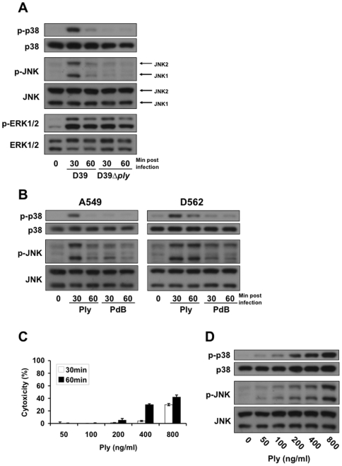
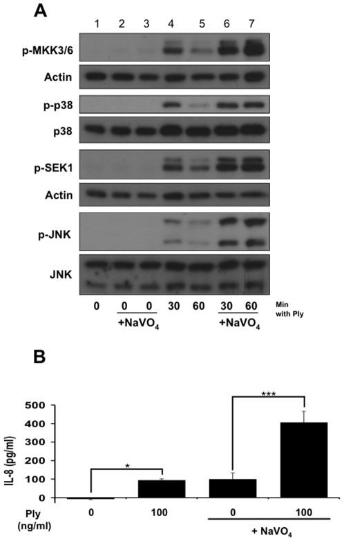
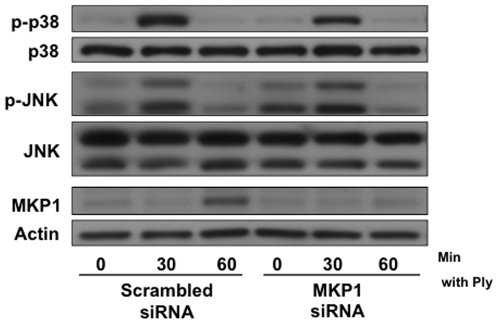
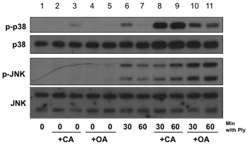
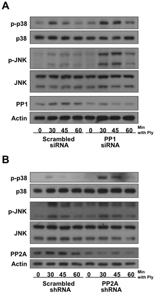
Diverse bacterial species produce pore-forming toxins (PFT) that can puncture eukaryotic cell membranes. Host cells respond to sublytic concentrations of PFT through conserved intracellular signaling pathways, including activation of mitogen-activated protein kinases (MAPK), which are critical to cell survival. Here we demonstrate that in respiratory epithelial cells p38 and JNK MAPK were phosphorylated within 30 min of exposure to pneumolysin, the PFT from Streptococcus pneumoniae. This activation was tightly regulated, and dephosphorylation of both MAPK occurred within 60 min following exposure. Pretreatment of epithelial cells with inhibitors of cellular phosphatases, including sodium orthovanadate, calyculin A, and okadaic acid, prolonged and intensified MAPK activation. Specific inhibition of MAPK phosphatase-1 did not affect the kinetics of MAPK activation in PFT-exposed epithelial cells, but siRNA-mediated knockdown of serine/threonine phosphatases PP1 and PP2A were potent inhibitors of MAPK dephosphorylation. These results indicate an important role for PP1 and PP2A in termination of epithelial responses to PFT and only a minor contribution of dual-specificity phosphatases, such as MAPK phosphatase-1, which are the major regulators of MAPK signals in other cell types. Epithelial regulation of MAPK signaling in response to membrane disruption involves distinct pathways and may require different strategies for therapeutic interventions.


Conflict of interest statement

Competing Interests: A.J.R. is Section Editor for Infectious Diseases at PLoS ONE.

Figures

Figure 1
Figure 1. Subcytolytic pore-formation by S. pneumoniae leads to temporary activation of epithelial MAP kinases.
(A) Confluent monolayers of A549 cells were stimulated for the indicated times with 4×10⁴ cfu/ml sonicated S. pneumoniae D39 or its isogenic Ply-deficient mutant, D39ply. Cells were subsequently lysed, and total and phosphorylated MAPK detected by western blot. Stimulation with D39, but not D39ply, leads to temporary phosphorylation of p38 and JNK MAPKs. (B) Stimulation of A549 and D562 respiratory epithelial cells for the indicated times with 200 ng/ml of purified Ply toxin but not its toxoid, PdB, induces MAPK activation. (C) A549 Cell lysis was measured by LDH assay of cell supernatants with triplicate samples. (D) Dose-dependent MAPK phosphorylation in A549 cells treated with indicated concentrations of Ply for 30 min.
Figure 2
Figure 2. Inactivation of the epithelial innate immune response to pore-formation is mediated by phosphatase activity.
(A) Confluent monolayers of A549 cells were stimulated for the indicated times with 200 ng/ml of purified Ply after pretreatment with either 100 µM sodium orthovanadate (lanes 6, 7) or vehicle control (lanes 4, 5) for 30 min. As corresponding negative controls, cells were left untreated (lane 1) or treated with sodium orthovanadate alone for 60 and 90 min (lanes 2 and 3, respectively). Sodium orthovanadate leads to enhanced Ply-induced phosphorylation of p38, JNK, and their respective upstream kinases, MKK3/6 and SEK1. (B) Pretreatment of A549s with 100 µM sodium orthovanadate (30 min) leads to significantly higher production of Ply-induced IL-8 than vehicle control (*  =  ≤0.05, ***  =  ≤0.001 by ANOVA with Tukey post-test).
Figure 3
Figure 3. Regulation of the MAPK response to pore-formation is MKP1 independent.
(A) A549 cells were transfected with 2.5 µg of MKP1 or scrambled siRNA per 1×10⁶ cells and stimulated 24 hrs post-transfection with 200 ng/ml of purified Ply for the indicated times. Transfection with MKP1 siRNA inhibited MKP1 expression but did not have an effect on Ply-induced MAPK phosphorylation.
Figure 4
Figure 4. Inhibition of serine/threonine phosphatases leads to increased Ply-induced epithelial MAPK phosphorylation.
Confluent monolayers of A549 cells were stimulated for the indicated times with 100 ng/ml of purified Ply after pretreatment with either 5 nM calyculin A for 30 mins (lanes 8, 9), 250 nM okadaic acid for 60 mins (lanes 10, 11), or vehicle control (lanes 6, 7) for 30 mins. As corresponding negative controls, cells were left untreated (lane 1) or treated with calyculin A alone for 30 and 60 mins (lanes 2 and 3, respectively) or okadaic acid alone for 60 and 90 mins (lanes 4 and 5, respectively). In both cases, pretreatment with serine/threonine phosphatase inhibitors leads to prolonged and enhanced p38 and JNK phosphorylation in response to Ply.
Figure 5
Figure 5. PP1 and PP2A mediate inactivation of the epithelial MAPK response to pore-formation.
(A)A549 cells were transfected with 2 µg PP1 or scrambled siRNA per 1×10⁶ cells and stimulated 54 hrs post-transfection with 100 ng/ml of purified Ply. Transfection with PP1 siRNA leads to reduced PP1 expression and a corresponding increase in Ply-induced p38 and JNK phosphorylation. (B) A549 cells were transfected with 6 µg PP2Aα/β or control shRNA per 1×10⁶ cells and stimulated 60 hrs post-transfection with 100 ng/ml of purified Ply. Transfection with PP2Aα/β shRNA leads to reduced PP2A expression and a moderate increase in p38 phosphorylation.

References

    1. Iacovache I, van der Goot FG, Pernot L. Pore formation: an ancient yet complex form of attack. Biochim Biophys Acta. 2008;1778:1611–1623. - PubMed
    1. Gelber SE, Aguilar JL, Lewis KL, Ratner AJ. Functional and phylogenetic characterization of Vaginolysin, the human-specific cytolysin from Gardnerella vaginalis. J Bacteriol. 2008;190:3896–3903. - PMC - PubMed
    1. Ratner AJ, Hippe KR, Aguilar JL, Bender MH, Nelson AL, et al. Epithelial cells are sensitive detectors of bacterial pore-forming toxins. J Biol Chem. 2006;281:12994–12998. - PMC - PubMed
    1. Huffman DL, Abrami L, Sasik R, Corbeil J, van der Goot FG, et al. Mitogen-activated protein kinase pathways defend against bacterial pore-forming toxins. Proc Natl Acad Sci U S A. 2004;101:10995–11000. - PMC - PubMed
    1. Bischof LJ, Kao CY, Los FC, Gonzalez MR, Shen Z, et al. Activation of the unfolded protein response is required for defenses against bacterial pore-forming toxin in vivo. PLoS Pathog. 2008;4:e1000176. - PMC - PubMed

Publication types

Substances