

Maggot secretions skew monocyte-macrophage differentiation away from a pro-inflammatory to a pro-angiogenic type
- PMID: 19956650
- PMCID: PMC2778998
- DOI: 10.1371/journal.pone.0008071
Maggot secretions skew monocyte-macrophage differentiation away from a pro-inflammatory to a pro-angiogenic type
Abstract
Background: Maggots of the blowfly Lucilia sericata are used for the treatment of chronic wounds. Earlier we reported maggot secretions to inhibit pro-inflammatory responses of human monocytes. The aim of this study was to investigate the effect of maggot secretions on the differentiation of monocytes into pro-inflammatory (MØ-1) and anti-inflammatory/pro-angiogenic macrophages (MØ-2) as these cells play a central role in wound healing.
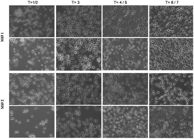
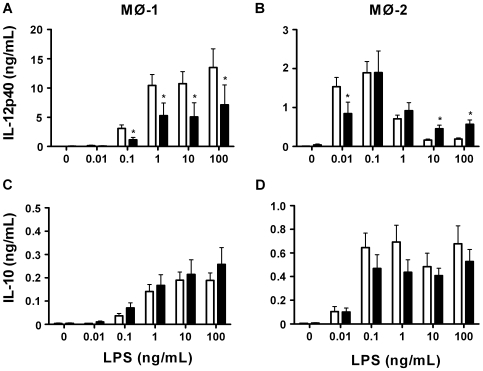
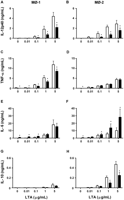
Methodology/principal findings: Freshly isolated monocytes were incubated with secretions and GM-CSF or M-CSF for 6 days and then stimulated with LPS or LTA for 18 h. The expression of cell surface molecules and the levels of cytokines, chemokines and growth factors in supernatants were measured. Our results showed secretions to affect monocyte-macrophage differentiation leading to MØ-1 with a partial MØ-2-like morphology but lacking CD163, which is characteristic for MØ-2. In response to LPS or LTA, secretions-differentiated MØ-1 produced less pro-inflammatory cytokines (TNF-alpha, IL-12p40 and MIF) than control cells. Similar results were observed for MØ-2 when stimulated with low concentrations of LPS. Furthermore, secretions dose-dependently led to MØ-1 and MØ-2 characterized by an altered chemokine production. Secretions led to MØ-2, but not MØ-1, producing enhanced levels of the growth factors bFGF and VEGF, as compared to control cells. The expression of cell-surface receptors involved in LPS/LTA was enhanced by secretions, that of CD86 and HLA-DR down-regulated, while receptors involved in phagocytosis remained largely unaffected.
Conclusions: Maggot secretions skew the differentiation of monocytes into macrophages away from a pro-inflammatory to a pro-angiogenic type.
Conflict of interest statement
Figures
Similar articles
-
Maggot secretions suppress pro-inflammatory responses of human monocytes through elevation of cyclic AMP.Diabetologia. 2009 Sep;52(9):1962-70. doi: 10.1007/s00125-009-1432-6. Epub 2009 Jul 3. Diabetologia. 2009. PMID: 19575178 Free PMC article.
-
Paracrine regulation of vascular endothelial growth factor--a expression during macrophage-melanoma cell interaction: role of monocyte chemotactic protein-1 and macrophage colony-stimulating factor.J Interferon Cytokine Res. 2005 Nov;25(11):674-83. doi: 10.1089/jir.2005.25.674. J Interferon Cytokine Res. 2005. PMID: 16318581
-
Nicotinamide: a vitamin able to shift macrophage differentiation toward macrophages with restricted inflammatory features.Innate Immun. 2015 Nov;21(8):813-26. doi: 10.1177/1753425915602545. Epub 2015 Sep 18. Innate Immun. 2015. PMID: 26385774
-
Human placental mesenchymal stem cells (pMSCs) play a role as immune suppressive cells by shifting macrophage differentiation from inflammatory M1 to anti-inflammatory M2 macrophages.Stem Cell Rev Rep. 2013 Oct;9(5):620-41. doi: 10.1007/s12015-013-9455-2. Stem Cell Rev Rep. 2013. PMID: 23812784
-
Harnessing medicinal maggot excretions/secretions for the ocular surface: A mini review.Cont Lens Anterior Eye. 2025 Apr 15:102422. doi: 10.1016/j.clae.2025.102422. Online ahead of print. Cont Lens Anterior Eye. 2025. PMID: 40240210 Review.
Cited by
-
Next Generation Sequencing Identifies Five Major Classes of Potentially Therapeutic Enzymes Secreted by Lucilia sericata Medical Maggots.Biomed Res Int. 2016;2016:8285428. doi: 10.1155/2016/8285428. Epub 2016 Mar 28. Biomed Res Int. 2016. PMID: 27119084 Free PMC article.
-
Practical context of enzymatic treatment for wound healing: A secreted protease approach (Review).Biomed Rep. 2020 Jul;13(1):3-14. doi: 10.3892/br.2020.1300. Epub 2020 Apr 27. Biomed Rep. 2020. PMID: 32440346 Free PMC article. Review.
-
The influence of ezetimibe on classical and alternative activation pathways of monocytes/macrophages isolated from patients with hypercholesterolemia.Naunyn Schmiedebergs Arch Pharmacol. 2014 Aug;387(8):733-42. doi: 10.1007/s00210-014-0982-4. Epub 2014 Apr 30. Naunyn Schmiedebergs Arch Pharmacol. 2014. PMID: 24781446 Free PMC article.
-
The in vitro and in vivo effects of Lucilia sericata larval secretions on Leishmania major.J Parasit Dis. 2023 Jun;47(2):363-368. doi: 10.1007/s12639-023-01574-x. Epub 2023 Apr 5. J Parasit Dis. 2023. PMID: 37193496 Free PMC article.
-
Pharmacological Properties of the Medical Maggot: A Novel Therapy Overview.Evid Based Complement Alternat Med. 2018 May 3;2018:4934890. doi: 10.1155/2018/4934890. eCollection 2018. Evid Based Complement Alternat Med. 2018. PMID: 29853956 Free PMC article. Review.
References
-
- Boulton AJ, Vileikyte L, Ragnarson-Tennvall G, Apelqvist J. The global burden of diabetic foot disease. Lancet. 2005;366:1719–1724. - PubMed
-
- Jeffcoate WJ, Harding KG. Diabetic foot ulcers. Lancet. 2003;361:1545–1551. - PubMed
-
- Jeffcoate WJ, Chipchase SY, Ince P, Game FL. Assessing the outcome of the management of diabetic foot ulcers using ulcer-related and person-related measures. Diabetes Care. 2006;29:1784–1787. - PubMed
-
- Oyibo SO, Jude EB, Tarawneh I, Nguyen HC, Armstrong DG, et al. The effects of ulcer size and site, patient's age, sex and type and duration of diabetes on the outcome of diabetic foot ulcers. Diabet Med. 2001;18:133–138. - PubMed
-
- Apelqvist J, Larsson J, Agardh CD. Long-term prognosis for diabetic patients with foot ulcers. J Intern Med. 1993;233:485–491. - PubMed
Publication types
MeSH terms
Substances
LinkOut - more resources
Full Text Sources
Research Materials
Miscellaneous
