Review
. 2009 Nov;5(11):e1000621.
doi: 10.1371/journal.ppat.1000621. Epub 2009 Nov 26.

Virus movements on the plasma membrane support infection and transmission between cells

Affiliations
Review

Virus movements on the plasma membrane support infection and transmission between cells

Christoph J Burckhardt et al. PLoS Pathog. 2009 Nov.

Abstract

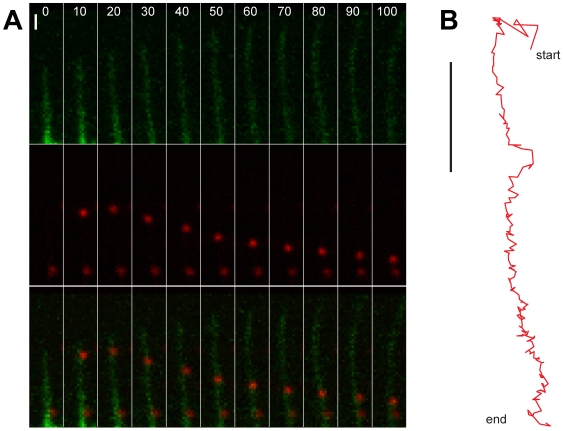
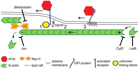
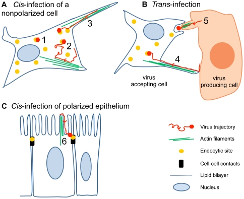
How viruses are transmitted across the mucosal epithelia of the respiratory, digestive, or excretory tracts, and how they spread from cell to cell and cause systemic infections, is incompletely understood. Recent advances from single virus tracking experiments have revealed conserved patterns of virus movements on the plasma membrane, including diffusive motions, drifting motions depending on retrograde flow of actin filaments or actin tail formation by polymerization, and confinement to submicrometer areas. Here, we discuss how viruses take advantage of cellular mechanisms that normally drive the movements of proteins and lipids on the cell surface. A concept emerges where short periods of fast diffusive motions allow viruses to rapidly move over several micrometers. Coupling to actin flow supports directional transport of virus particles during entry and cell-cell transmission, and local confinement coincides with either nonproductive stalling or infectious endocytic uptake. These conserved features of virus-host interactions upstream of infectious entry offer new perspectives for anti-viral interference.


Conflict of interest statement

The authors have declared that no competing interests exist.

Figures

Figure 1
Figure 1. Diffusional motions cover larger surface areas than directed drifts and confined motions.
Viruses have been observed to undergo three types of motion, random diffusion (cyan), retrograde drifts (also called retrograde flow, red), and confined motions (black) (see Table 1 and main text). (A and B) show the heterogeneity of two typical trajectories of adenovirus serotype 2 particles on human embryonic retinoblasts. The motion patterns were recorded by confocal microscopy at 25 Hz acquisition frequency and automatically classified by a machine-based learning algorithm . Nonclassified motions are depicted in dark blue.
Figure 2
Figure 2. Viral surfing on a growing filopodium.
(A) Red fluorescent human adenovirus type 2 particles (red puncta in middle and lower rows) and actin were imaged by spinning disc confocal microscopy on human embryonic retinoblast 911 cells stably expressing GFP-actin (green structures in upper and lower rows). Note that the upper particle attached to a filopodium at time point 10 s, and engaged in a drifting motion towards the cell body (lower side of the images, not shown). During this movement, the filopodial actin structures expanded away from the cell body. A second virus particle bound to the same filopodium remained stationary up to 100 s, indicating that it was not coupled to the actin flow. Bar  = 2 µm. (B) Trajectory profile of the drifting particle from (A) acquired by automated tracking of 2 Hz images. Note that this particle covered approximately 7 µm in 90 s from the start point (10 s) to the end point (100 s) with an average speed of 0.08 µm/s Bar  = 2 µm.
Figure 3
Figure 3. Principles of virus coupling to retrograde actin flow.
Retrograde flow of filamentous actin (F-actin) is maintained by two machineries. One is actin filament polymerization at the plus end of the filament, for example, near the tip of a filopodium, and depolymerization at the opposite minus end. Depolymerization of F-actin by cytochalasin D (CytD), inhibition of actin polymerization by latrunculin B (LatB), or stabilization by jasplakinolide (Jas) inhibit retrograde flow of F-actin, virus drifts on filopodia, and also infection. The second machinery is based on the myosin II (Myo II) motor, which pulls actin filaments to the cell body. Myo II is anchored in the actin mesh at the cell body and cortex. Inhibition of Myo II by blebbistatin inhibits actin retrograde flow, virus drifts, and infection. The linkage of viruses to retrograde flow can occur through viral transmembrane receptors directly or indirect to F-actin (1), or require signalling downstream of virus binding and receptor clustering (2). Another mechanism is by the partitioning of receptors into specialized membrane domains, such as lipid rafts that transiently link to actin retrograde flow (3).
Figure 4
Figure 4. Infectious lateral mobility of viruses on the cell surface.
(A) Cis-infection by virus targeting to endocytic hot spots. Reovirus, for example, depends on clathrin-coated pits that form near the virus (yellow dots, scenario 1). Other viruses, such as influenza virus, induce their own clathrin-coated pits . Polyomaviruses , papillomaviruses , or dengue virus may use various types of motions to scan the surface for preexisting coated pits or caveolae (2). Retroviruses ,, papilomavirus , vaccinia virus ,, adenovirus , and polyomaviruses use directional drifts from the distal tips of filopodia to the cell body (3). (B) Trans-infection by cell surface movements. Cell-to-cell transmission of extracellular retroviruses or herpesviruses can occur in virological synapses and cytonemes from the surface of a donor cell to an acceptor cell , (4). Vaccinia virus egress is driven by actin comet tails that form underneath an extracellular virus, and thereby propel the virus towards an acceptor cell (5). (C) Virus infection of epithelia. Coxsackievirus B, an enterovirus of the picornavirus family, is targeted to cell–cell contacts (black bars), where it interacts with the endocytic machinery (yellow dot) (6). Retroviruses move along microvilli to reach the cell body, where they may be endocytozed or fuse with the plasma membrane .

References

    1. Liberali P, Ramo P, Pelkmans L. Protein kinases: starting a molecular-systems view of endocytosis. Annu Rev Cell Dev Biol. 2008;24:501–523. - PubMed
    1. Sherer NM, Lehmann MJ, Jimenez-Soto LF, Horensavitz C, Pypaert M, et al. Retroviruses can establish filopodial bridges for efficient cell-to-cell transmission. Nat Cell Biol. 2007;9:310–315. - PMC - PubMed
    1. Sattentau Q. Avoiding the void: cell-to-cell spread of human viruses. Nat Rev Microbiol. 2008;6:815–826. - PubMed
    1. Greber UF, Way M. A super highway to virus infection. Cell. 2006;124:741–754. - PubMed
    1. Brandenburg B, Zhuang X. Virus trafficking - learning from single-virus tracking. Nat Rev Microbiol. 2007;5:197–208. - PMC - PubMed

Publication types