. 2009 Nov 23;4(11):e7949.
doi: 10.1371/journal.pone.0007949.

Multiple functions of Nm23-H1 are regulated by oxido-reduction system

Affiliations

Multiple functions of Nm23-H1 are regulated by oxido-reduction system

Eunsun Lee et al. PLoS One. .

Abstract

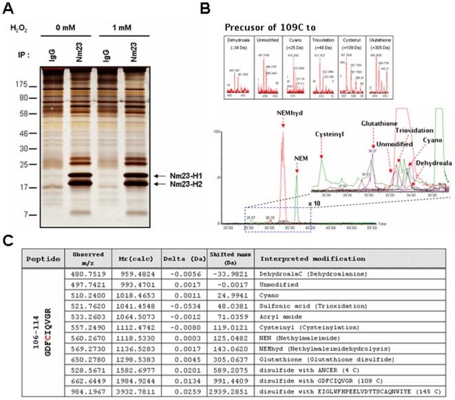
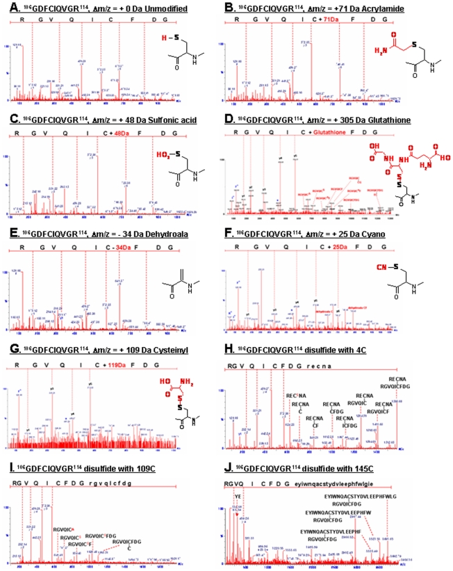
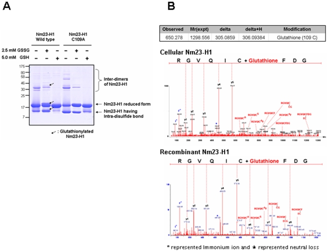
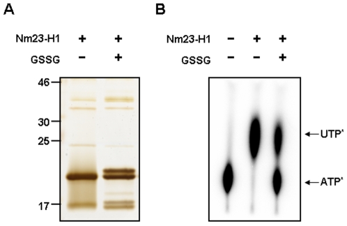
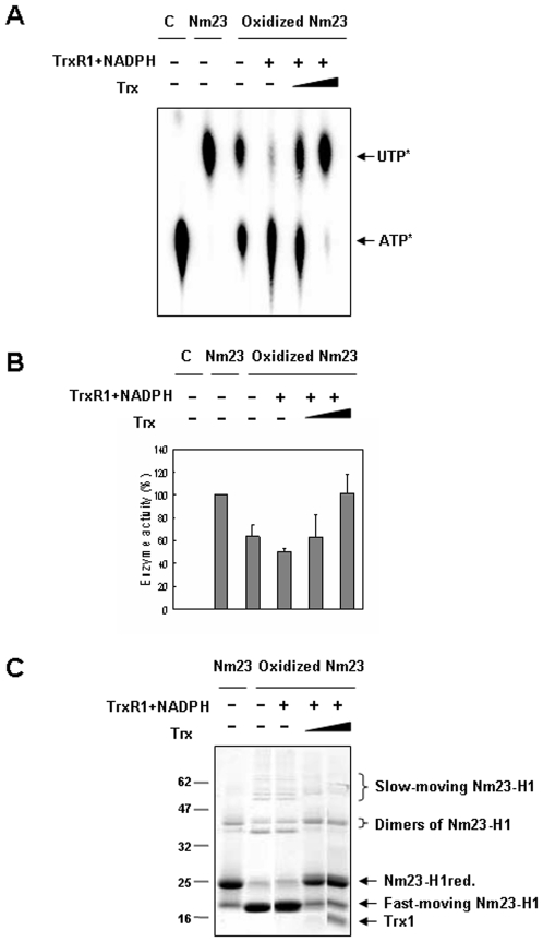
Nucleoside diphosphate kinase (NDPK, Nm23), a housekeeping enzyme, is known to be a multifunctional protein, acting as a metastasis suppressor, transactivation activity on c-myc, and regulating endocytosis. The cellular mechanisms regulating Nm23 functions are poorly understood. In this study, we identified the modifications and interacting proteins of Nm23-H1 in response to oxidative stress. We found that Cys109 in Nm23-H1 is oxidized to various oxidation states including intra- and inter-disulfide crosslinks, glutathionylation, and sulfonic acid formation in response to H(2)O(2) treatment both in vivo and in vitro. The cross-linking sites and modifications of oxidized Nm23-H1 were identified by peptide sequencing using UPLC-ESI-q-TOF tandem MS. Glutathionylation and oxidation of Cys109 inhibited the NDPK enzymatic activity of Nm23-H1. We also found that thioredoxin reductase 1 (TrxR1) is an interacting protein of Nm23-H1, and it binds specifically to oxidized Nm23-H1. Oxidized Nm23 is a substrate of NADPH-TrxR1-thioredoxin shuttle system, and the disulfide crosslinking is reversibly reduced and the enzymatic activity is recovered by this system. Oxidation of Cys109 in Nm23-H1 inhibited its metastatic suppressor activity as well as the enzymatic activities. The mutant, Nm23-H1 C109A, retained both the enzymatic and metastasis suppressor activities under oxidative stress. This suggests that key enzymatic and metastasis suppressor functions of Nm23-H1 are regulated by oxido-reduction of its Cys109.


Conflict of interest statement

Competing Interests: The authors have declared that no competing interests exist.

Figures

Figure 1
Figure 1. Post-translational modifications of cellular Nm23-H1 in MCF-7 breast cancer cells.
(A) MCF-7 breast cancer cells, non-invasive cell lines highly expressing Nm23-H1, were exposed to control and 1 mM H2O2 for 1 h at 37°C. Endogenous Nm23-H1 and Nm23-H2 were purified by immunoprecipitation using anti-Nm23-H1 antibody. These immuno-complexes were separated on 12% SDS-PAGE under non-reducing condition and detected with silver-staining. (B) Post-translational modifications in active cysteine containing tryptic peptide of Nm23-H1, 106GDFCIQVGR114 (993.4701 Da) were identified by nanoLC-ESI-q-TOF tandem MS. Bottom panel is a total ion chromatogram containing various modifications of peptide 106GDFCIQVGR114, and dotted box of this chromatogram was magnified to right panel. Precursor ion of each modified peptide was shown in each box. (C) Table presents the predicted and detected molecular mass and interpreted modifications at Cys-109.
Figure 2
Figure 2. Peptide sequencing using MS/MS analysis of tryptic peptide from modified peptide at Cys-109.
(A) Unmodified peptide 106GDFCIQVGR114; (B) Acrylamide adduct of free SH; (C) Oxidized C109 to sulfonic acid (-SO3H), +48 Da at C109; (D) Glutathionylated C109; (E) Dehydroalanine (Δm/z = −33.9821 Da); (F) Formation of Cys-S-CN, +25 Da; (G) Cysteinylation (109CysS-SCys, Δm/z = +119.0 Da); (H) inter-disulfide C109; (I,J) Intra-disulfide bond with Cys4 peptide (109CysS-S-AN4CER, Δm/z = +589.2075 Da) and C145 (109CysS-S-ELGLWFHPEELVDYTS145CAQNWIYE, Δm/z = +2939.2851 Da).
Figure 3
Figure 3. MS/MS spectra studies of tryptic peptide reveal glutathionylation of Nm23-H1.
(A) Purified recombinant proteins, wild type Nm23-H1 and C109A mutant, were treated with and without 2.5 mM oxidized glutathione (GSSG), 5 mM reduced glutathione (GSH) at 37°C for 1 h. Each sample was separated on 12% SDS-PAGE under non-reducing condition and detected with coomassie blue staining. Only glutathionylation of wild Nm23-H1, not C109A mutant, was easily observed. (B) Tandem MS spectra of glutathionylated cellular and recombinant Nm23-H1 were compared. Both mass spectra profile was identical, only abundance of glutathionylated peptide is higher in recombinant protein.
Figure 4
Figure 4. Glutathionylation of Nm23-H1 abolishes NDP kinase enzymatic activity.
(A) Recombinant wild type Nm23-H1 was treated with 5.0 mM glutathione for 30 min at 37°C. Each sample was separated on 12% SDS-PAGE under non-reducing condition and detected with silver staining. (B) NDP kinase activity of each sample was measured as described in Experimental procedure. Glutathionylated fraction of sample lost their NDP kinase activity.
Figure 5
Figure 5. Oxidized Nm23-H1 interacts with thioredoxin reductase 1.
(A,B) Hela cells transiently transfected with Flag-Nm23-H1 were exposed to 5 mM H2O2 for 1 h at 37°C and recovered for 6 h without H2O2. Endogenous Nm23-H1s were purified by immunoprecipitation using anti-Flag antibody. These immuno-complexes were separated on 12% SDS-PAGE under non-reducing condition and detected with silver-staining (A) and with western analysis using anti-Flag antibody (B). (C) Protein interacting with oxidized Nm23 as indicated arrow in Figure 4A was identified with MALDI-TOF MS as thioredoxin reductase (TrxR). (D) Thioredoxin reductase interacting with oxidized Nm23-H1 was confirmed by western analysis using anti-TrxR antibody.
Figure 6
Figure 6. Oxidized Nm23 is a substrate of NADPH-TrxR-Trx system.
(A) Suggested scheme of oxido-reduction regulation of Nm23-H1 by TrxR-Trx-NADPH system. (B) TrxR assay was performed in solution containing 50 mM sodium phosphate, pH 7.0, 50 mM KCl, 1 mM EDTA, 100 µM Nm23-H1, 200 µM NADPH, 80 µM rat TrxR1, 50 µM Trx1, and monitored the absorbance change at 340 nm. Absorbance decreases by conversion of NADPH to NADP were measured as TR activity. It turns out that oxidized Nm23-H1 is a substrate of TrxR-Trx-NADPH system.
Figure 7
Figure 7. Impaired NDP kinase enzyme activity of oxidized Nm23-H1 is restored by of NADPH-TrxR-Trx system.
(A,B) Recombinant Nm23-H1 proteins (100 µM) were oxidized and reincubated with 200 µM NADPH, 80 µM rat TrxR1 and various concentration of Trx (0, 10 and 50 µM) same as in Figure 5. Enzymatic activities were measured on TLC plate (A) and quantitatively analyzed (B). (A) Same samples were separated on % SDS-PAGE under non-reducing condition.
Figure 8
Figure 8. NDP kinase enzymatic activity of Nm23-H1 is required for its ability to suppress metastasis.
(A) Comparison of Nm23-H1 expression level and invasion potential between MDA-MB-231, invasive breast cancer cell line, and MCF-7, non invasive breast cancer cell line. Upper panel shows the expression level of Nm23-H1 and α-tubulin as a control, and lower panel, invasive potential in matrigel assay. (B) Comparison of invasion potential of MCF-7 cells reducing Nm23-H1 expression level by siRNA. Upper panel shows the expression level of Nm23-H1 and α-tubulin as a control, and lower panel, invasive potential in matrigel assay. (C) Invasion potential of MDA-MB-231 cells transiently transfected with pFlag-CMV-2, pFlag-Nm23-H1 or pFlag-Nm23-H1 C109A mutant were examined. Western analysis of cell lysates using indicated antibody were employed after separation on 12% SDS-PAGE. Cells overexpressing pFlag-CMV-2, pFlag-Nm23-H1, pFlag-Nm23-H1 C109A mutant, or pFlag-Nm23-H1 H118F mutant were seeded on matrigel-pre-coated upper chamber (3×10⁴ cells/150 µL) and invasion potentials were measured (left panel). The recombinant wild type Nm23-H1 or C109A mutant were treated with 5 mM H2O2 or 20 mM NEM for 1 h at 37°C, and NDP kinase activity were measured (right panel).
Figure 9
Figure 9. Oxidative stress inhibits the metastasis suppression potential of Nm23-H1.
Invasion potential of MDA-MB-231 cells transiently transfected with pFlag-CMV-2, pFlag-Nm23-H1 or pFlag-Nm23-H1 C109A mutant were examined. Western analysis of cell lysates using indicated antibody were employed after separation on 12% SDS-PAGE. Cells overexpressing pFlag-CMV-2, pFlag-Nm23-H1, pFlag-Nm23-H1 C109A mutant or pFlag-Nm23-H1 H118F mutant were exposed to 0.5 mM H2O2 for 1 h (B) or none (A). And they were seeded on matrigel-pre-coated upper chamber (3×10⁴ cells/150 µL) and invasion potentials were measured.

Similar articles

Cited by

References

    1. Rosengard AM, Krutzsch HC, Shearn A, Biggs JR, Barker E, et al. Reduced Nm23/Awd protein in tumour metastasis and aberrant Drosophila development. Nature. 1989;342:177–180. - PubMed
    1. Nakayama T, Ohtsuru A, Nakao K, Shima M, Nakata K, et al. Expression in human hepatocellular carcinoma of nucleoside diphosphate kinase, a homologue of the nm23 gene product. J Natl Cancer Inst. 1992;84:1349–1354. - PubMed
    1. Steeg PS, Bevilacqua G, Kopper L, Thorgeirsson UP, Talmadge JE, et al. Evidence for a novel gene associated with low tumor metastatic potential. J Natl Cancer Inst. 1988;80:200–204. - PubMed
    1. Steeg PS. Metastasis suppressors alter the signal transduction of cancer cells. Nat Rev Cancer. 2003;3:55–63. - PubMed
    1. Leone A, Flatow U, VanHoutte K, Steeg PS. Transfection of human nm23-H1 into the human MDA-MB-435 breast carcinoma cell line: effects on tumor metastatic potential, colonization and enzymatic activity. Oncogene. 1993;8:2325–2333. - PubMed

Publication types