. 2009 Nov 24;4(11):e7967.
doi: 10.1371/journal.pone.0007967.

Characterization of mucosal Candida albicans biofilms

Affiliations

Characterization of mucosal Candida albicans biofilms

Anna Dongari-Bagtzoglou et al. PLoS One. .

Abstract

C. albicans triggers recurrent infections of the alimentary tract mucosa that result from biofilm growth. Although the ability of C. albicans to form a biofilm on abiotic surfaces has been well documented in recent years, no information exists on biofilms that form directly on mucosal surfaces. The objectives of this study were to characterize the structure and composition of Candida biofilms forming on the oral mucosa. We found that oral Candida biofilms consist of yeast, hyphae, and commensal bacteria, with keratin dispersed in the intercellular spaces. Neutrophils migrate through the oral mucosa and form nests within the biofilm mass. The cell wall polysaccharide beta-glucan is exposed during mucosal biofilm growth and is more uniformly present on the surface of biofilm organisms invading the oral mucosa. We conclude that C. albicans forms complex mucosal biofilms consisting of both commensal bacterial flora and host components. These discoveries are important since they can prompt a shift of focus for current research in investigating the role of Candida-bacterial interactions in the pathogenesis of mucosal infections as well as the role of beta-glucan mediated signaling in the host response.


Conflict of interest statement

Competing Interests: JPV is employed by Biothera which provided the monoclonal antibody against beta-glucan. Biothera makes this antibody available to all interested investigators via a Material Transfer Agreement. A beta-glucan antibody is also commercially available and can be purchased by investigators. This does not alter adherence to PLoS ONE policies on sharing data and materials.

Figures

Figure 1
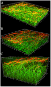
Figure 1. C. albicans presence in “white plaque” lesions formed on the tongue of mice with oropharyngeal candidiasis.
C. albicans-challenged mice were sacrificed after 5 days of oral exposure to the GFP-expressing strain MRL51. Panel A depicts the dorsal aspect of a tongue from an uninfected control. Panel B depicts the white plaque lesions formed on the tongue of an infected mouse. Panel C depicts a three dimensional reconstruction of a live biofilm as visualized via confocal microscopy.
Figure 2
Figure 2. Display of β-glucan during different stages of C. albicans mucosal biofilm growth.
Panels depict representative confocal images of tissue sections from mice with oropharyngeal candidiasis (A–D) or from a three-dimensional in vitro model of the oral mucosa (F). Panels A–D and F depict sections stained for β-glucan with a BFDiv monoclonal antibody (red), C. albicans with a polyclonal anti-Candida Ab (green) and counterstained with the nucleic acid stain TO-PRO-3, which stains tissue cells blue. Panel E depicts a section stained with an IgM isotype control antibody for the BFDiv stain. Notice that β-glucan becomes more uniformly present on the surface of fungal cells invading the tongue mucosa (arrows). Scale bar = 20 µm.
Figure 3
Figure 3. β-glucan and extracellular material staining in C. albicans biofilms forming on glass.
Panel A depicts a 2h biofilm and panel B depicts a 48h biofilm. Biofilms of the GFP-expressing C. albicans strain (green) were stained for β-glucan with a BFDiv monoclonal antibody (red) and the extracellular material was stained with ConA-Alexa 350 (blue). In 2h biofilms there is partial co-localization of the BFDiv mAb and ConA (pink). BFDiv stains parts of the fungal cell, but not the germinating buds, and ConA stains the entire fungal cell surface (3A). In 48h biofilms deposits of cell-dissociated ECM stained with ConA but not with BFDiv (3B, arrows). Scale bar = 20 µm.
Figure 4
Figure 4. β-glucan and extracellular material staining during C. albicans SC5314 in vitro biofilm growth on glass.
Panels depict 3D reconstructions of confocal stacks of images of 24h (A), 48h (B) and 72h (C) biofilms of C. albicans grown on cover slips and stained for β-glucan with BFDiv mAb (red) and ConA-Alexa 488 (green). Notice that regardless of biofilm thickness β-glucan is localized in the growing end of the biofilm (arrows).
Figure 5
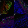
Figure 5. Cytokeratin presence in C. albicans biofilms formed on the tongue of mice with oropharyngeal candidiasis.
Tissue sections in panels A, B and C were stained with anti-cytokeratin mAb (red) and anti-Candida pAb (green). Host cell nuclei were visualized with TO-PRO-3 (blue). Panel D represents an isotype control (IgG) stain. Arrows indicate areas where fungal cells are surrounded by keratin. Scale bar = 20 µm.
Figure 6
Figure 6. Neutrophils form aggregates in tongue biofilms of C.albicans-infected mice.
Panels A–D depict tissue sections stained with an anti-mouse neutrophil mAb (red), an anti-Candida Ab (green) and the nucleic acid stain TO-PRO-3 (blue). Panel E depicts a confocal image of a negative control stain (primary anti-neutrophil mAb was omitted). Arrows indicate the presence of neutrophils directly juxtaposed to, or within the biofilm mass. Scale bar = 20 µm.
Figure 7
Figure 7. Presence of bacteria in mucosal biofilms of mice with oropharyngeal candidiasis.
Panels A and B depict tissue sections stained with an anti-Candida pAb (green) and the nucleic acid stain Syto59 (red). Panel B is a 3.5× zoom image of the marked area in Panel A. Notice the close association of C. albicans and bacterial cells (arrows). Panel C depicts a tissue section stained with an anti-Candida antibody (green), processed for fluorescence in situ hybridization (FISH) with the all bacteria-specific oligonucleotide probe EUB388 (red) and counterstained with the nucleic acid stain Hoechst 33258 (blue). Notice the presence of bacteria (pink) throughout the mucosal biofilms (arrows). Scale bar = 20 µm.
Figure 8
Figure 8. Identification of specific components of bacterial commensal flora in mucosal biofilms of mice with oropharyngeal candidiasis.
Panels A-C depict a tongue tissue section stained with an anti-Candida antibody (green) and processed for fluorescence in situ hybridization (FISH) with the all bacteria-specific oligonucleotide probe EUB388 (blue) and the Lactobacillus and Enterococcus sp.-specific probe LAB158 (red). Panels B and C are a 3.5× zoom image of the marked areas in Panel A. Bacteria positive with the LAB158 probe appear pink due to co-localization with the all bacteria specific probe EUB338 (arrows). Panels D and F depict a tongue tissue section stained with an anti-Candida antibody (green) and processed for FISH with the all bacteria-specific probe EUB388 (blue) and the Staphylococcus sp.-specific probe STA697 (red). Panels E and F are a 3.5× zoom image of the marked areas in Panel D. Bacteria positive with the STA697 probe appear pink due to co-localization with the EUB338 probe (arrows). Scale bar = 20 µm.

References

    1. Costerton JW, Stewart PS, Greenberg EP. Bacterial biofilms: A common cause of persistent infections. Science. 1999;284:1318–1322. - PubMed
    1. Hall-Stoodley L, Hu FZ, Gieseke A, Nistico L, Nguyen D, et al. Direct detection of bacterial biofilms on the middle-ear mucosa of children with chronic otitis media. JAMA. 2006;296:202–211. - PMC - PubMed
    1. Post JC, Hiller NL, Nistico L, Stoodley P, Ehrlich GD. The role of biofilms in otolaryngologic infections: update 2007. Curr Opin Otolaryngol Head Neck Surg. 2007;15:347–351. - PubMed
    1. Odds FC. Activity of Cilofungin (Ly121019) against Candida species invitro. J Antimicrobial Chem. 1988;22:891–897. - PubMed
    1. Seneviratne CJ, Jin L, Samaranayake LP. Biofilm lifestyle of Candida: a mini review. Oral Dis. 2008;14:582–590. - PubMed

Publication types