

Brain temperature by Biosensor Imaging of Redundant Deviation in Shifts (BIRDS): comparison between TmDOTP5- and TmDOTMA-
- PMID: 19957287
- PMCID: PMC2843767
- DOI: 10.1002/nbm.1461
Brain temperature by Biosensor Imaging of Redundant Deviation in Shifts (BIRDS): comparison between TmDOTP5- and TmDOTMA-
Abstract
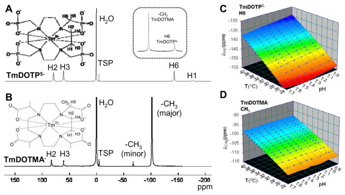
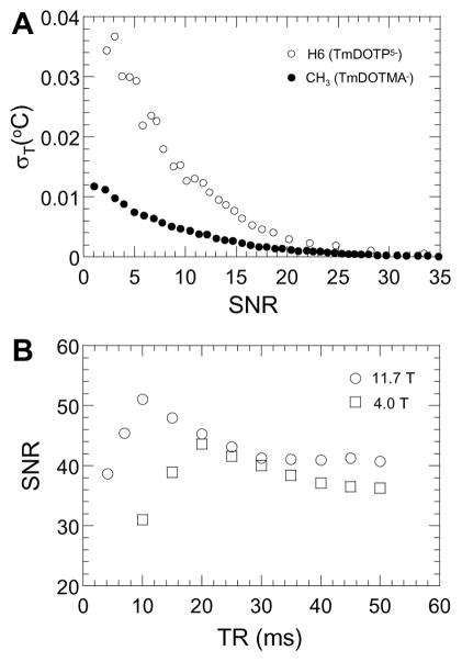
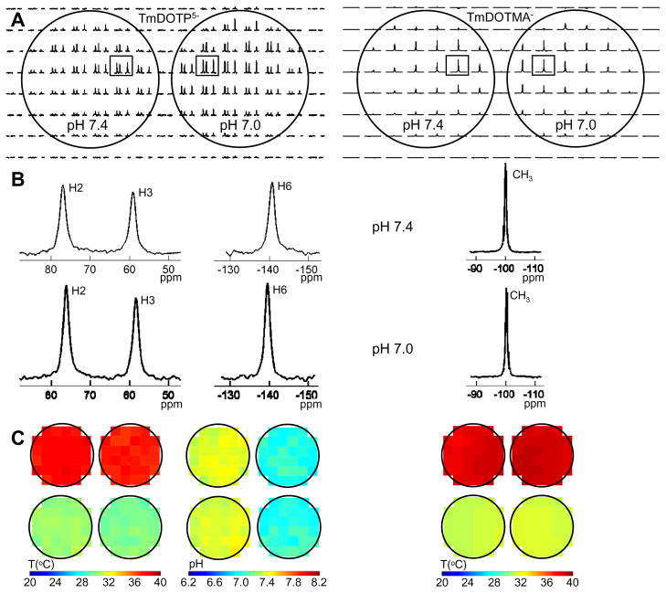
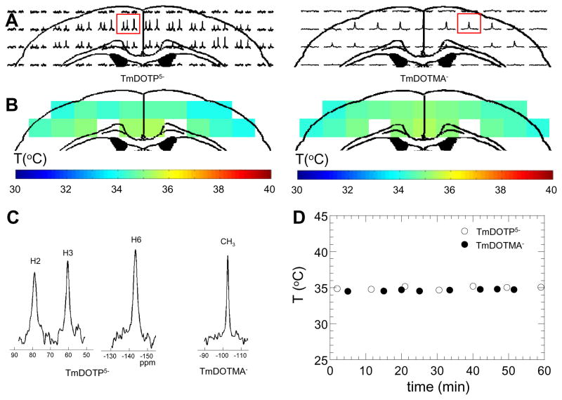
Chemical shifts of complexes between paramagnetic lanthanide ions and macrocyclic chelates are sensitive to physiological variations (of temperature and/or pH). Here we demonstrate utility of a complex between thulium ion (Tm(3+)) and the macrocyclic chelate 1,4,7,10-tetramethyl 1,4,7,10-tetraazacyclododecane-1,4,7,10-tetraacetate (or DOTMA(4-)) for absolute temperature mapping in rat brain. The feasibility of TmDOTMA(-) is compared with that of another Tm(3+)-containing biosensor which is based on the macrocyclic chelate 1,4,7,10-tetraazacyclododecane- 1,4,7,10-tetrakis(methylene phosphonate) (or DOTP(8-)). In general, the in vitro and in vivo results suggest that Biosensor Imaging of Redundant Deviation in Shifts (BIRDS) which originate from these agents (but exclude water) can provide temperature maps with good accuracy. While TmDOTP(5-) emanates three major distinct proton resonances which are differentially sensitive to temperature and pH, TmDOTMA(-) has a dominant pH-insensitive proton resonance from a -CH(3) group to allow higher signal-to-noise ratio (SNR) temperature assessment. Temperature (and pH) sensitivities of these resonances are practically identical at low (4.0T) and high (11.7T) magnetic fields and at nominal repetition times only marginal SNR loss is expected at the lower field. Since these resonances have extremely short relaxation times, high-speed chemical shift imaging (CSI) is needed to detect them. Repeated in vivo CSI scans with BIRDS demonstrate excellent measurement stability. Overall, results with TmDOTP(5-) and TmDOTMA(-) suggest that BIRDS can be reliably applied, either at low or high magnetic fields, for functional studies in rodents.
2009 John Wiley & Sons, Ltd.
Figures
Similar articles
-
In vivo three-dimensional molecular imaging with Biosensor Imaging of Redundant Deviation in Shifts (BIRDS) at high spatiotemporal resolution.NMR Biomed. 2013 Nov;26(11):1589-95. doi: 10.1002/nbm.2995. Epub 2013 Jul 24. NMR Biomed. 2013. PMID: 23881869 Free PMC article.
-
Simultaneous measurements of temperature and pH in vivo using NMR in conjunction with TmDOTP5-.NMR Biomed. 2000 Dec;13(8):460-6. doi: 10.1002/nbm.676. NMR Biomed. 2000. PMID: 11252031
-
Noninvasive MR thermometry using paramagnetic lanthanide complexes of 1,4,7,10-tetraazacyclodoecane-alpha,alpha',alpha'',alpha'''-tetramethyl-1,4,7,10-tetraacetic acid (DOTMA4-).Magn Reson Med. 2005 Feb;53(2):294-303. doi: 10.1002/mrm.20345. Magn Reson Med. 2005. PMID: 15678553
-
Brain temperature and pH measured by (1)H chemical shift imaging of a thulium agent.NMR Biomed. 2009 Feb;22(2):229-39. doi: 10.1002/nbm.1312. NMR Biomed. 2009. PMID: 19130468 Free PMC article.
-
Ytterbium chelated to 1,4,7,10-tetraazacyclododecane-1,4,7-triacetic acid,10-orthoaminoanilide.2011 Nov 26 [updated 2012 Jan 5]. In: Molecular Imaging and Contrast Agent Database (MICAD) [Internet]. Bethesda (MD): National Center for Biotechnology Information (US); 2004–2013. 2011 Nov 26 [updated 2012 Jan 5]. In: Molecular Imaging and Contrast Agent Database (MICAD) [Internet]. Bethesda (MD): National Center for Biotechnology Information (US); 2004–2013. PMID: 22238803 Free Books & Documents. Review.
Cited by
-
Extracellular pH mapping of liver cancer on a clinical 3T MRI scanner.Magn Reson Med. 2020 May;83(5):1553-1564. doi: 10.1002/mrm.28035. Epub 2019 Nov 5. Magn Reson Med. 2020. PMID: 31691371 Free PMC article.
-
Distribution of temperature changes and neurovascular coupling in rat brain following 3,4-methylenedioxymethamphetamine (MDMA, "ecstasy") exposure.NMR Biomed. 2015 Oct;28(10):1257-66. doi: 10.1002/nbm.3375. Epub 2015 Aug 19. NMR Biomed. 2015. PMID: 26286889 Free PMC article.
-
Mapping Extracellular pH of Gliomas in Presence of Superparamagnetic Nanoparticles: Towards Imaging the Distribution of Drug-Containing Nanoparticles and Their Curative Effect on the Tumor Microenvironment.Contrast Media Mol Imaging. 2017 Nov 22;2017:3849373. doi: 10.1155/2017/3849373. eCollection 2017. Contrast Media Mol Imaging. 2017. PMID: 29362558 Free PMC article.
-
Reduced removal of waste products from energy metabolism takes center stage in human brain aging.Sci Rep. 2025 Mar 8;15(1):8127. doi: 10.1038/s41598-025-90342-3. Sci Rep. 2025. PMID: 40057554 Free PMC article.
-
Towards longitudinal mapping of extracellular pH in gliomas.NMR Biomed. 2016 Oct;29(10):1364-72. doi: 10.1002/nbm.3578. Epub 2016 Jul 29. NMR Biomed. 2016. PMID: 27472471 Free PMC article.
References
-
- Ginsberg MD, Busto R. Combating hyperthermia in acute stroke: a significant clinical concern. Stroke. 1998;29(2):529–534. - PubMed
-
- Thompson HJ, Tkacs NC, Saatman KE, Raghupathi R, McIntosh TK. Hyperthermia following traumatic brain injury: a critical evaluation. Neurobiol Dis. 2003;12(3):163–173. - PubMed
-
- Yager JY, Armstrong EA, Jaharus C, Saucier DM, Wirrell EC. Preventing hyperthermia decreases brain damage following neonatal hypoxic-ischemic seizures. Brain Res. 2004;1011(1):48–57. - PubMed
-
- van der Zee J. Heating the patient: a promising approach? Ann Oncol. 2002;13(8):1173–1184. - PubMed
Publication types
MeSH terms
Substances
Grants and funding
LinkOut - more resources
Full Text Sources
Other Literature Sources
