

A multi-species comparative structural bioinformatics analysis of inherited mutations in alpha-D-mannosidase reveals strong genotype-phenotype correlation
- PMID: 19958498
- PMCID: PMC2788387
- DOI: 10.1186/1471-2164-10-S3-S33
A multi-species comparative structural bioinformatics analysis of inherited mutations in alpha-D-mannosidase reveals strong genotype-phenotype correlation
Abstract
Background: Lysosomal alpha-mannosidase is an enzyme that acts to degrade N-linked oligosaccharides and hence plays an important role in mannose metabolism in humans and other mammalian species, especially livestock. Mutations in the gene (MAN2B1) encoding lysosomal alpha-D-mannosidase cause improper coding, resulting in dysfunctional or non-functional protein, causing the disease alpha-mannosidosis. Mapping disease mutations to the structure of the protein can help in understanding the functional consequences of these mutations and thus indirectly, the finer aspects of the pathology and clinical manifestations of the disease, including phenotypic severity as a function of the genotype.
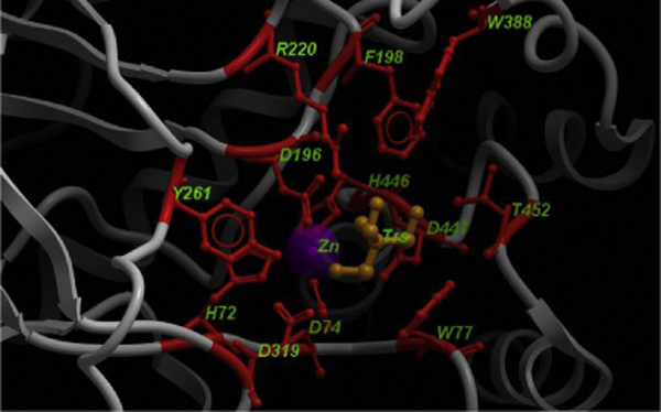
Results: A comprehensive homology modeling study of all the wild-type and inherited mutations of lysosomal alpha-mannosidase in four different species, human, cow, cat and guinea pig, reveals a significant correlation between the severity of the genotype and the phenotype in alpha-mannosidosis. We used the X-ray crystallographic structure of bovine lysosomal alpha-mannosidase as template, containing only two disulphide bonds and some ligands, to build structural models of wild-type structures with four disulfide linkages and all bound ligands. These wild-type models were then used as templates for disease mutations. All the truncations and substitutions involving the residues in and around the active site and those that destabilize the fold led to severe genotypes resulting in lethal phenotypes, whereas the mutations lying away from the active site were milder in both their genotypic and phenotypic expression.
Conclusion: Based on the co-location of mutations from different organisms and their proximity to the enzyme active site, we have extrapolated observed mutations from one species to homologous positions in other organisms, as a predictive approach for detecting likely alpha-mannosidosis. Besides predicting new disease mutations, this approach also provides a way for detecting mutation hotspots in the gene, where novel mutations could be implicated in disease. The current study has identified five mutational hot-spot regions along the MAN2B1 gene. Structural mapping can thus provide a rational approach for predicting the phenotype of a disease, based on observed genotypic variations.
Figures


Similar articles
-
A comparative structural bioinformatics analysis of inherited mutations in β-D-Mannosidase across multiple species reveals a genotype-phenotype correlation.BMC Genomics. 2011 Nov 30;12 Suppl 3(Suppl 3):S22. doi: 10.1186/1471-2164-12-S3-S22. Epub 2011 Nov 30. BMC Genomics. 2011. PMID: 22369051 Free PMC article.
-
amamutdb.no: A relational database for MAN2B1 allelic variants that compiles genotypes, clinical phenotypes, and biochemical and structural data of mutant MAN2B1 in α-mannosidosis.Hum Mutat. 2015 Jun;36(6):581-6. doi: 10.1002/humu.22787. Epub 2015 Apr 9. Hum Mutat. 2015. PMID: 25762455
-
Purification of feline lysosomal alpha-mannosidase, determination of its cDNA sequence and identification of a mutation causing alpha-mannosidosis in Persian cats.Biochem J. 1997 Dec 15;328 ( Pt 3)(Pt 3):863-70. doi: 10.1042/bj3280863. Biochem J. 1997. PMID: 9396732 Free PMC article.
-
Lysosomal alpha-mannosidase and alpha-mannosidosis.Front Biosci (Landmark Ed). 2017 Jan 1;22(1):157-167. doi: 10.2741/4478. Front Biosci (Landmark Ed). 2017. PMID: 27814608 Review.
-
Alpha-Mannosidosis: Therapeutic Strategies.Int J Mol Sci. 2018 May 17;19(5):1500. doi: 10.3390/ijms19051500. Int J Mol Sci. 2018. PMID: 29772816 Free PMC article. Review.
Cited by
-
Mucopolysaccharidosis Type I and α-Mannosidosis-Phenotypically Comparable but Genetically Different: Diagnostic and Therapeutic Considerations.Biomedicines. 2025 May 14;13(5):1199. doi: 10.3390/biomedicines13051199. Biomedicines. 2025. PMID: 40427026 Free PMC article. Review.
-
A meta-analysis of genome-wide association studies to identify candidate genes associated with feed efficiency traits in pigs.J Anim Sci. 2025 Jan 4;103:skaf010. doi: 10.1093/jas/skaf010. J Anim Sci. 2025. PMID: 39847436 Free PMC article.
-
A comparative structural bioinformatics analysis of inherited mutations in β-D-Mannosidase across multiple species reveals a genotype-phenotype correlation.BMC Genomics. 2011 Nov 30;12 Suppl 3(Suppl 3):S22. doi: 10.1186/1471-2164-12-S3-S22. Epub 2011 Nov 30. BMC Genomics. 2011. PMID: 22369051 Free PMC article.
-
Structure-Function Relationships of LDL Receptor Missense Mutations Using Homology Modeling.Protein J. 2019 Aug;38(4):447-462. doi: 10.1007/s10930-019-09860-5. Protein J. 2019. PMID: 31401775
-
Immune Infiltration Associated MAN2B1 Is a Novel Prognostic Biomarker for Glioma.Front Oncol. 2022 Feb 2;12:842973. doi: 10.3389/fonc.2022.842973. eCollection 2022. Front Oncol. 2022. PMID: 35186771 Free PMC article.
References
-
- Ranganathan S, Male DA, Ormsby RJ, Giannakis E, Gordon DL. Pinpointing the putative heparin/sialic acid-binding residues in the 'sushi' domain 7 of factor H: a molecular modeling study. Pac Symp Biocomput. 2000. pp. 155–167. - PubMed
-
- Ormsby RJ, Ranganathan S, Tong JC, Griggs KM, Dimasi DP, Hewitt AW, Burdon KP, Craig JE, Hoh J, Gordon DL. Functional and structural implications of the complement factor H Y402H polymorphism associated with age-related macular degeneration. Invest Ophthalmol Vis Sci. 2008;49:1763–1770. doi: 10.1167/iovs.07-1297. - DOI - PubMed
Publication types
MeSH terms
Substances
LinkOut - more resources
Full Text Sources
Other Literature Sources
Miscellaneous
