. 2010 Feb 17;165(4):1261-74.
doi: 10.1016/j.neuroscience.2009.11.056. Epub 2009 Dec 1.

Ror1-Ror2 complexes modulate synapse formation in hippocampal neurons

Affiliations

Ror1-Ror2 complexes modulate synapse formation in hippocampal neurons

S Paganoni et al. Neuroscience. .

Abstract

Ror1 and Ror2, a small family of tyrosine kinase receptors, have been implicated in multiple aspects of brain development in C. elegans and X. laevis. More recently, we have shown that these receptors modulate the rate of neurite elongation in cultured rat hippocampal neurons. However, no information is available regarding a potential role of these receptors in other developmental milestones in mammalian central neurons. Neither is the identity known of the Ror ligand(s) and/or the signal transduction pathway(s) in which they participate. Here we report that the down regulation of either Ror1 or Ror2 led to a significant decrease in synapse formation in cultured hippocampal neurons. Simultaneous targeting of Ror proteins, however, did not result in an additive phenotype. Our results also indicated that Ror1 and Ror2 physically interact in the mouse brain, suggesting that they might function as heterodimers in central neurons. In addition, these Ror complexes interacted with Wnt-5a mediating its effects on synaptogenesis. Together, these data suggest that Ror proteins play a key role in Wnt-5a-activated signaling pathways leading to synapse formation in the mammalian CNS.


Figures
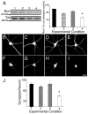
Figure 1
Figure 1. Effect of Ror1 down regulation by RNAi on synapse formation in cultured hippocampal neurons
(A) Western blot analysis of whole cell extracts prepared from control (lane 1), mutated Ror1 siRNA- (lane 2), scrambled Ror1 siRNA- (lane 3) and Ror1 siRNA- (lane 4) transfected hippocampal cultures. Densitometric analysis of the Ror1 immunoreactive bands is shown on the right. Values represent the mean ± S.E.M for 3 independent experiments normalized using tubulin as an internal control. The numbers correspond to the percent of Ror1 protein present in the siRNA-transfected cultures as compared to the levels detected in the untreated samples (100%). *Differs from untransfected, mutated siRNA- and scrambled siRNA-transfected cultures, P<0.01. (B–I) Seven days in culture control (B, F), mutated Ror1 siRNA- (C, G), scrambled Ror1 siRNA- (D, H) and Ror1 siRNA- (E, I) transfected hippocampal neurons were double stained with tubulin (B–E) and synaptophysin (F–I) antibodies. Note the reduction in the number of synaptophysin immunoreactive dots in the Ror1 siRNA-transfected cultures (I) as compared to the control ones (F, G, H). (J) Quantitative analysis of the number of synapses formed by neurons cultured under the experimental conditions described above. The numbers represent the mean ± S.E.M for 3 independent experiments. * Differs from all other experimental conditions, P< 0.01. Scale bar: 20 μm.
Figure 2
Figure 2. Ror1 suppression by antisense oligonucleotide treatment in cultured hippocampal neurons
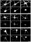
(A–F) Seven days in culture control (A, D), Ror1 sense- (B, E) and Ror1 antisense- (C, F) treated hippocampal neurons were double stained with tubulin (A–C) and Ror1 (D–F) antibodies. Ror1 was readily detectable in control (D) and sense-treated (E) neurons. Only faint immunoreactivity was left in the neurons treated with Ror1 antisense oligonucleotides (F). (G–L) Seven days in culture control (G, J), Ror1 sense- (H, K) and Ror1 antisense- (I, L) treated hippocampal neurons were double stained with tubulin (G–I) and synaptophysin (J–L) antibodies. Fewer synaptophysin immunoreactive spots were detected when Ror1 expression was targeted by the addition of Ror1 antisense oligonucleotides as compared to controls (compare L to J and K). (M–R) Seven days in culture control (M, P), Ror1 sense- (N, Q) and Ror1 antisense- (O, R) treated hippocampal neurons were double stained with MAP2 (M–O) and synapsin I (P–R) antibodies. Fewer synapsin I immunoreactive dots (arrowheads) were present in the targeted cultures as compared to controls (R). Note also the diffuse synapsin I immunoreactivity in some of the axons growing on top of MAP2 immunoreactive processes in antisense-treated neurons (arrow in R). Scale bars: 20 μm.
Figure 3
Figure 3. Ror1 and Ror2 interaction in HEK cells and in the mammalian central nervous system
(A–D) HEK cells were transfected with Ror1-Flag and Ror2-HA constructs. Immunolabeling with anti-Flag (A) and anti-HA (C) antibodies showed that Ror1 and Ror2 co-localized when co-expressed in HEK cells (A, C, D). (B) Sister cultures were immunoprecipitated with Ror1 antibody. Whole cell lysates (WCL) and immunoprecipitates (IP) were separated by SDS-PAGE and analyzed by Western blotting using HA and Ror1 antibodies. Note that an HA immunoreactive band was detected in the immunoprecipitates prepared from the cultures where Ror1-Flag and Ror2-HA had been co-expressed. (E) Whole brain lysates from E16 mouse embryos were subjected to immunoprecipitation with Ror1 antibody. Controls included samples where the primary antibody was omitted or replaced with normal rabbit IgGs. Samples were analyzed by Western blotting using Ror1 and Ror2 antibodies. When Ror1 was immunoprecipitated from mouse embryonic brains, Ror2 immunoreactive bands were detected in the precipitate. IP: immunoprecipitate; WCL: whole cell lysate. Scale bar: 20 μm.
Figure 4
Figure 4. Wnt-5a interaction with Ror proteins in vitro and in the mouse brain
(A) HEK cells were transfected with mouse Ror1-Flag, Ror2-Flag and Wnt-5a-HA plasmids as indicated. Whole cell lysates (WCL) were analyzed by Western blotting to show expression of the transfected constructs. Lysates were immunoprecipitated with HA antibody and probed for Ror1 and Ror2. No Ror1 band was detected in any of the precipitates (IP). Conversely, a Ror2 immunoreactive band was present in the IP samples prepared from the cultures that had been double transfected with Ror2 and Wnt-5a, but not in the ones prepared from HEK cells that had been transfected with either Ror2 or Wnt-5a alone. (B) Whole brain lysates from E16 mouse embryos were subjected to immunoprecipitation with Ror1 antibodies. In control samples, the primary antibody was omitted. Precipitates were probed for Ror1, Ror2 and Wnt-5a. In the Ror1 immunoprecipitates, Ror2 and Wnt-5a immunoreactive bands were detected. (C) HEK cells were transfected with mouse Ror2 tagged with either Flag or HA epitopes as indicated. Ror2 was pulled down using anti-HA antibodies. Whole cell lysates (WCL) and immunoprecipitates (IP) were immunoblotted with HA and Flag antibodies. Note that Ror2-Flag was detected in the immunoprecipitates when co-expressed with Ror2-HA. IP: immunoprecipitate; WCL: whole cell lysate.
Figure 5
Figure 5. Determination of Wnt-5a levels in hippocampal neuron- and astrocyte- conditioned culture medium
(A & B) Quantitative analysis of Wnt-5a levels in culture medium obtained from hippocampal neurons (A) and astrocytes (B) cultures maintained up to 14 days. Note that the Wnt-5a concentration was significantly higher in astrocyte-conditioned medium when compared to hippocampal neuron-conditioned one.
Figure 6
Figure 6. Wnt-5a induced synapse formation in cultured hippocampal neurons
(A & B) Quantitative analysis of Wnt-5a levels in culture medium obtained from control (C) and Wnt-5a-transfected (T) astrocyte culture maintained for 7 days. Immunoblot membranes were reacted with Wnt-5a and HA antibodies. Values represent the mean ± S.E.M for 3 independent experiments. *Differs from controls, P<0.01. (C–F) Seven days in culture hippocampal neurons maintained in the presence of untransfected (C &D) and Wnt-5a-transfected astrocyte cultures were double stained with tubulin (C & E) and synaptophysin (D & F) antibodies. Note the increase in the number of synaptophysin immunoreactive dots in hippocampal neurons co-cultured with Wnt-5a-transfected astrocytes (F) as compared to the control ones (D). Scale bar: 20 μm.
Figure 7
Figure 7. Down regulation of Ror1 and/or Ror2 expression prevented Wnt-5a induced synapse formation in cultured hippocampal neurons
(A–H) Seven days in culture control (A & B), antisense Ror1- (C & D), antisense Ror2- (E & F), and antisense Ror1/Ror2- (G & H) treated hippocampal neurons maintained in the presence of Wnt-5a-transfected astrocyte cultures were double stained with tubulin (A, C, E, G) and synaptophysin (B, D, F, H) antibodies. Note the decreased in the number of synaptophysin immunoreactive dots in Ror-depleted hippocampal neurons co-cultured with Wnt-5a-transfected astrocytes (D, F, H) as compared to the control ones (B). Scale bar: 20 μm.

Similar articles

Cited by

References

    1. Ahmad-Annuar A, Ciani L, Simeonidis L, Herreros J, Ben Fredj N, Rosso SB, Hall A, Brickley S, Salinas PC. Signaling across the synapse: a role for Wnt and disheveled in presynaptic assembly and neurotransmitter relase. J Cell Biol. 2006;174:127–139. - PMC - PubMed
    1. Al-Shawi R, Ashton SV, Underwood C, Simons JP. Expression of the Ror1 and Ror2 receptor tyrosine kinase genes during mouse development. Dev Genes Evol. 2001;211:161–171. - PubMed
    1. Baker JC, Beddington RS, Harland RM. Wnt signaling in Xenopus embryos inhibits bmp4 expression and activates neural development. Genes Dev. 1999;13:3149–3159. - PMC - PubMed
    1. Bhanot P, Brink M, Samos CH, Hsieh JC, Wang Y, Macke JP, Andrew D, Nathans J, Nusse R. A new member of the frizzled family from Drosophila functions as a Wingless receptor. Nature. 1996;382:225–230. - PubMed
    1. Billiard J, Way DS, Seestaller-Wehr LM, Moran RA, Mangine A, Bodine PV. The Orphan Receptor Tyrosine Kinase Ror2 Modulates Canonical Wnt Signaling in Osteoblastic Cells. Mol Endocrinol. 2005;19:90–101. - PubMed

Publication types