. 2009 Nov;19(4):615-24.
doi: 10.1016/j.nic.2009.09.001.

Quantitative sodium MR imaging and sodium bioscales for the management of brain tumors

Affiliations

Quantitative sodium MR imaging and sodium bioscales for the management of brain tumors

Keith R Thulborn et al. Neuroimaging Clin N Am. 2009 Nov.

Abstract

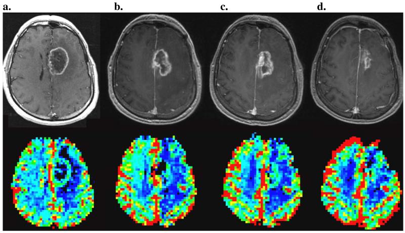
Treatment of high-grade primary brain tumors is based on experience from multicenter trials. However, the prognosis has changed little in 3 decades. This suggests that there is a fundamental oversight in treatment. This article provides an imaging perspective of how regional responses of primary brain tumors may be examined to guide a flexible treatment plan. Sodium imaging provides a measurement of cell density that can be used to measure regional cell kill. Such a bioscales of regionally and temporally sensitive biologic-based parameters may be helpful to guide tumor treatment. These suggestions are speculative and still being examined, but are presented to challenge the medical community to be receptive to changes in the standard of care when that standard continues to fail.


Figures

Figure 1
Figure 1
Representative MR images from the 3.0 Tesla clinical examination of a patient with a large right hemispheric high-grade glioneuroma prior to treatment. Images are from the following acquisition sequences: (top left) non-contrast-enhanced T1-weighted 3D inversion recovery gradient echo, (top middle) contrast-enhanced T1-weighted 3D inversion recovery gradient echo, (top right) magnetic susceptibility-sensitive 2D gradient echo, (bottom left) 3D quantitative 3D sodium image at a nominal resolution of 5 ×5 × 5 mm3 acquired in under 10 minutes, (bottom middle) relative cerebral blood volume map from a dynamic susceptibility contrast MR perfusion study, and (bottom right) 2D T2-weighted FLAIR propeller. The interpretation is a large, heterogeneous mass with cystic and hemorrhagic components and markedly increased (red) relative cerebral blood volume centered in the right parietotemporal region. The lesion has been biopsied. Surgical resection was not considered as an option.
Figure 2
Figure 2
Functional MRI performed for presurgical planning of a contrast-enhancing abnormality appearing the surgical bed years after treatment for a brain tumor in the left frontal lobe. The activation map from the fMRI using the reading language comprehension paradigm is presented superimposed over the contrast-enhanced 3D T1-weighted inversion recovery gradient echo images (3D SPGR +C, top row) and over the T2-weighted spin-echo, echo-planar images (AX SE EPI, bottom row) in the axial (left column), coronal (middle column) and sagittal (right column) planes. The planes are cross-referenced with colored lines (blue = axial, green = coronal, yellow = sagittal). Broca’s area is cross-referenced (intersection of planes) and is anterior and inferior to the lesion but in close proximity to it so that the surgeon was aware of the potential of producing an aphasia. This location of function was confirmed by intraoperative cortical mapping. The lesion was primarily radiation necrosis as predicted by the low rCBV.
Figure 3
Figure 3
The radiation treatment planning fuses the CT and MRI studies (left), especially the perfusion study for high-grade tumors, to generate a radiation distribution (middle) that covers the tumor while minimizing the dose to normal brain. The contour of the head is drawn as the blue outline. The base dose (middle left) is supplemented by an additional boost to the tumor (middle right). The radiation plan (red) can then be superimposed over the contrast-enhanced MR image. The large enhancing tumor in the right temporal lobe is well covered but there is still considerable exposure to the rest of the brain despite targeted treatment.
Figure 4
Figure 4
Pseudoprogression is demonstrated in a patient across their early treatment in which the tumor shows variation in enhancement (top row) and the relative cerebral blood volume (rCBV) map (bottom row). (a) Prior to surgery and radiation treatment, there is a thin rim of enhancement and increased rCBV. (b) After resection, radiation and low dose chemotherapy, there is increasingly thickened enhancement that then subsequently resolves while the rCBV shows continuous monotonically decreasing rCBV. (b,c,d) Follow-up studies were done every 2-3 monthly after radiation treatment was completed.
Figure 5
Figure 5
Sodium imaging and its derived metabolic bioscales in a normal subject. (a) 4 partitions from a 3D sodium imaging dataset, (b) TSC map derived from (a), (c) CVF map derived by applying Equation 2 to the TSC map and (d) calibration phantom image used to calibrate the sodium signal intensity into a TSC value. These measurements were performed at 3.0 Tesla at a resolution of 5 × 5 × 5 mm3 in less than 10 minutes.
Figure 6
Figure 6
(a) Linear calibration curve for the conversion of the intensity (arbitrary scale) from the sodium MR signal to a tissue sodium concentration (mM). The phantom has the same electrical loading on the RF coil as the human head and is also corrected for static magnetic field and RF excitation field inhomogeneities. Similar calibration curves are obtained at 3.0 and 9.4 Tesla. (b) An image through the phantom showing the three calibration tubes with different concentrations (30, 70, 110 mM).
Figure 7
Figure 7
(a) Sodium images showing the high sodium concentration in the large tumor (arrow) in the right hemisphere of the same patient as in Figure 1. The color bioscale shows TSC from 0 to 150mM with normal brain shown as green (cell density = 0.8) and the tumor as pink indicating a lower cell density. (cell density ~ 0.4-0.6). These images were acquired at 9.4 Tesla at a nominal resolution of 3 × 3 × 3 mm3 in less than 10 minutes.
Figure 8
Figure 8
Conventional proton MR images of a patient after resection of a right temporal GBM. (top left) T1-weighted gradient echo image without gadolinium contrast enhancement, (top middle) T1-weighted gradient echo image with gadolinium contrast enhancement, (top right) magnetic susceptibility-sensitive T2-weighted gradient echo image, (bottom left) rCBV map showing residual areas of increased rCBV, (bottom middle) apparent diffusion coefficient map and (bottom right) T2-weighted FLAIR image.
Figure 9
Figure 9
Realigned sodium images showing the superimposed accumulated radiation dose (red) for the same patient in Figures 3 and 8. The left image is prior to radiation and each subsequent image to the right is another week of radiation therapy with the far right image being after completion of treatment. The images have been co-registered in post-processing to ensure alignment. The realignment is performed within k-space to avoid blurring that would occur if performed in image space, an important advantage of 3D datasets. The two tubes anterior to the brain are calibration phantoms placed within the field of view to demonstrate reproducible signal intensity. These can be used for quantification but with less accuracy than obtained with a separate calibration phantom.
Figure 10
Figure 10
Voxel-wise classification of temporal responses of TSC during radiation therapy covering the center of the surgical bed of the lesion of the patient shown in Figures 3, 8 and 9. The 5 categories of responses: A: contralateral hemisphere with normal TSC values (green star), B: no response with elevated TSC (red solid triangle); C: desirable response with cell death shown by increasing TSC (orange solid circle); D: decreasing edema with elevated TSC decreasing back towards normal TSC (blue solid diamond) and E: elevated TSC decreasing but towards tumor TSC values (magenta solid square). Each voxel can be classified into these responses and related to the subsequent follow-up change in rCBV that revealed recurrence but many weeks later. Response patterns B (red triangle) and E (magenta square) are hypothesized to be predictive of recurrence as they are incomplete responses.

Similar articles

Cited by

References

    1. Ries LAG, Harkins D, Krapcho M, Mariotto A, Miller BA, Feuer EJ, Clegg L, Eisner MP, Horner MJ, Howlader N, Hayat M, Hankey BF, Edwards BK, editors. SEER Cancer Statistics Review, 1975-2003. National Cancer Institute; Bethesda, MD: 2006. http://seer.cancer.gov/csr/1975_2003/, based on November 2005 SEER data submission, posted to the SEER web site.
    1. Fabi A, Russillo M, Metro G, Vidiri A, Di Giovanni S, Cognetti F. Pseudoprogression and MGMT status in glioblastoma patients: implications in clinical practice. Anticancer Res. 2009 Jul;29(7):2607–10. - PubMed
    1. Hall EJ, Cox JD. In: Radiation Oncology. 6th Ed. Williams TM, Cox JD, editors. pp. 1–57. Chapter 1.
    1. Cairncross G, Berkey B, Shaw E, Jenkins R, et al. Phase III trial of chemotherapy plus radiotherapy compared with radiotherapy alone for pure and mixed anaplastic oligodendroglioma: Intergroup radiation therapy oncology group trial 9402. J Clin Oncol. 2006;24(18):2707–2714. - PubMed
    1. Gilbert MR, Lang FF. Anaplastic oligodendroglial tumors: a tale of two trials. J Clin Oncol. 2006;24(18):2689–2690. - PubMed

Publication types

MeSH terms