Review
. 2010 Mar;35(3):169-78.
doi: 10.1016/j.tibs.2009.10.004. Epub 2009 Dec 1.

Role of RNA structure in regulating pre-mRNA splicing

Affiliations
Review

Role of RNA structure in regulating pre-mRNA splicing

M Bryan Warf et al. Trends Biochem Sci. 2010 Mar.

Abstract

Pre-mRNA splicing involves removing non-coding introns from RNA transcripts. It is carried out by the spliceosome, along with other auxiliary factors. In general, research in splicing has focused on the sequences within the pre-mRNA, without considering the structures that these sequences might form. We propose that the role of RNA structure deserves more consideration when thinking about splicing mechanisms. RNA structures can inhibit or aid binding of spliceosomal components to the pre-mRNA, or can increase splicing efficiency by bringing important sequences into close proximity. Recent reports have identified proteins and small molecules that can regulate splicing by modulating RNA structures, thereby expanding our knowledge of the mechanisms used to regulate splicing.


Figures

Figure 1
Figure 1. Model of spliceosomal assembly
Exons are represented by blue boxes and introns are depicted as a black line. The branch-point adenosine and 5’ and 3’ splice sites are noted. Spliceosomal assembly is seen to be a sequential process, at least in vitro, as different proteins and protein–RNA complexes (known as snRNPs, green) load onto the pre-mRNA. Note that, in E complex, the proteins SF1, U2AF65 and U2AF35 (orange) recognize sequences in the 3’ end of the intron and help recruit the U2 snRNP to the intron in A complex
Figure 2
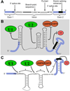
Figure 2. Structures that directly regulate splicing
The primary mechanisms through which RNA structures can either repress or aid in splice site selection are described in this figure. A) Diagram of the canonical consensus sequences within a pre-mRNA which are important for splicing. B) Representative diagram of structures that inhibit splicing. Shown are stem-loops that repress binding of the U1 snRNP (green) to the 5’ splice site, the U2 snRNP to the branch-point, U2AF65 and U2AF35 (orange) to the 3’ splice site and an SR protein (pink) to a sequence within the exon. C) Representative diagram of structures that aid splicing. Depicted is a structure that brings the 5’ and 3’ splice sites into closer proximity, a stem that brings the 3’ splice site and branch-point into closer proximity, a stem that masks a cryptic 3’ splice site (denoted as YAG*) and a stem that properly displays an enhancer sequence in the exon that an SR protein binds.
Figure 3
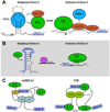
Figure 3. Proteins that regulate splicing by modulating RNA structure
Three mechanisms that proteins use to regulate splicing by modulating RNA structure are described in this figure. A) MBNL1 (turquoise) inhibits U2AF65 (orange) binding and U2 (green) recruitment to the 3’ splice site of intron 4 in the cardiac troponin T pre-mRNA. MBNL1 and U2AF65 compete by binding mutually exclusive RNA structures. B) The DEAD-box RNA helicase p72 (purple) is hypothesized to increase exon 4 inclusion in the CD44 pre-mRNA by destabilizing a stem that encompasses the 5’ splice site following exon 4. C) hnRNP A1 (light blue) and PTB (lime green) are hypothesized to cause skipping of exons by “looping out” of the exon. hnRNP A1 causes skipping of exon 7B of its own pre-mRNA by both directly inhibiting binding of the U1 snRNP to the 5’ splice site following the exon, and by helping to recruit a protein whose identity is unknown (?; gold) to the 3’ splice site directly upstream of the exon. This unidentified factor inhibits U2AF65/U2AF35 binding to the 3’ splice site. PTB represses inclusion of exon N1 in the Src pre-mRNA. PTB did not affect binding of the U1 snRNP to the 5’ splice site, but did inhibit U2AF65/U2AF35 binding to the downstream 3’ splice site.
Figure 4
Figure 4. Thiamine pyrophosphate (TPP) binds a riboswitch that affects splicing for a negative feedback loop in its own biosynthesis pathway
Thiamine C synthase (THIC; blue) is a protein in the biosynthesis pathway for TPP (turquoise), which is a vitamin B1 coenzyme. High levels of TPP lead to inhibition of splicing of an intron in the 3’ UTR of the THIC pre-mRNA. Splicing stabilizes the pre-mRNA, whereas the unspliced pre-mRNA is degraded more quickly. TPP binds a riboswitch that encompasses the 3’ splice site of the intron, and represses splicing. This action provides a negative feedback loop to inhibit THIC protein production when adequate levels of TPP have been produced.

References

    1. Pan Q, et al. Deep surveying of alternative splicing complexity in the human transcriptome by high-throughput sequencing. Nat Genet. 2008;40(12):1413–1415. - PubMed
    1. Wahl MC, Will CL, Luhrmann R. The spliceosome: design principles of a dynamic RNP machine. Cell. 2009;136(4):701–718. - PubMed
    1. Pedersen JS, et al. Identification and classification of conserved RNA secondary structures in the human genome. PLoS Comput Biol. 2006;2(4):e33. - PMC - PubMed
    1. Chen Y, et al. RNA secondary structure and compensatory evolution. Genes Genet Syst. 1999;74(6):271–286. - PubMed
    1. Bentley DL. Rules of engagement: co-transcriptional recruitment of pre-mRNA processing factors. Curr Opin Cell Biol. 2005;17(3):251–256. - PubMed

Publication types

LinkOut - more resources