. 2009 Dec 22;106(51):21930-5.
doi: 10.1073/pnas.0811794106. Epub 2009 Dec 3.

Vitamin B12 deficiency reduces proliferation and promotes differentiation of neuroblastoma cells and up-regulates PP2A, proNGF, and TACE

Affiliations

Vitamin B12 deficiency reduces proliferation and promotes differentiation of neuroblastoma cells and up-regulates PP2A, proNGF, and TACE

Shyue-fang Battaglia-Hsu et al. Proc Natl Acad Sci U S A. .

Abstract

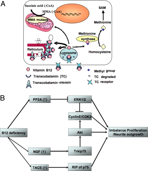
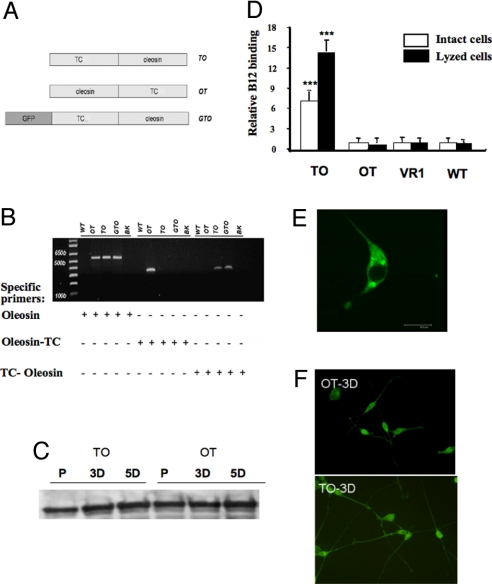
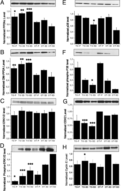
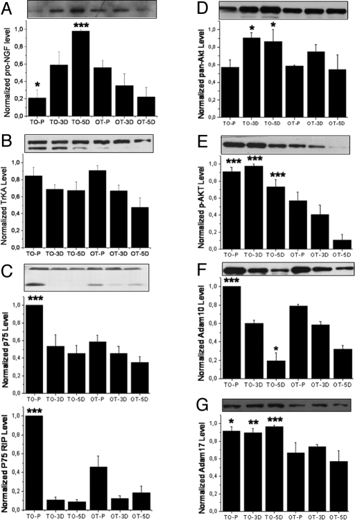
Vitamin B12 (cobalamin, Cbl) is indispensable for proper brain development and functioning, suggesting that it has neurotrophic effects beside its well-known importance in metabolism. The molecular basis of these effects remains hypothetical, one of the reasons being that no efficient cell model has been made available for investigating the consequences of B12 cellular deficiency in neuronal cells. Here, we designed an approach by stable transfection of NIE115 neuroblastoma cells to impose the anchorage of a chimeric B12-binding protein, transcobalamin-oleosin (TO) to the intracellular membrane. This model produced an intracellular sequestration of B12 evidenced by decreased methyl-Cbl and S-adenosylmethionine and increased homocysteine and methylmalonic acid concentrations. B12 deficiency affected the proliferation of NIE115 cells through an overall increase in catalytic protein phosphatase 2A (PP2A), despite its demethylation. It promoted cellular differentiation by improving initial outgrowth of neurites and, at the molecular level, by augmenting the levels of proNGF and p75(NTR). The up-regulation of PP2A and pro-nerve growth factor (NGF) triggered changes in ERK1/2 and Akt, two signaling pathways that influence the balance between proliferation and neurite outgrowth. Compared with control cells, a 2-fold increase of p75(NTR)-regulated intramembraneous proteolysis (RIP) was observed in proliferating TO cells (P < 0.0001) that was associated with an increased expression of two tumor necrosis factor (TNF)-alpha converting enzyme (TACE) secretase enzymes, Adam 10 and Adam 17. In conclusion, our data show that B12 cellular deficiency produces a slower proliferation and a speedier differentiation of neuroblastoma cells through interacting signaling pathways that are related with increased expression of PP2A, proNGF, and TACE.


Conflict of interest statement

The authors declare no conflict of interest.

Figures

Fig. 1.
Fig. 1.
Experimental model of vitamin B12 cellular sequestration and proposed effects on the growth and the differentiation of neuronal cultured cells (adapted from ref. 8). (A) The schematic of the TO experimental model is shown. (B) Vitamin B12 deficiency up-regulates PP2Ac, proNGF, and TACE. These reduce proliferation and promote differentiation of neuroblastoma cells by modulating the interacting downstream pathways including ERK1/2, Akt/PKB, and p75NTR.
Fig. 2.
Fig. 2.
Expression of transcobalamin-fused constructs in NIE115 cells. (A) The schematics of the plasmids TO, OT, and GTO are shown. (B) The incorporation of the three plasmids in NIE115 cells in corresponding stable cell line was evaluated by PCR technique (see Materials and Methods for the specific primers). (C) The relatively constant contents of the tyrosine hydroxylase in TO and OT cells indicate that these transfected cells remain as valid neuronal model (typical result). (D) Radiolabeled B12 (30,000 cpm at 300 μCi/μg) was added to ≈106 cells and then incubated for 20 min at either 37 °C (intact cell) or 4 °C (lysed cell). Compared with OT, VR1, and WT, the expression of TO leads to an increased B12-binding capacity in either intact or lysed cells (P < 0.001). The VR1 expressing cells serve as a control to normalize the B12 binding to the TO, OT, and WT cells. (typical result out of five runs performed in three repeats. Mean and SE are indicated). (E) The confocal image of the NIE-115 cells transfected with GTO shows that the expressed fusion protein is targeted to reticulum (see supplemental figure for TO- and OT-tranfected cells). (F) Three days after initiating the differentiation of the NIE115 cells, more extensive neurite outgrowth was evident in TO cells. Here, cells were immuno-labeled with Adam17 antibody. The secondary antibody was coupled to Alexa Fluor 488 for visualization. P, proliferation; 3D, 3 days after differentiation; 5D, 5 days after differentiation; WT, wild-type NIE115 cells; BK, blank without DNA template; TC, transcobalamin.
Fig. 3.
Fig. 3.
Changes in B12-related parameters in TO cells. (A) A higher concentration of Hcy in culture medium, higher content of MMA and lower content of succinic acid in cell extract was observed in TO, compared with OT and WT cells (P < 0.001). (B and C) TO cells had a statistically significant reduction in their SAM/SAH ratio (P < 0.001), with a weaker change in SAM (P = 0.010) and no significant modification in SAH cellular level (P = 0.116). (C) 57Colabeled Cbl (≈300 μCi/μg) at a concentration of 30,000 cpm/μL was incorporated into culture medium (30,000 cpm/mL culture medium) for 3 days. The various forms of vitamin B12 in whole cell extracts were analyzed by HPLC (described in ref. 3). The total amount of radioactivity taken by each cell lines was considered as 100%. Absolute amount of radioactivity in 100 000 × g pellet and supernatant of TO and OT extracts is showed in B. Significantly less Me-Cbl, Ado-Cbl and OH2-Cbl were formed in TO cells, as evidenced in C (P < 0.001). CN-Cbl, cyano-cobalamin; Me-Cbl, methyl-cobalamin; Ado-Cbl, adenosylcobalamin; OH2-Cbl, aquo-cobalamin; SAM, S-adenosylmethionine; SAH, S-adenosylhomocysteine; Hcy, homocysteine. (A–C) Typical result out of five runs performed in three repeats. Mean and SE are indicated.
Fig. 4.
Fig. 4.
B12 deficiency leads to slower growth and earlier differentiation. (A) A significant reduced rate of proliferation was associated with the vitamin B12-deficient TO cells, compared with WT and OT cells. When adding 200 μM B12 in culture medium, the growth rate of TO cells became similar to that of WT and OT cells cultivated in normal B12 concentration (number of viable cells was counted by trypan blue exclusion method). (B and C) upon change to differentiation medium, within 48 h TO cells showed a more significant outgrowth of neurite than WT and OT cells; this was evident in total neurite number (B) and total neurite length (C). (D) TO cells had an increased level of PSD 95 at day 5 of differentiation. See Materials and Methods for the analysis of neurite using NeuroJ program. The neurite number and the neurite length were the sum of all cells examined in the optical field (constant density of 130–150 cells). (A) typical result out of three runs performed in three repeats. (B and C) Results from five independent determinations. Mean and SE are indicated.
Fig. 5.
Fig. 5.
Western blots for proteins relevant to neuronal cell growth. (A) The level of total protein PP2Ac (phosphatase 2A, catalytic subunit) increase in all three cultured states of the TO cells (P = 0.0281, 0.0012, and <0.0001 for P, 3D, and 5D cells, respectively). (B) The level of demethylated PP2Ac also increased in TO cells, with P = 0.0012, 0.0082, and 0.0006 for P, 3D, and 5D cells, respectively. (C and D) No difference of total ERK1/2 (C) could be observed between the TO and the OT cells; phospho-ERK1/2 level (D) was significantly reduced in TO cells (for total ERK1/2, P = 0.4226, 0.3992, and 0.8563 and for phospho-ERK1/2, P = 0.01181, P < 0.0001, and P < 0.0001 for P, 3D, and 5D cells, respectively). (E and F) for both total p38 (E) and phospho-p38 (F), an increase was visible at the 5D stage of the cells (for p38, P = 0.1087, 0.6742, and 0.02038 and for P-p38, P = 0.2513, 0.6881, and 0.0013, respectively for P, 3D, and 5D cells). (G) The total amount of cyclin-dependent protein kinase 2 (CDK2) appeared reduced in TO cells in differentiated cells (for P, 3D, and 5D cells: P = 0.2032, 0.0005, and 0.0004, respectively), reflecting the growth retardation associated with the TO cells. (H) A significant decrease was detected in cyclin E at 3D (for proliferating cells, P = 0.394; for 3D cells, P = 0.0283; for 5D cells, P = 0.709). (A–H) Typical result out of three runs performed in three repeats. Densitometric analysis was normalized to an arbitrary value of 1.0 that represented the maximal value recorded within each experiment series. Mean and SE are indicated.
Fig. 6.
Fig. 6.
Western blots for proteins relevant to neurite outgrowth. (A) The level of proNGF increased very rapidly in TO cells upon differentiation instead of decrease, as in the case of OTcells (6A: P = 0.03, 0.302, <0.001 for P, 3D, and 5D cells, respectively). (B) For TrkA neurotrophin receptor, only the 140-kDa band was quantified because it represents the mature form of the functional NGF receptor. No difference could be detected between TO and OT cells (P = 0.612, 0.841, and 0.222 for the P, 3D, and 5D cells, respectively). However, in TO cells the 110-kDa form of the TrkA (underglycosylated immature precursor) was up-regulated. (C) The level of neurotrophin receptor p75NTR and its RIP appeared to increase dramatically in proliferating TO cells (upper band: P < 0.001, 0.443, and 0.203; lower band RIP: P < 0.001, 0.734, and 0.245, respectively for P, 3D, and 5D cells). (D and E) The level of total Akt (D) and S473 phospho-Akt (E) were in general increased in TO cells, especially the S473 phosphorylated form of Akt (for pan-Akt: P = 0.789, 0.033, and 0.042 for P, 3D, and 5D cells respectively; for p-Akt: P < 0.001, <0.0001, and <0.0001 for P, 3D, and 5D cells, respectively). (F) The level of Adam 17 increase in TO cells in all states examined (P = 0.0111, 0.002, and <0.001, for P, 3D, and 5D cells, respectively). (G) The level of Adam 10 increased in proliferating but not differentiated TO cells (P < 0.001, 0.5931, and 0.0576 for P, 3D, and 5D cells, respectively). (A–C) Typical result out of three runs performed in three repeats. Densitometric analysis as in Fig. 5. Mean and SE are indicated.

References

    1. Russell JSR, Batten FE, Collier J. Subacute combined degeneration of the spinal cord. Brain. 1900;23:39–110.
    1. Gillette Guyonnet S, et al. IANA task force on nutrition and cognitive decline with aging. J Nutr Health Aging. 2007;11:132–152. - PubMed
    1. Pons L, Guy M, Lambert D, Hatier R, Guéant J. Transcytosis and coenzymatic conversion of [(57)Co]cobalamin bound to either endogenous transcobalamin or exogenous intrinsic factor in caco-2 cells. Cell Physiol Biochem. 2000;10:135–148. - PubMed
    1. McLean GR, et al. Antibodies to transcobalamin II block in vitro proliferation of leukemic cells. Blood. 1997;89:235–242. - PubMed
    1. Hsieh K, Huang AH. Lipid-rich tapetosomes in Brassica tapetum are composed of oleosin-coated oil droplets and vesicles, both assembled in and then detached from the endoplasmic reticulum. Plant J. 2005;43:889–899. - PubMed

Publication types