. 2009 Dec 22;106(51):21813-8.
doi: 10.1073/pnas.0912126106. Epub 2009 Dec 3.

Ca2+ dysregulation in Ryr1(I4895T/wt) mice causes congenital myopathy with progressive formation of minicores, cores, and nemaline rods

Affiliations

Ca2+ dysregulation in Ryr1(I4895T/wt) mice causes congenital myopathy with progressive formation of minicores, cores, and nemaline rods

Elena Zvaritch et al. Proc Natl Acad Sci U S A. .

Abstract

Ryr1(I4895T/wt) (IT/+) mice express a knockin mutation corresponding to the human I4898T EC-uncoupling mutation in the type 1 ryanodine receptor/Ca(2+) release channel (RyR1), which causes a severe form of central core disease (CCD). IT/+ mice exhibit a slowly progressive congenital myopathy, with neonatal respiratory stress, skeletal muscle weakness, impaired mobility, dorsal kyphosis, and hind limb paralysis. Lesions observed in myofibers from diseased mice undergo age-dependent transformation from minicores to cores and nemaline rods. Early ultrastructural abnormalities include sarcomeric misalignment, Z-line streaming, focal loss of cross-striations, and myofibrillar splitting and intermingling that may arise from defective myofibrillogenesis. However, manifestation of the disease phenotype is highly variable on a Sv129 genomic background. Quantitative RT-PCR shows an equimolar ratio of WT and mutant Ryr1 transcripts within IT/+ myofibers and total RyR1 protein expression levels are normal. We propose a unifying theory in which the cause of core formation lies in functional heterogeneity among RyR1 tetramers. Random combinations of normal and either leaky or EC-uncoupled RyR subunits would lead to spatial differences in Ca(2+) transients; the resulting heterogeneity of contraction among myofibrils would lead to focal, irreversible tearing and shearing, which would, over time, enlarge to form minicores, cores, and nemaline rods. The IT/+ mouse line is proposed to be a valid model of RyR1-related congenital myopathy, offering high potential for elucidation of the pathogenesis of skeletal muscle disorders arising from impaired EC coupling.


Conflict of interest statement

The authors declare no conflict of interest.

Figures
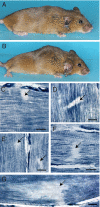
Fig. 1.
Fig. 1.
Morphological and histological abnormalities in IT/+ mice. (A) Twelve-month-old WT and (B) IT/+ female littermates. The IT/+ mouse exhibits dorsal kyphosis and hind limb paresis. Note the flattened posture and outstretched hind limbs that fail to lift the hindquarters. (C–G) NADH-TR-stained longitudinal sections of IT/+ soleus myofibers from 12-month- (C–E) and 20-month-old (F and G) mice showing minicores and cores. In C–E, arrows show minicores occupying eccentric (C), peripheral (E), and central (D) positions. In D, the minicore (arrow) shows intense peripheral staining revealing the distorted cross-striation of focally contracted myofibrils. In F and G, the arrows show cores extending longitudinally over multiple sarcomeres. (Scale bars, 10 mm.)
Fig. 2.
Fig. 2.
Toluidine blue-stained sections from 6-month- (A) and 18-month-old (B) mice. (A) A compact, well-demarcated minicore (arrow) in a single fiber, surrounded by fibers with normal cross-striation. The arrowhead designates a blood vessel. The Inset provides an enlarged image of the minicore. (B) A myofiber featuring a central core area with a severe loss of cross striation. Arrows point to rod-like inclusions colocalizing within the core and running the length of the fiber. (Scale bars, 10 μm.)
Fig. 3.
Fig. 3.
Ultrastructural abnormalities in longitudinal sections of soleus myofibers from 6-week- (A and B) and 6-month-old (C and D) IT/+ mice. (A) A full-width section of a type 2 fiber showing myofibrillar splitting (arrows) and intermingling (bracket). The boxed area shows a focal loss of myofibrillar organization over several sarcomeres. (B) A transitional area between a normally structured area and a compacted core area in a type 1 fiber. The core lesion shows Z-line streaming and focal loss of a Z-disk (arrowheads). Note that intermyofibrillar spaces are reduced and mitochondria and SR are absent from this area (Upper Left diagonal), which is sharply delineated from a contiguous, normally structured region (Lower Right diagonal). Mitochondria in the unaffected area (asterisks) have a normal disposition and appearance. (C) A type 2 fiber shows a well-demarcated lesion with gradual loss of sarcomeric organization from a structured (Upper Right) to an unstructured area (Center). Double-headed arrows show the variability of sarcomeric lengths in the core area. The arrowheads show how loss of sarcomeric register is transmitted to adjacent regions. (D) The central part of an structured core in a type 1 fiber. Z-lines (arrows) are wavy and disintegrating, intermyofibrillar space is reduced, and mitochondria and SR are absent. [Scale bars, 5 μm (A), 2 μm (B and C), and 1 μm (D).]
Fig. 4.
Fig. 4.
Ultrastructural abnormalities in longitudinal sections of a type 1 fiber from an 18-month-old IT/+ mouse. (A) Core-like lesions with rods streaming the length of a full-width myofiber. (B) An unstructured core area with rods, which is contiguous with an area of relatively well-structured myofibrils, illustrating the origin of a “cornucopia” of rods in the Z-line. (C) A higher magnification of a rod from B, revealing its lattice-like structure. [Scale bars, 5 μm (A), 2 μm (B), and 1 μm (D).]
Fig. 5.
Fig. 5.
(A) Quantitative RT-PCR analysis of the relative abundance of allelic Ryr1 transcripts in soleus muscle from 2-month-old IT/+ mice. Representative results of the cDNA analysis of two IT/+ mice (het 1 and het 2) and a normal littermate (WT) are shown. (B) Western blot analysis of the expression of Ca2+ regulatory proteins in microsomes from soleus muscle of WT and IT/+ mice. The amount of total protein (μg) loaded per lane is shown Below the panels.

References

    1. Sharma MC, Jain D, Sarkar C, Goebel HH. Congenital myopathies–a comprehensive update of recent advancements. Acta Neurol Scand. 2009;119:281–292. - PubMed
    1. Sewry CA, Jimenez-Mallebrera C, Muntoni F. Congenital myopathies. Curr Opin Neurol. 2008;21:569–575. - PubMed
    1. Dubowitz V, Sewry CA. Muscle Biopsy: A Practical Approach. 3rd Edition. Amsterdam: Elsevier; 2007.
    1. Bethlem J, Arts WF, Dingemans KP. Common origin of rods, cores, miniature cores, and focal loss of cross-striations. Arch Neurol. 1978;35:555–566. - PubMed
    1. Vallat JM, et al. Coexistence of minicores, cores, and rods in the same muscle biopsy. A new example of mixed congenital myopathy. Acta Neuropathol. 1982;58:229–232. - PubMed

Publication types

MeSH terms

LinkOut - more resources