

Telomere shortening reduces regenerative capacity after acute kidney injury
- PMID: 19959722
- PMCID: PMC2834551
- DOI: 10.1681/ASN.2009010072
Telomere shortening reduces regenerative capacity after acute kidney injury
Abstract
Telomeres of most somatic cells progressively shorten, compromising the regenerative capacity of human tissues during aging and chronic diseases and after acute injury. Whether telomere shortening reduces renal regeneration after acute injury is unknown. Here, renal ischemia-reperfusion injury led to greater impairment of renal function and increased acute and chronic histopathologic damage in fourth-generation telomerase-deficient mice compared with both wild-type and first-generation telomerase-deficient mice. Critically short telomeres, increased expression of the cell-cycle inhibitor p21, and more apoptotic renal cells accompanied the pronounced damage in fourth-generation telomerase-deficient mice. These mice also demonstrated significantly reduced proliferative capacity in tubular, glomerular, and interstitial cells. These data suggest that critical telomere shortening in the kidney leads to increased senescence and apoptosis, thereby limiting regenerative capacity in response to injury.
Figures
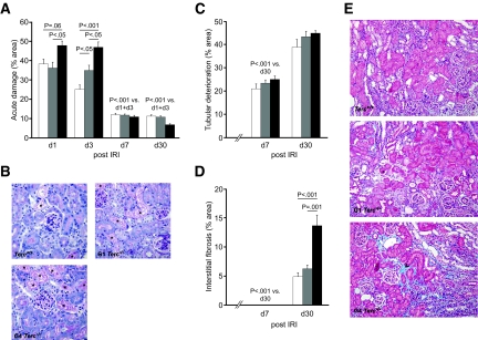
 , G1 Terc−/−; ■, G4 Terc−/−. Data are means ± SEM; significances are indicated. Magnification, ×200.
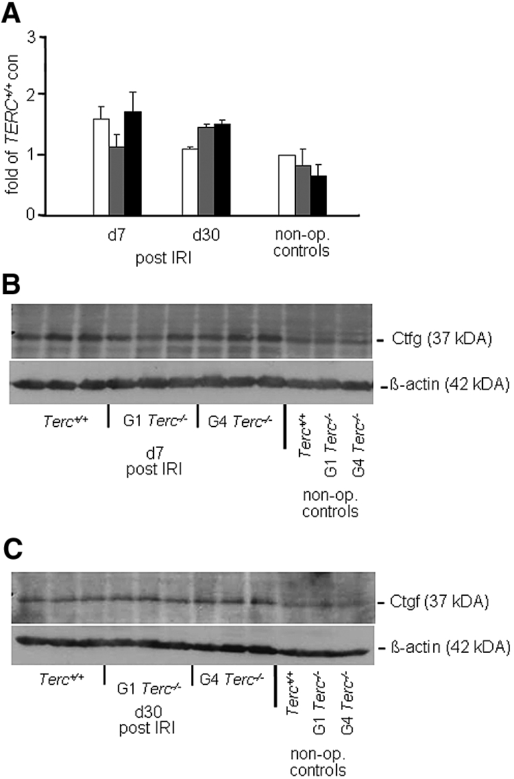 , G1 Terc−/−; ■, G4 Terc−/−. Data are means ± SEM; significances are indicated.
 , G1 Terc−/−; ■, G4 Terc−/−. Data are means ± SEM; significances are indicated. Figure 3 is also provided in color as Supplemental Figure 3.
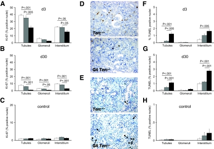 , G1 Terc−/−; ■, G4 Terc−/−. Data are means ± SEM; significances are indicated. Magnification, ×200.
Comment in
-
Telomere shortening and regenerative capacity after acute kidney injury.J Am Soc Nephrol. 2010 Feb;21(2):202-4. doi: 10.1681/ASN.2009121270. Epub 2010 Jan 14. J Am Soc Nephrol. 2010. PMID: 20075064 No abstract available.
References
-
- Lindeman RD, Goldman R: Anatomic and physiologic age changes in the kidney. Exp Gerontol 21: 379–406, 1986 - PubMed
-
- Goyal VK: Changes with age in the human kidney. Exp Gerontol 17: 321–331, 1982 - PubMed
-
- Melk A, Schmidt BM, Takeuchi O, Sawitzki B, Rayner DC, Halloran PF: Expression of p16INK4a and other cell cycle regulator and senescence associated genes in aging human kidney. Kidney Int 65: 510–520, 2004 - PubMed
-
- Schmitt R, Coca S, Kanbay M, Tinetti ME, Cantley LG, Parikh CR: Recovery of kidney function after acute kidney injury in the elderly: A systematic review and meta-analysis. Am J Kidney Dis 52: 262–271, 2008 - PubMed
-
- US Renal Data System: USRDS 2007 Annual Data Report: Atlas of Chronic Kidney Disease and End-Stage Renal Disease in the United States, Bethesda, National Institutes of Health, National Institute of Diabetes and Digestive and Kidney Diseases, 2007
Publication types
MeSH terms
Substances
Grants and funding
LinkOut - more resources
Full Text Sources
Other Literature Sources
Medical
Molecular Biology Databases
