. 2010 Jan;176(1):108-21.
doi: 10.2353/ajpath.2010.090256. Epub 2009 Dec 3.

Small cytoskeleton-associated molecule, fibroblast growth factor receptor 1 oncogene partner 2/wound inducible transcript-3.0 (FGFR1OP2/wit3.0), facilitates fibroblast-driven wound closure

Affiliations

Small cytoskeleton-associated molecule, fibroblast growth factor receptor 1 oncogene partner 2/wound inducible transcript-3.0 (FGFR1OP2/wit3.0), facilitates fibroblast-driven wound closure

Audrey Lin et al. Am J Pathol. 2010 Jan.

Abstract

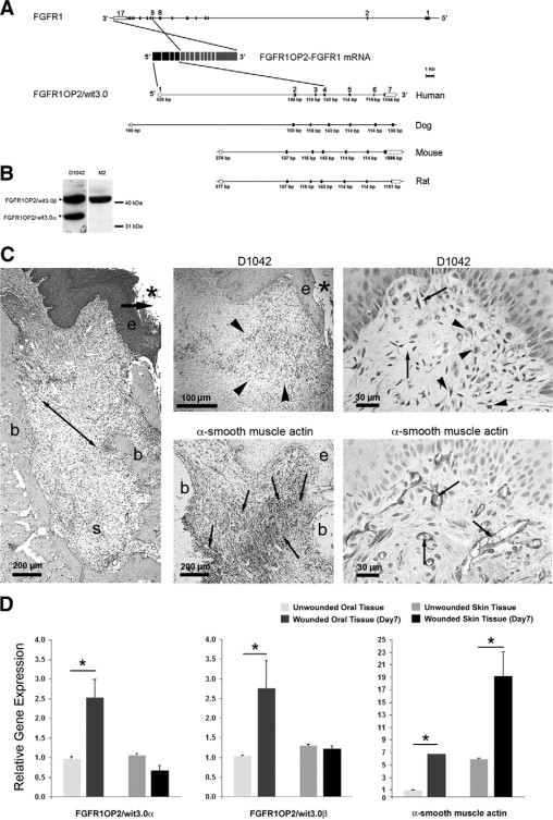
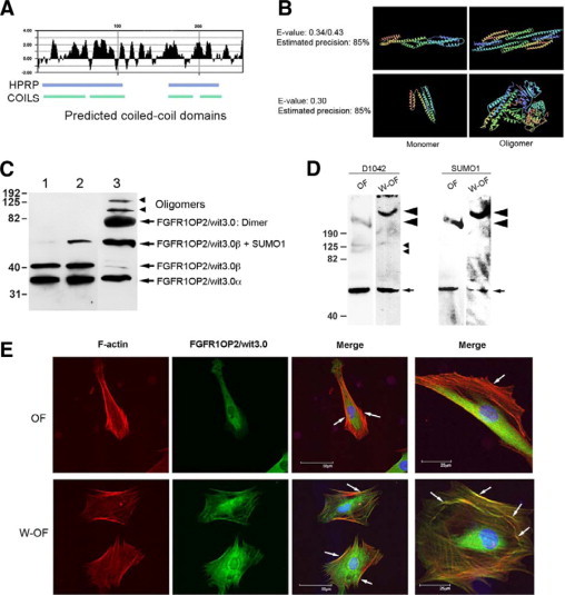
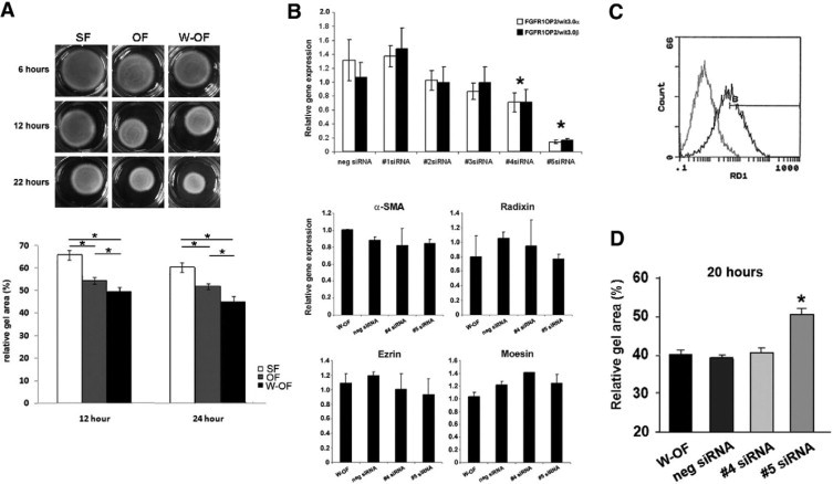
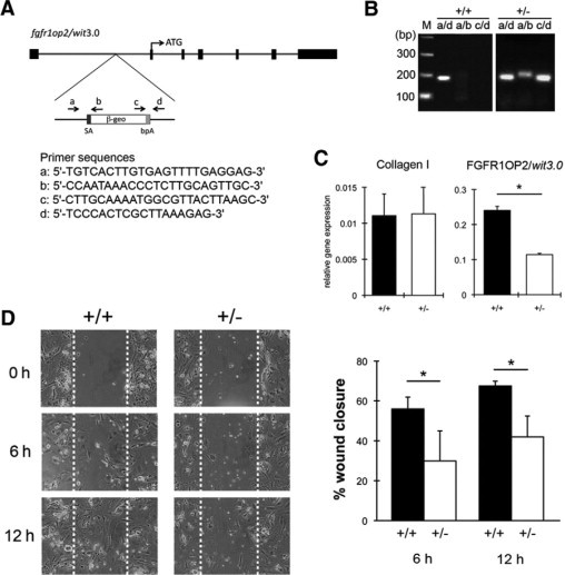
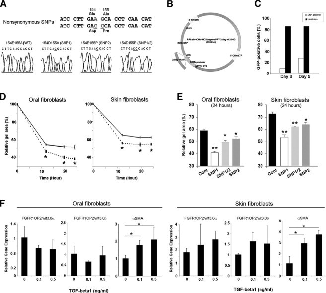
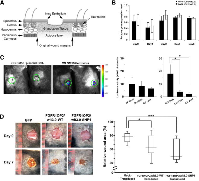
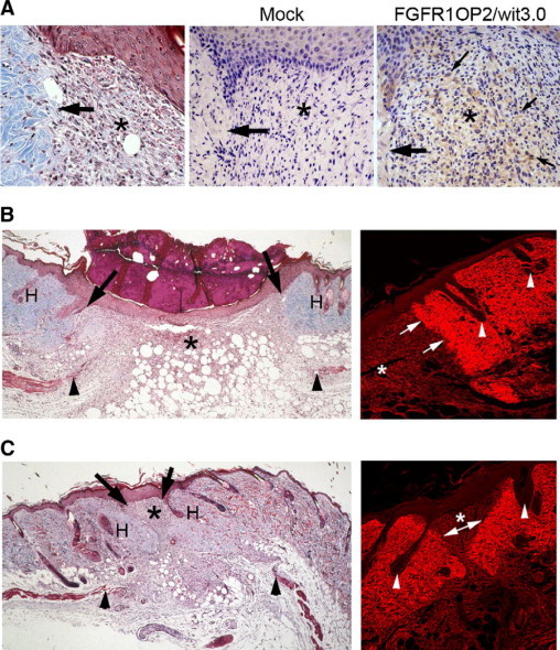
Wounds created in the oral cavity heal rapidly and leave minimal scarring. We have examined a role of a previously isolated cDNA from oral wounds encoding wound inducible transcript-3.0 (wit3.0), also known as fibroblast growth factor receptor 1 oncogene partner 2 (FGFR1OP2). FGFR1OP2/wit3.0 was highly expressed in oral wound fibroblasts without noticeable up-regulation of alpha-smooth muscle actin. In silico analyses, denaturing and nondenaturing gel Western blot, and immunocytology together demonstrated that FGFR1OP2/wit3.0 were able to dimerize and oligomerize through coiled-coil structures and appeared to associate with cytoskeleton networks in oral wound fibroblasts. Overexpression of FGFR1OP2/wit3.0 increased the floating collagen gel contraction of naïve oral fibroblasts to the level of oral wound fibroblasts, which was in turn attenuated by small-interfering RNA knockdown. The FGFR1OP2/wit3.0 synthesis did not affect the expression of collagen I as well as procontractile peptides such as alpha-smooth muscle actin, and transforming growth factor-beta1 had no effect on FGFR1OP2/wit3.0 expression. Fibroblastic cells derived from embryonic stem cells carrying FGFR1OP2/wit3.0 (+/-) mutation showed significant retardation in cell migration. Thus, we postulate that FGFR1OP2/wit3.0 may regulate cell motility and stimulate wound closure. FGFR1OP2/wit3.0 was not up-regulated during skin wound healing; however, when treated with FGFR1OP2/wit3.0 -expression vector, the skin wound closure was significantly accelerated, resulting in the limited granulation tissue formation. Our data suggest that FGFR1OP2/wit3.0 may possess a therapeutic potential for wound management.


Figures

Figure 1
Figure 1
FGFR1OP2/wit3.0 and α-SMA in oral wound. A: Gene structures of FGFR1 and FGFR1OP2/wit3.0. In a case report of acute myeloid leukemia, the exons 1 to 4 of FGFR1OP2/wit3.0 were trans-inserted to the intron between exons 8 and 9 of FGFR1, and the resulting chimerical peptide formed a ligand-independent dimer through the FGFR1OP2/wit3.0-derived coiled-coil structure. The native FGFR1OP2/wit3.0 gene contains seven highly conserved exons. B: Western blot analysis of monospecific polyclonal antibody (D1042) recognized 41 kDa and 32 kDa bands, corresponding to FGFR1OP2/wit3.0β and FGFR1OP2/wit3.0α, respectively, in NIH fibroblasts. The transfected 3XFLAG-FGFR1OP2/wit3.0β fusion peptide was recognized by M2 monoclonal antibody against 3XFLAG epitope at 41 kDa. C: Oral open wound (asterisk) created by tooth extraction showed progressive wound contraction (arrows) by wound margin approximation (e: proliferation/migration front of oral epithelium; b: alveolar bone; s: tooth extraction socket, H&E staining). D1042 antibody stained a highly restricted zone of oral connective tissue (arrowheads) immediately adjacent to the migration/proliferation front of oral epithelium (e). α-SMA (small arrows) was identified along the ligament-like structure (double arrowhead in H&E stained section) connecting alveolar bones (b). D1042 stained the cytoplasma of fibroblasts with cuboidal shapes (arrowheads), whereas α-SMA was found in vascular smooth muscles (small arrows). D: Real-time RT-PCR revealed that FGFR1OP2/wit3.0α and FGFR1OP2/wit3.0β were up-regulated in oral wound tissues but not in skin wound tissues, whereas α-SMA was up-regulated in both oral and skin wound tissues albeit at different levels (*P < 0.05).
Figure 2
Figure 2
FGFR1OP2/wit3.0 coiled-coil structure and its association with cytoskeletal network in oral wound fibroblasts. A: Hydrophylicity plot of FGFR1OP2/wit3.0β and the coiled-coil domains predicted by the Human Protein Reference Database and COILS. B: The Imperial College Protein Homology/Analogy Recognition Engine predicted that three-dimensional structures of FGFR1OP2/wit3.0β would share similar structures of a-spectrin (top) and apoliprotein A-I (bottom), both molecules were known to form dimers and oligomers. C: In denatured Western blot analysis, D1042 antibody depicted FGFR1OP2/wit3.0α and FGFR1OP2/wit3.0β in NIH3T3 fibroblasts (lane 1), NIH3T3 fibroblasts overexpressing FGFR1OP2/wit3.0β (lane 2), and rat oral wound fibroblasts (lane 3). In oral wound fibroblasts, FGFR1OP2/wit3.0 molecules formed dimers and oligomers that were not affected by the reducing agent, β-melcaptoethanol. D: Nondenature gel Western blot analysis of cytoplasmic peptide of naïve oral fibroblasts (OFs) and oral wound fibroblasts (W-OFs) with D1042 indicated a single band at 50 kDa, which was found modified by SUMO-1 (small arrow). The D1042 positive FGFR1OP2/wit3.0 oligomers at 100 kDa and 160 kDa (small arrowheads) were visible in OFs but not in W-OFs. The involvement of FGFR1OP2/wit3.0 in a large molecular moiety of over 190 kDa (large arrowhead) was also noted in both OFs and W-OFs; however, the size of the large molecular moiety and D1042 signal intensity increased in W-OFs. E: Immunocytology of OFs and W-OFs with D1042 antibody (FGFR1OP2/wit3.0: green) as well as phallotoxins (F-actin: red) and DAPI (nuclei: blue). FGFR1OP2/wit3.0 appeared to aggregate in perinucleic cytoplasma. Whereas stress fibers were formed in thick bundles in OF (white arrows), cuboidal shaped W-OFs formed a thin web of cytoskeleton networks (white arrows). Scale bar = 25 μm.
Figure 3
Figure 3
SiRNA knockdown of FGFR1OP2/wit3.0 attenuated collagen gel contraction of oral wound fibroblasts. A: Floating collagen gel contraction assay showed the increased gel contraction by oral wound fibroblasts (W-OFs) as compared with naïve oral fibroblasts (OFs) and skin fibroblasts (SFs) at 12 hours and 24 hours (*P < 0.05). B: Efficacy of five siRNA constructs was evaluated by real time RT-PCR (*P < 0.05 against negative control). The treatment by siRNAs number 4 and number 5 did not affect the expression of contractile and procontractile molecules: α-SMA, radixin, ezrin, and moesin. C: FACS analysis demonstrated the establishment of the optimal transfection protocol for primary oral wound fibroblasts achieving >80% transfection rates (black line) compared with untransfected control (gray line). D: Floating collagen gel contraction assay of oral wound fibroblasts showed that FGFR1OP2/wit3.0 siRNA number 5 significantly attenuated gel contraction (*P < 0.05).
Figure 4
Figure 4
FGFR1OP2/wit3.0 (+/−) mutation reduced the migration ability of mouse ES cell-derived fibroblastic cells. A: Gene Trap mutation was accomplished by inserting a fusion construct of β-galactosidase and neomysin resistant genes (β-geo), which contains splice acceptance sequence (SA) and bovine fibroblast growth factor poly A signal (bpA) into the first intron of mouse fgfrop2/wit3.0 allele. B: Genotype of wild-type (+/+) and FGFROP2/wit3.0 heterogyzous null mutant (+/−) mouse ES cells was determined by PCR. Primers a and d flanked the β-geo construct (Figure 4A) giving rise to the wild-type PCR product (a/d), whereas the combinations of primers a and b (a/b) and c and d (c/d) recognized the β-geo insert. C: Fibroblastic cells derived from wild-type (+/+) and FGFR1OP2/wit3.0 (+/−) mutant ES cells expressed the comparative mRNA level of collagen 1 α 1 chain, whereas the FGFR1OP2/wit3.0 mRNA level was significantly decreased in the mutant cells (*P < 0.5). D:In vitro scratch test revealed that FGFR1OP2/wit3.0 (+/−) mutation significantly decreased fibroblastic cell migration.
Figure 5
Figure 5
FGFR1OP2/wit3.0β overexpression increased collagen gel contraction by both oral and skin fibroblasts. A: Nonsynonymous SNPs in human FGFR1OP2/wit3.0β sequence. B: A schematic structure of lentiviral vector carrying humanized FGFR1OP2/wit3.0β (wild-type, SNP1, SNP2, or SNP1/2) and internal ribosome entry site-green fluorescent protein. C: FACS analysis of the transduction rate of the lentiviral vector for primary oral fibroblasts assessed by green fluorescent protein. D: Floating collagen gel contraction assay revealed that both oral and skin fibroblasts transduced by FGFR1OP2/wit3.0β lentiviral vectors (dotted lines) increased gel contraction rates. E: SNP1, SNP2, and SNP1/2 of FGFR1OP2/wit3.0β increased collagen gel contraction rates. In both oral and skin fibroblasts, SNP1 FGFR1OP2/wit3.0β showed the largest gel contraction rate. F: The treatment of TGF-β1 did not modulate the expression of FGFR1OP2/wit3.0 in oral and skin fibroblasts, whereas it increased the α-SMA expression. *P < 0.05, **P < 0.01.
Figure 6
Figure 6
FGFR1OP2/wit3.0β induced skin wound closures in vivo. A: Schematic diagram of full-thickness wound created in mouse dorsal skin. B: Unlike in oral wounds, the mRNA levels of FGFR1OP2/wit3.0α and FGFR1OP2/wit3.0β remained unchanged in mouse skin excisional wounds during the healing period of day 0 to day 6 (P > 0.05). C: The single-view three-dimensional optical imaging depicting the luciferase reporter gene derived biofluorescence at the mouse dorsal skin excisional wound with plasmid DNA or lentiviral vectors mixed with CG SM50. The luciferase assay measuring the transfection/transduction efficiency of plasmid DNA or lentiviral vectors mixed with collagen gel carrier (Cell Prime, CP) or CG. Lentiviral vectors mixed with CG showed the highest transfection/transduction efficiency. D: Mouse dorsal skin excisional wound treated with wild-type or SNP1 FGFR1OP2/wit3.0β lentiviral vectors mixed with CG carrier. The wound areas contraction measured at day 7 indicated the significant closure of wounds treated with FGFR1OP2/wit3.0β or SNP1 FGFR1OP2/wit3.0β. *P < 0.05, ***P < 0.001.
Figure 7
Figure 7
Decreased granulation tissue and scarring after FGFR1OP2/wit3.0β treatment. A: Histological specimen of mouse dorsal skin showed a clear demarcation of excisional wounding (arrow) flanking the highly cellular granulation tissue (asterisk, left; 7 days after wounding Goldner's trichrome staining). Immunostaining with D1042 was negative in the Mock-transducation group using green fluorescent protein lentiviral vector. FGFR1OP2/wit3.0β lentiviral vector treatment resulted in the strong D1042 immunostaining in fibroblasts of the granulation tissue juxtaposing the excisinal wound site (arrow). B: Mock-treated mouse skin excisional wound healing (day 7). While the blunted panniculus carnosus layer (arrowheads) remained patent, the granulation tissues asterisk were formed between the dermis wound edges (arrows) and covered by new epithelial keratinocytes. The expenditures of skin such as hair follicles (H) were not observed in the granulation tissue (left; Goldner's trichrome staining). Confocal laser scanning micrograph of skin wound stained by Sirius Red showed that collagen fibers were strongly stained in the dermis containing hair follicles (arrowheads). The dermis wound edge was clearly visible (arrows) after 7 days of wounding. The granulation tissue asterisk contained less organized collagen fibers (right). C: Histological specimen of mouse dorsal skin excisional wound treated with FGFR1OP2/wit3.0β. The blunted panniculus carnosus layer indicates the original wound edge (arrowheads). The dermis was significantly approximated toward the wound center (arrows) leaving a small area of granulation tissue asterisk covered by the keratinocyte layer. The hair follicles (H) were present in the wound area following the dermis closure (left; Goldner's trichrome staining). Confocal laser scanning micrograph of the Sirius Red stained mouse skin wound treated with FGFR1OP2/wit3.0β. The dermis wound edges (arrows) were approximated toward the wound center, surrounding a small granulation tissue (asterisk, right).

Similar articles

Cited by

References

    1. Martin P. Wound healing: aiming for perfect skin regeneration. Science. 1997;276:75–81. - PubMed
    1. Singer AJ, Clark RAF. Cutaneous wound healing. New Eng J Med. 1999;341:738–746. - PubMed
    1. Desmouliere A, Chaponnier C, Gabbiani G. Tissue repair, contraction, and the myofibroblast. Wound Repair Regen. 2005;13:7–12. - PubMed
    1. Darenfed H, Mandato CA. Wound-induced contractile ring: a model for cytokinesis. Biochem Cell Biol. 2005;83:711–720. - PubMed
    1. Hinz B. Formation and function of the myofibroblast during tissue repair. J Invest Dermatol. 2007;127:526–537. - PubMed

Publication types

MeSH terms