. 2009 Dec;119(12):3544-55.
doi: 10.1172/JCI40093.

Nonpathogenic SIV infection of African green monkeys induces a strong but rapidly controlled type I IFN response

Affiliations

Nonpathogenic SIV infection of African green monkeys induces a strong but rapidly controlled type I IFN response

Béatrice Jacquelin et al. J Clin Invest. 2009 Dec.

Abstract

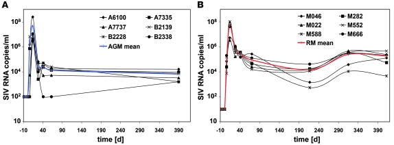
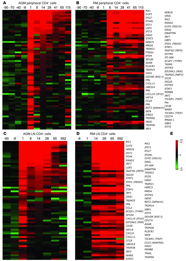
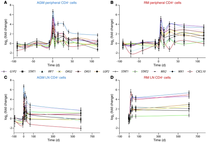
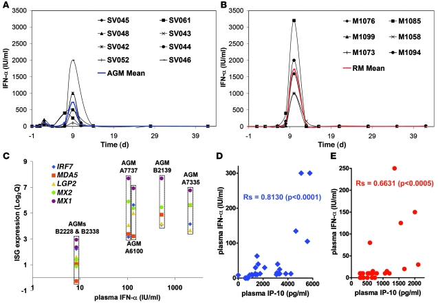
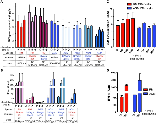
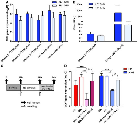
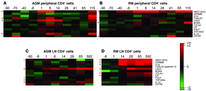
African green monkeys (AGMs) infected with the AGM type of SIV (SIVagm) do not develop chronic immune activation and AIDS, despite viral loads similar to those detected in humans infected with HIV-1 and rhesus macaques (RMs) infected with the RM type of SIV (SIVmac). Because chronic immune activation drives progressive CD4+ T cell depletion and immune cell dysfunctions, factors that characterize disease progression, we sought to understand the molecular basis of this AGM phenotype. To this end, we longitudinally assessed the gene expression profiles of blood- and lymph node-derived CD4+ cells from AGMs and RMs in response to SIVagm and SIVmac infection, respectively, using a genomic microarray platform. The molecular signature of acute infection was characterized, in both species, by strong upregulation of type I IFN-stimulated genes (ISGs). ISG expression returned to basal levels after postinfection day 28 in AGMs but was sustained in RMs, especially in the lymph node-derived cells. We also found that SIVagm induced IFN-alpha production by AGM cells in vitro and that low IFN-alpha levels were sufficient to induce strong ISG responses. In conclusion, SIV infection triggered a rapid and strong IFN-alpha response in vivo in both AGMs and RMs, with this response being efficiently controlled only in AGMs, possibly as a result of active regulatory mechanisms.


Figures

Figure 1
Figure 1. Plasma viral loads.
Plasma SIV RNA copy numbers (cut off value: 0.5 × 10² to 1 × 10² copies/ml of plasma) in (A) AGMs and (B) RMs. Designations for individual animals are shown. The mean is indicated in blue for AGMs and in red for RMs.
Figure 2
Figure 2. Type I ISG expression in blood and LN CD4+ cells.
The genes of the ISG cluster that were significantly regulated (P < 0.05) in at least 1 of the 2 species are represented here as heatmaps. Mean values of the log2Q of type I ISG expression in peripheral (A and B, respectively) and LN CD4+ cells (C and D, respectively) from 6 AGMs and 6 RMs are shown. Many of these genes can also be induced by IFN-II. (E) The color scheme indicates the log2Q. Gene expressions shown in red were upregulated, and those shown in green were downregulated.
Figure 3
Figure 3. Expression profiles of representative type I ISGs.
Microarray gene expression profiles of 11 ISGs are shown for CD4+ cells in (A and B) blood and (C and D) LN from (A and C) AGMs and (B and D) RMs. Statistics associated with these profiles are provided in Supplemental Table 1.
Figure 4
Figure 4. Systemic IFN-α levels and correlation with type I ISG expression and plasma IP-10 quantities.
Levels of bioactive IFN-α in plasma of (A) 8 additional AGMs and (B) 6 additional RMs. The detection level of the assay was 2 IU/ml. (C) Expression levels of 5 upregulated ISGs at day 1 p.i. were plotted against respective plasma IFN-α levels for the 6 AGMs included in the microarray analysis. Correlation between IFN-α and IP-10 plasma levels in (D) AGMs (blue) and (E) RMs (red) during acute infection (days 1–28). The Spearman coefficient (Rs) is indicated.
Figure 5
Figure 5. In vitro induction of type I ISG and IFN-α.
Peripheral CD4+ cells of uninfected AGMs (blue) and RMs (red) were cultured in the presence of the CD4 cell fraction in a 2-chamber system. (A and B) Cells were incubated for 12, 18, and 24 hours with SIV or 1,500 IU/ml of human recombinant IFN-α. Viral infectious titers used are indicated. (C and D) Cells were incubated with a range of doses of recombinant IFN-α. (A and C) For MX1 gene expression, the mean ± SEM of log2Q is shown. (B and D) Means of the amounts of bioactive IFN-α produced in supernatants are represented (±SEM). The means were calculated from 3 independent experiments with cells from 3 different animals of each species.
Figure 6
Figure 6. In vitro restimulation of AGM cells.
(A and B) As in Figure 5, peripheral CD4+ cells of 5 uninfected and 6 chronically infected AGMs were cultured in the presence of the CD4 cell fraction for 18 hours with SIV or IFN-α. (A) MX1 gene expression (mean ± SEM of log2Q). (B) Mean ± SEM of bioactive IFN-α produced in supernatants. The difference in IFN-α production between SIV+ and SIV AGMs was not significant. (C and D) Repeated treatments with recombinant IFN-α were conducted on PBMCs from 3 healthy AGMs and 2 healthy RMs as described in Methods and shown in C. (D) MX1 gene expression. **P < 0.01, ***P < 0.001.
Figure 7
Figure 7. Microarray results for immunosuppressive genes in blood and LN CD4+ cells of SIVagm-infected AGMs and SIVmac-infected RMs.
Heatmaps of expression profiles of immunosuppressive genes are shown for AGM and RM peripheral CD4+ cells (A and B, respectively) and LN CD4+ cells (C and D, respectively). The color scheme indicates the log2Q. Gene expressions shown in red were upregulated and those shown in green were downregulated with respect to baseline levels. Red asterisks indicate probes found to be significantly increased, and green asterisks those significantly decreased after infection as compared with before infection (P < 0.05).

Comment in

Similar articles

Cited by

References

    1. Giorgi J.V., et al. Shorter survival in advanced human immunodeficiency virus type 1 infection is more closely associated with T lymphocyte activation than with plasma virus burden or virus chemokine coreceptor usage. J. Infect. Dis. 1999;179:859–870. doi: 10.1086/314660. - DOI - PubMed
    1. Sousa A.E., Carneiro J., Meier-Schellersheim M., Grossman Z., Victorino R.M. CD4 T cell depletion is linked directly to immune activation in the pathogenesis of HIV-1 and HIV-2 but only indirectly to the viral load. J. Immunol. 2002;169:3400–3406. - PubMed
    1. Deeks S.G., et al. Immune activation set point during early HIV infection predicts subsequent CD4+ T-cell changes independent of viral load. Blood. 2004;104:942–947. doi: 10.1182/blood-2003-09-3333. - DOI - PubMed
    1. Hazenberg M.D., et al. Persistent immune activation in HIV-1 infection is associated with progression to AIDS. AIDS. 2003;17:1881–1888. doi: 10.1097/00002030-200309050-00006. - DOI - PubMed
    1. van Asten L., et al. Pre-seroconversion immune status predicts the rate of CD4 T cell decline following HIV infection. AIDS. 2004;18:1885–1893. doi: 10.1097/00002030-200409240-00004. - DOI - PubMed

Publication types

MeSH terms