. 2010 Jan 20;29(2):505-14.
doi: 10.1038/emboj.2009.352. Epub 2009 Dec 3.

How baculovirus polyhedra fit square pegs into round holes to robustly package viruses

Affiliations

How baculovirus polyhedra fit square pegs into round holes to robustly package viruses

Xiaoyun Ji et al. EMBO J. .

Abstract

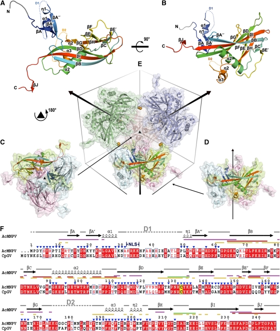
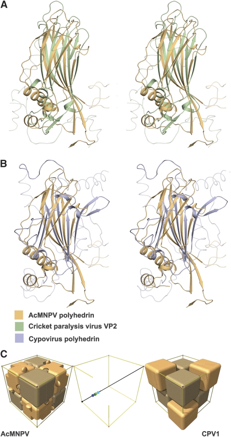
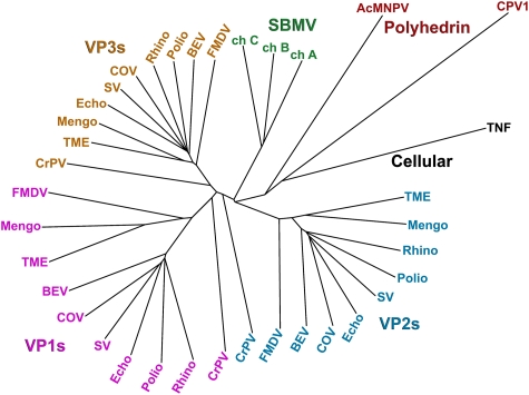
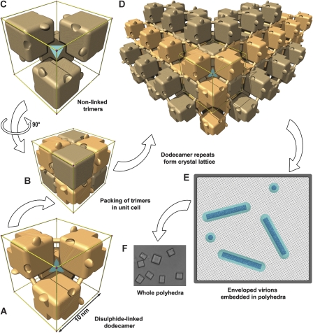
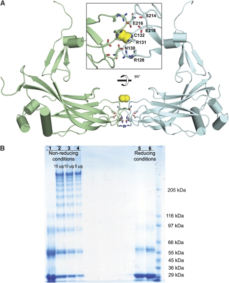
Natural protein crystals (polyhedra) armour certain viruses, allowing them to survive for years under hostile conditions. We have determined the structure of polyhedra of the baculovirus Autographa californica multiple nucleopolyhedrovirus (AcMNPV), revealing a highly symmetrical covalently cross-braced robust lattice, the subunits of which possess a flexible adaptor enabling this supra-molecular assembly to specifically entrap massive baculoviruses. Inter-subunit chemical switches modulate the controlled release of virus particles in the unusual high pH environment of the target insect's gut. Surprisingly, the polyhedrin subunits are more similar to picornavirus coat proteins than to the polyhedrin of cytoplasmic polyhedrosis virus (CPV). It is, therefore, remarkable that both AcMNPV and CPV polyhedra possess identical crystal lattices and crystal symmetry. This crystalline arrangement must be particularly well suited to the functional requirements of the polyhedra and has been either preserved or re-selected during evolution. The use of flexible adaptors to generate a powerful system for packaging irregular particles is characteristic of the AcMNPV polyhedrin and may provide a vehicle to sequester a wide range of objects such as biological nano-particles.


Conflict of interest statement

The authors declare that they have no conflict of interest.

Figures

Figure 1
Figure 1
Overview of structure. (A, B) The structure of AcMNPV polyhedrin, in two orthogonal views, shown as cartoon representations coloured from blue at the N-terminus to red at the C-terminus. Residues 3–7, which were built as poly-ala are drawn in grey. Disordered segments D1 (residues 32–48) and D2 (residues 174–186) are shown as dots. (C, D) Cartoon and surface representations of polyhedrin trimers shown in two orientations aligned to (E). Subunits are coloured by chain with one in each trimer coloured as in (A). (E) Dodecameric unit of four trimers linked by disulphide bonds (represented as orange spheres). The unit cell and three-fold axes are drawn to facilitate observation. (F) Amino-acid sequence conservation between the type species of the alphaBVs (AcMNPV) and betaBVs (CpGV). Conserved residues drawn in red boxes, similar residues in red type. The secondary structure assignment for AcMNPV is drawn above. Conserved cysteine is highlighted in yellow. Interactions involved in various stages of oligomization are mapped as lines coloured thus; purple for trimer, green for dodecamer, red for interactions through C-terminal and yellow for other interactions in the unit cell. The position of the nuclear localization signal (NLS) is indicated and residues exposed to the cavity are marked by blue triangles. The sequences were aligned with ClustalW (Chenna et al, 2003) and visualized using ESPript (Gouet et al, 1999).
Figure 2
Figure 2
Structural superpositions. Stereo views showing the alignment of (A) AcMNPV polyhedrin (orange) with cricket paralysis virus VP2 (green) (Tate et al, 1999) and (B) AcMNPV polyhedrin (orange) with CPV1 polyhedrin (purple). The program SHP (Stuart et al, 1979) was used to perform the superpositions. (C) Schematic representation of the packing of both AcMNPV and CPV1 polyhedrin trimers (shown as blocks) within their unit cells. The trimeric units are positioned rather differently on the three-fold body diagonal—this is represented in the central panel in which the centres of the trimers of differing polarity are reflected into the (0,0,0) to (½, ½, ½) portion. The AcMNPV trimer centres (shown in shades of green) lie close to (¼, ¼, ¼), whereas the CPV1 trimers (centres shown in shades of blue) are offset by about 6 Å. This accounts for the differences seen in the outer panels.
Figure 3
Figure 3
Phylogenetic tree showing the relationship between the polyhedrin proteins of AcMNPV and CPV1, against picorna-like virus capsid proteins. Superpositions were performed using SHP (Stuart et al, 1979) and the phylogenetic tree calculated with PHYLIP (Felsenstein, 1989). The following abbreviations are used: AcMNPV, Autographa californica baculovirus; BEV, bovine enterovirus; ch, chain; COV, coxsackievirus A3; CPV1, cytoplasmic polyhedrosis virus type 1; CrPV, cricket paralysis virus; Echo, echovirus; FMDV, foot-and-mouth disease virus; Mengo, mengo virus; Polio, poliovirus; Rhino, rhinovirus; SBMV, southern bean mosaic virus; SV, swine vesicular disease virus; TME, theiler murine encephalomyelitis virus; TNF, tumour necrosis factor. Note that CPV1 polyhedrin is closest to AcMNPV polyhedrin, which is, however, closer to the picornavirus capsid structures than CPV1.
Figure 4
Figure 4
Schematic representations of polyhedra organization. Polyhedrin trimers are depicted as simplified cubic blocks, with the C-terminal hooks and pockets present. To clarify interpretation, the edges of the unit cell are shown in gold and a cyan tetrahedron symbolizes the cell centre. Within a unit cell, disulphide-linked trimers with one polarity are coloured light beech (A) and those with the opposite polarity are coloured light brown (C). (B) All eight trimers in the unit cell. The disulphide bond connecting adjoining trimers is shown as a dowel. (D) The crystal lattice is built up from repeats of the dodecameric unit. (E) Sketch of a cross-section through a polyhedron. The lattice spacing of the unit cells is illustrated as a dot pattern into which are embedded nucleocapsids (dark blue) surrounded by an envelope (cyan). (F) Light microscopy image of G25D mutant AcMNPV polyhedra.
Figure 5
Figure 5
Inter-subunit disulphide bond. (A) Cartoon diagram of two disulphide-bonded polyhedrin related by a two-fold rotation forming an extended dimer. Structures are coloured by chains and the helices are shown in cylinders. Residues involved in intermolecular salt bridges are drawn as sticks and disulphides are shown as yellow spheres in two orthogonal views. (B) SDS–PAGE of AcMNPV polyhedra under reducing and non-reducing conditions when dissolved in bicarbonate buffer, pH 10.5. Polyhedra were incubated at 80°C for 2 h to inactivate the protease associated with polyhedra before exposure to alkaline buffer. Samples were electrophoresed on 4–8% gradient SDS gels with 1 min (lanes 2–4 and 6) and 5 min (lanes 1 and 5) heating at 100°C.

References

    1. Abrahams JP, Leslie AG (1996) Methods used in the structure determination of bovine mitochondrial F1 ATPase. Acta Crystallogr D Biol Crystallogr 52(Pt 1): 30–42 - PubMed
    1. Ackermann HW, Smirnoff WA (1983) A morphological investigation of 23 baculoviruses. J Invert Pathol 41: 269–280
    1. Adams PD, Grosse-Kunstleve RW, Hung LW, Ioerger TR, McCoy AJ, Moriarty NW, Read RJ, Sacchettini JC, Sauter NK, Terwilliger TC (2002) PHENIX: building new software for automated crystallographic structure determination. Acta Crystallogr D Biol Crystallogr 58(Pt 11): 1948–1954 - PubMed
    1. Anduleit K, Sutton G, Diprose JM, Mertens PP, Grimes JM, Stuart DI (2005) Crystal lattice as biological phenotype for insect viruses. Protein Sci 14: 2741–2743 - PMC - PubMed
    1. Bamford DH, Grimes JM, Stuart DI (2005) What does structure tell us about virus evolution? Curr Opin Struct Biol 15: 655–663 - PubMed

Publication types

LinkOut - more resources