. 2010 Jan 20;29(2):387-97.
doi: 10.1038/emboj.2009.337. Epub 2009 Dec 3.

Enforcing temporal control of maternal mRNA translation during oocyte cell-cycle progression

Affiliations

Enforcing temporal control of maternal mRNA translation during oocyte cell-cycle progression

Karthik Arumugam et al. EMBO J. .

Abstract

Meiotic cell-cycle progression in progesterone-stimulated Xenopus oocytes requires that the translation of pre-existing maternal mRNAs occur in a strict temporal order. Timing of translation is regulated through elements within the mRNA 3' untranslated region (3' UTR), which respond to cell cycle-dependant signalling. One element that has been previously implicated in the temporal control of mRNA translation is the cytoplasmic polyadenylation element (CPE). In this study, we show that the CPE does not direct early mRNA translation. Rather, early translation is directed through specific early factors, including the Musashi-binding element (MBE) and the MBE-binding protein, Musashi. Our findings indicate that although the cyclin B5 3' UTR contains both CPEs and an MBE, the MBE is the critical regulator of early translation. The cyclin B2 3' UTR contains CPEs, but lacks an MBE and is translationally activated late in maturation. Finally, utilizing antisense oligonucleotides to attenuate endogenous Musashi synthesis, we show that Musashi is critical for the initiation of early class mRNA translation and for the subsequent activation of CPE-dependant mRNA translation.


Conflict of interest statement

The authors declare that they have no conflict of interest.

Figures
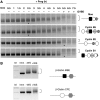
Figure 1
Figure 1
The cytoplasmic polyadenylation element (CPE) combinatorial code does not correctly predict cyclin B2 mRNA polyadenylation. (A) Oocytes treated with or without progesterone for the indicated times were analyzed for initiation of polyadenylation of multiple endogenous mRNAs by RNA ligation-coupled PCR from the same cDNA preparation at each time point. Oocytes reached GVBD50 (see Materials and methods) at 5.5 h and were segregated into those that had not (−) or had (+) completed GVBD. An increase in the size of the mRNA population is indicative of polyadenylation (bracketed). An asterisk indicates when polyadenylation of each mRNA initiated as evidenced by a shift in mRNA population size above the basal size in immature oocytes (indicated by a dotted line). On the right of the panel, a schematic representation of each mRNA 3′ untranslated region (UTR) shows the position of consensus Musashi-binding sites (solid black square), consensus CPEs (white circle), non-consensus CPEs (diagonal bar circle) and consensus polyadenylation hexanucleotides (grey hexagon). (B) β-globin reporter 3′ UTRs were generated by inserting a consensus Musashi-binding site (solid black square) or a consensus CPE (white circle) 5′ of the polyadenylation hexanucleotide (grey hexagon). Separate pools of oocytes were injected with the indicated mRNA reporter constructs, incubated for approximately 16 h and then left untreated or stimulated with progesterone. When 50% of the progesterone-treated oocyte population reached GVBD, oocytes were segregated based on whether they had (+WS) or had not (−WS) completed GVBD and RNA isolated. RNA was also isolated from time matched immature oocyte samples. The polyadenylation status of the reporter mRNAs analyzed by RNA-ligation coupled PCR using appropriate forward primers. It should be noted that in the absence of inserted elements, the β-globin 3′ UTR does not undergo progesterone-dependant polyadenylation (Hyman and Wormington, 1988). The experiment was repeated three times with identical results.
Figure 2
Figure 2
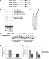
Early translational activation of the cyclin B5 3′ untranslated region (UTR) requires a Musashi-binding element (MBE). (A) Schematic showing the regulatory elements of cyclin B5 3′ UTR fused to a Firefly luciferase reporter (see legend to Figure 1 for symbol definitions). The β-globin 3′ UTR lacks progesterone-responsive elements and so serves as an unregulated control luciferase reporter mRNA. (B) RNA EMSA utilizing a biotin-labelled cyclin B1 3′ UTR and reticulocyte expressing either the GST moiety alone or GST fused to the C-terminal RNA binding domain of CPEB (ΔN-CPEB). A specific cyclin B1 RNA binding complex is formed with ΔN-CPEB but not GST (arrowhead). This complex can be effectively competed with unlabelled wild-type (wt) cyclin B5 3′ UTR and the Musashi-binding mutant form of the cyclin B5 3′ UTR (mbm). By contrast, no competition for ΔN-CPEB binding is seen with a cytoplasmic polyadenylation element (CPE)-disrupted cyclin B5 3′ UTR (CPE mut). (C) GST Western blot demonstrates equivalent levels amount of GST and GST-ΔN-CPEB protein expression in the reticulocyte lysates used in (B). (D) Wild-type (wt) or Musashi-binding mutant (mbm) cyclin B5 3′ UTRs were fused to a GST open reading frame and the resulting transcribed RNA injected into immature oocytes. The polyadenylation status of the reporter RNAs was assessed by RNA ligation PCR where an increase in the size of the mRNA population is indicative of polyadenylation (bracketed). Imm, immature oocytes. Whether oocytes had (+) or had not (−) completed GVBD is indicated above each time point. (E) Oocytes were co-injected with a Renilla luciferase mRNA and a Firefly luciferase RNA under the control of either the wild-type (wt) or polyadenylation hexanucleotide mutant cyclin B5 UTR and incubated for 16 h before progesterone treatment. When the oocytes reached GVBD50, oocytes were segregated based on whether they had or had not completed GVBD. For these experiments, three pools of five oocytes were harvested for each analysis from the oocytes, which had not yet completed GVBD (and so are considered to be in the early phases of maturation). In addition, three pools of five oocytes were harvested from time matched immature oocyte samples. The triplicate sets of Firefly:Renilla ratios of the cyclin B5 UTR constructs from progesterone-stimulated oocytes were normalized to the Firefly:Renilla ratios for the same 3′ UTR constructs in immature oocytes (ratio P:I). The data presented are the mean±SEM from three independent experiments. (F) Oocytes were co-injected with a Renilla luciferase mRNA and a Firefly luciferase RNA under the control of either the wild-type (wt) or Musashi binding mutant (mbm) cyclin B5 UTR. Luciferase analyses were performed as described in (E) and the data are presented from two independent experiments with the SEM shown for each individual experiment. We confirmed that the reporter mRNAs were expressed to similar levels (Supplementary Figure S3).
Figure 3
Figure 3
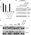
Antisense oligonucleotides targeting endogenous Musashi1 and Musashi2 mRNAs inhibit oocyte maturation. (A) Immature oocytes were injected with water, 200 ng control antisense oligonucleotide (Control AS) or a combination of 100 ng xMsi1 and 100 ng xMsi2 antisense oligonucleotides (xMsi1+xMsi2 AS) and incubated for 16 h. The oocytes were then stimulated with progesterone and GVBD scored over the course of the experiment. (B) Pools of 10 oocytes from each experimental condition in panel (A) were harvested when control AS-injected oocytes reached GVBD50. Time-matched immature and progesterone-treated samples were analyzed for MAP kinase and cdc2 activation. In these analyses, phosphorylated MAP kinase (pMAPK) indicates activation and loss of phosphorylated (and inhibited) cdc2 (pcdc2) indicates activation. Ringo and CPEB protein levels were also assessed in the same samples by western blotting, with tubulin serving as an internal control for protein loading. The data are representative of three separate experiments. (C) Oocytes were injected with water, 200 ng control antisense oligonucleotide (AS), or a combination of 100 ng xMsi1 and 100 ng xMsi2 AS and incubated for 16 h before progesterone stimulation. Pools of 10 oocytes were harvested for each condition when control AS-injected oocytes reached GVBD50. Similar to (A), the xMsi1/2 AS-injected oocytes failed to mature in response to progesterone stimulation. The polyadenylation of endogenous cyclin B5, Mos, TATA BP2, cyclin B2 and cyclin B1 mRNAs was determined using RNA ligation-coupled PCR from the same cDNA preparations at each time point. Progesterone-dependant polyadenylation of each mRNA is indicated (bracket). The experiment was repeated three times with similar results.
Figure 4
Figure 4
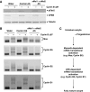
Musashi, but CPEB, controls the activation of early class mRNAs and enforces temporal regulation of maternal mRNA polyadenylation and translation. (A) Oocytes were separately injected with 100 ng control antisense oligonucleotide (AS) or a combination of 50 ng xMsi1 and 50 ng xMsi2 AS and incubated for 16 h before progesterone stimulation. The injected oocytes were then split into pools and left untreated (Con AS and Msi AS) or re-injected with the indicated GST fusion proteins: wild-type Musashi1 (GST–Msi); RNA binding mutant Musashi (GST–Msi bm) or CPEB (GST–CPEB). After addition of progesterone, oocyte GVBD was assessed after 7 h. The data represent three independent experiments with the SEM indicated. UI, uninjected oocytes treated with progesterone. (B) Lysates from the experiment in (A) were assessed for expression of the GST fusion proteins (upper panel). The anticipated full length 70 kD wild-type (wt) and RNA binding mutant (bm) Musashi and 90 kD CPEB proteins were detected at similar levels. Minor CPEB breakdown products were also observed in these samples. Active MAPK kinase (pMAPK) and inactive cdc2 (pcdc2) were assessed as described in the legend to Figure 3. In these experiments, some active MAPK was detected in Msi AS injected oocytes, but the levels were below the enhanced level seen in control that had completed GVBD. No activation of cdc2 was observed in Msi AS or Msi AS oocytes expressing Msi bm or CPEB. A tubulin western blot of the same lysates illustrates equivalent loading of protein in all lanes. Whether the oocytes did (+) or did not (−) complete GVBD after progesterone stimulation is indicated. (C) Total RNA was prepared from pools of five oocytes harvested from immature or progesterone-stimulated samples described in (A) above when control AS-injected oocytes reached GVBD50. Progesterone-stimulated oocytes were segregated based on whether they had or had not completed GVBD (+ and −, respectively). The polyadenylation of endogenous cyclin B5 and cyclin B1 mRNAs was determined using RNA ligation-coupled PCR from the same cDNA preparations at each time point.
Figure 5
Figure 5
A regulatory hierarchy governs activation of early and late class mRNAs. Oocytes were injected with water or antisense oligonucleotides (AS) as indicated and incubated for 16 h before Cyclin B Δ87 protein injection. Injection of cyclin B Δ87 induced cdc2 activation and GVBD in water, control AS- and Musashi AS-injected oocytes. In these experiments, GVBD50 occurred 2–3 h after injection. Cyclin B Δ87 injected oocytes were harvested when all the oocytes had completed GVBD and compared with samples prepared from time matched, mock-injected oocytes (cyclin B Δ87+or – as indicated). (A) Lysates were analyzed for cdc2 and CPEB activation status by western blotting (see Figure 3 legend). A tubulin western blot of the same samples serves as an internal loading control. (B) Polyadenylation of endogenous mRNAs (brackets) was assessed by RNA ligation-coupled PCR of the same oocyte samples used in (A). An increase in size of the PCR products is indicative of polyadenylation. (C) A model illustrating the regulatory roles of the Musashi-binding element (MBE) and the cytoplasmic polyadenylation element (CPE) in establishing the temporal order of maternal mRNA translation. Musashi is necessary for both early class, CPE-independent mRNA translational activation as well as for subsequent CPE-dependant mRNA translation. See text for details.

Similar articles

Cited by

References

    1. AndrÈsson T, Ruderman JV (1998) The kinase Eg2 is a component of the Xenopus oocyte progesterone-activated signaling pathway. EMBO J 17: 5627–5637 - PMC - PubMed
    1. Ballantyne S, Daniel DLJ, Wickens M (1997) A dependent pathway of cytoplasmic polyadenylation reactions linked to cell cycle control by c-mos and CDK1 activation. Mol Biol Cell 8: 1633–1648 - PMC - PubMed
    1. Barkoff AF, Dickson KS, Gray NK, Wickens M (2000) Translational control of cyclin B1 mRNA during meiotic maturation: coordinated repression and cytoplasmic polyadenylation. Dev Biol 220: 97–109 - PubMed
    1. Castro A, Mandart E, Lorca T, Galas S (2003) Involvement of Aurora A kinase during meiosis I–II transition in Xenopus oocytes. J Biol Chem 278: 2236–2241 - PubMed
    1. Charlesworth A, Cox LL, MacNicol AM (2004) Cytoplasmic polyadenylation element (CPE)- and CPE-binding protein (CPEB)-independent mechanisms regulate early class maternal mRNA translational activation in xenopus oocytes. J Biol Chem 279: 17650–17659 - PMC - PubMed

Publication types

MeSH terms