. 2009 Dec 16;28(24):3820-31.
doi: 10.1038/emboj.2009.365.

Functional involvement of Tudor and dPRMT5 in the piRNA processing pathway in Drosophila germlines

Affiliations

Functional involvement of Tudor and dPRMT5 in the piRNA processing pathway in Drosophila germlines

Kazumichi M Nishida et al. EMBO J. .

Abstract

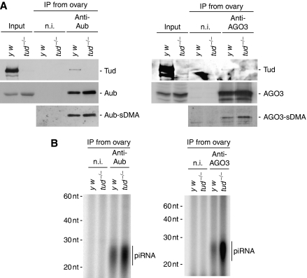
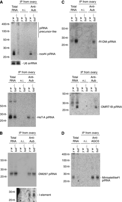
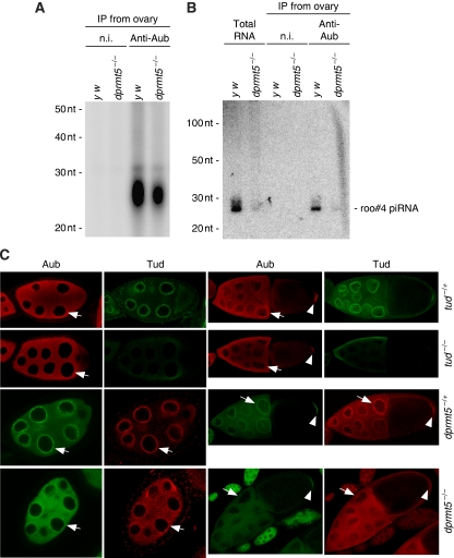
In Drosophila, the PIWI proteins, Aubergine (Aub), AGO3, and Piwi are expressed in germlines and function in silencing transposons by associating with PIWI-interacting RNAs (piRNAs). Recent studies show that PIWI proteins contain symmetric dimethyl-arginines (sDMAs) and that dPRMT5/Capsuleen/DART5 is the modifying enzyme. Here, we show that Tudor (Tud), one of Tud domain-containing proteins, associates with Aub and AGO3, specifically through their sDMA modifications and that these three proteins form heteromeric complexes. piRNA precursor-like molecules are detected in these complexes. The expression levels of Aub and AGO3, along with their degree of sDMA modification, were not changed by tud mutations. However, the population of transposon-derived piRNAs associated with Aub and AGO3 was altered by tud mutations, whereas the total amounts of small RNAs on Aub and AGO3 was increased. Loss of dprmt5 did not change the stability of Aub, but impaired its association with Tud and lowered piRNA association with Aub. Thus, in germline cells, piRNAs are quality-controlled by dPRMT5 that modifies PIWI proteins, in tight association with Tud.


Conflict of interest statement

The authors declare that they have no conflict of interest.

Figures

Figure 1
Figure 1
LC-MS/MS analysis of Aub modification. (A) Aub peptide identified by database searching. Letters with yellow background are search hits. Those with green background indicate amino acids with modifications; of those, M, R, and C indicate methionine with oxidation, arginine with sDMA, and cysteine with carbamidomethylation, respectively. Ac on the first methionine indicates that the amino acid is N-terminal acetylated. (B) MS spectrum of the Aub peptide, from M1 to R17 (M1–R17; m/z=501.5413, theoretical value=501.5415). (C) ETD MS/MS spectrum of the Aub peptide, M1–R17. Ion masses in the fragmentation well match those of C ions from the Aub peptide, spanning from alanine10 to glycine16 (A10–G16) A-2meR-G-2meR-G-2meR-G-R, where ‘2meR' indicates an arginine that is sDMA modified. Fragmentation peaks of the region, asparagine2 to isoleucine10 (N2–L9), are not assigned.
Figure 2
Figure 2
Tud is specifically associated with sDMA-modified Aub. (A) Pull-down assays from ovary lysates were performed using Aub peptides corresponding to methionine1 to asparagine25 (M1–N25). The sequences of the peptides are shown beneath the figure. ‘Aub peptide' and ‘Aub-sDMA peptide' were made without and with sDMA modification, respectively. Rs in red indicates Rs that were sDMA modified. A prominent protein of ∼280 kDa associated specifically with the Aub-sDMA peptide. MS analysis revealed that the protein corresponds to Tud. (B) Western blot analysis with anti-Tud antibodies of the protein pools associated with Aub peptides in (A) confirmed that the ∼280 kDa band is Tud. Spn-E was not detected in either protein pool. (C) Immunoprecipitation experiments were performed from ovary lysates of wt and dprmt5 mutants using anti-Aub antibodies. Non-immune IgG (n.i.) was used as a negative control. The immunopurified complexes were then probed with anti-Tud, anti-Aub, and SYM11, an antibody specifically recognizing sDMA-modified proteins. Tud was observed only in the Aub complex obtained from wt ovaries. Aub in dprmt5 mutants was not detected with SYM11, as has been reported earlier (Kirino et al, 2009). The signals marked with an asterisk in ‘Input' lanes were background. (D) The anti-Tud immunopurified complexes from wt ovary lysates were probed with anti-Tud, anti-Aub, and anti-AGO3 antibodies. Both Aub and AGO3 were detected in the complexes, suggesting the sDMA-dependent association of Tud with AGO3.
Figure 3
Figure 3
The sDMA-dependent association of Tud with AGO3. (A) Two AGO3 peptides, AGO3-1 and AGO3-2, which correspond to methionine1 to lysine25 (M1–K25) and to threonine58 to histidine82 (T58–H82), respectively, were synthesized with and without sDMA modification and pull-down assays were performed from ovary lysates as in Figure 2A. Rs in red indicates those Rs that were sDMA modified. The protein pools obtained were probed with anti-Tud and anti-Spn-E antibodies. Signals for Tud, but not for Spn-E, were detected in pools pulled-down with Aub-sDMA and AGO3-2-sDMA, but not with AGO3-1-sDMA peptides, indicating that the AGO3 region, T58–H82, is the binding domain for Tud. (B) Proteins bound with Aub, Aub-sDMA, AGO3-2, and AGO3-2-sDMA peptides were probed with anti-Aub and anti-AGO3 antibodies. Both proteins were detected specifically with the sDMA peptides. These results suggest that Tud is able to simultaneously associate with Aub and AGO3 to form a heteromeric complex. (C) The anti-AGO3 immunopurified complexes from wt ovary lysates were probed with anti-Tud, anti-Aub, and anti-AGO3 antibodies.
Figure 4
Figure 4
Mature piRNAs and piRNA precursor-like molecules contained in the Tud–Aub complex. (A) The immunoprecipitated complexes from ovary lysates using anti-Tud and anti-Aub antibodies were probed with anti-Tud and anti-Aub antibodies. Prior to the western blot analyses, the anti-Aub immunoprecipitate was diluted 1:2.5, 1:5, and 1:10. It should be noted that the ∼1:5 dilution of the Aub complex equalized the amounts of Aub in the anti-Tud and anti-Aub immunoprecipitated complexes. (B) RNA molecules isolated from the anti-Tud and anti-Aub immunoprecipitated complexes were probed with 32P-labelled DNA oligos that recognize roo piRNAs (antisense and sense on left and right panels, respectively). Although ∼1:5 dilution of the Aub complex equalized the amounts of Aub in both complexes, lower levels of roo piRNAs were detected in the anti-Tud immunoprecipitate (left panel), indicating that Aub, whereas bound to Tud, is associated with low levels of piRNAs. piRNA precursor-like signals (indicated as pre-piRNA-like) were observed in the anti-Tud immunoprecipitates with both sense and antisense probes, but sense roo piRNAs were not detected in Tud- or Aub-immunoprecipitated complexes (right). We speculate that the association of Tud with Aub and AGO3, through sDMA modification, recruits piRNA precursors to the complex, in a mechanism analogous to the recruitment of U snRNA into the SMN-Sm protein complex.
Figure 5
Figure 5
Effects of tud mutations on piRNA loading onto Aub and AGO3 in ovaries. (A) The anti-Aub (left) and anti-AGO3 (right) immunoprecipitates from wt (y w) and tud mutant ovaries were probed with anti-Tud, anti-Aub, andi-AGO3, and SYM11 (bottom panels). tud mutations did not change the stability or the sDMA modification of Aub or AGO3. (B) Small RNAs associated with Aub (left) and AGO3 (right) in wt and tud mutants were visualized by 32P-labelling. Immunoprecipitation of approximately equal amounts of Aub (left) and AGO3 (right) were checked by western blot analysis, as in (A). The total amounts of small RNAs associated with Aub and AGO3 in tud mutant ovaries were about two-fold and four-fold greater, respectively, compared with that in wt ovaries (this calculation was done with three individual sets of experimental data).
Figure 6
Figure 6
Northern blot analyses for piRNAs associated with Aub and AGO3. (A) Northern blot analyses of RNAs illustrated in Figure 5B. DNA oligo probes for a roo piRNA, roo#4 piRNA, and HeT-A piRNAs were used. Although the total amount of small RNAs associated with Aub in tud mutant ovaries was two-fold greater compared with that in wt ovaries (Figure 5B), roo#4 and HeT-A-derived piRNAs in the Aub complex in tud mutant ovaries were at much lower levels compared with those in wt ovaries. Interestingly, roo piRNA precursor-like signals were observed to accumulate in tud mutant ovaries. (B) An alkaline-treated RNA probe (∼200 nt) for I-element-derived piRNAs and a DNA oligo for DM297-derived piRNA were used. (C) DNA oligos for R1DM- and DMRT1B-derived piRNAs were used. (D) Northern blot analyses of RNAs illustrated in Figure 5B (right). A DNA oligo that recognized a piRNA, minisatellite#1, was used. Similar signals were detected in the AGO3 lanes of both wt and tud mutant.
Figure 7
Figure 7
Loss of dprmt5 function reduces the loading of piRNAs onto Aub in ovaries. (A) Small RNAs associated with Aub in wt and dprmt5 mutants were visualized by 32P-labelling. Immunoprecipitation of approximately equal amounts of Aub were checked by western blot analysis (as in Figure 2C). The total amount of small RNAs associated with Aub in dprmt5 mutant ovaries was approximately three-fold less compared with that in wt ovaries (the calculation was done with three individual sets of experimental data). (B) Northern blot analyses of RNAs illustrated in (A). A DNA oligo that recognized roo#4 piRNA was used. The signal was hardly detected in the dprmt5 mutant lanes. Like tud, loss of dprmt5 function seems to cause aberrant piRNA loading onto Aub, but the extent was lower than that in tud mutants. (C) Immunohistochemical analyses of tud and dprmt5 mutant egg chambers using anti-Aub and anti-Tud antibodies. In tud mutants, accumulation of Aub and Tud in the nurse cell nuage (indicated with an arrow) was severely impaired and the two proteins were distributed more evenly in the cytoplasm. In stage 10 egg chambers of dprmt5 and tud mutants, Aub accumulates to similar extents at the posterior pole; thus, Aub accumulation at the posterior pole (indicated with an arrow head) is not entirely dependent on both sDMA modification and association with Tud. Tud accumulation at the posterior pole in dprmt5 mutants was totally abolished as is seen in vls mutants (Anne and Mechler, 2005).

Similar articles

Cited by

References

    1. Anne J, Mechler BM (2005) Valois, a component of the nuage and pole plasm, is involved in assembly of these structures, and binds to Tudor and the methyltransferase Capsuléen. Development 132: 2167–2177 - PubMed
    1. Anne J, Ollo R, Ephrussi A, Mechler BM (2007) Arginine methyltransferase Capsuleen is essential for methylation of spliceosomal Sm proteins and germ cell formation in Drosophila. Development 134: 137–146 - PubMed
    1. Aravin AA, Hannon GJ, Brennecke J (2007) The Piwi-piRNA pathway provides an adaptive defense in the transposon arms race. Science 318: 761–764 - PubMed
    1. Arkov AL, Wang JY, Ramos A, Lehmann R (2006) The role of Tudor domains in germline development and polar granule architecture. Development 133: 4053–4062 - PubMed
    1. Bardsley A, McDonald K, Boswell RE (1993) Distribution of tudor protein in the Drosophila embryo suggests separation of functions based on site of localization. Development 119: 207–219 - PubMed

MeSH terms