. 2010 Jan 20;29(2):327-39.
doi: 10.1038/emboj.2009.358. Epub 2009 Dec 3.

Bactofilins, a ubiquitous class of cytoskeletal proteins mediating polar localization of a cell wall synthase in Caulobacter crescentus

Affiliations

Bactofilins, a ubiquitous class of cytoskeletal proteins mediating polar localization of a cell wall synthase in Caulobacter crescentus

Juliane Kühn et al. EMBO J. .

Abstract

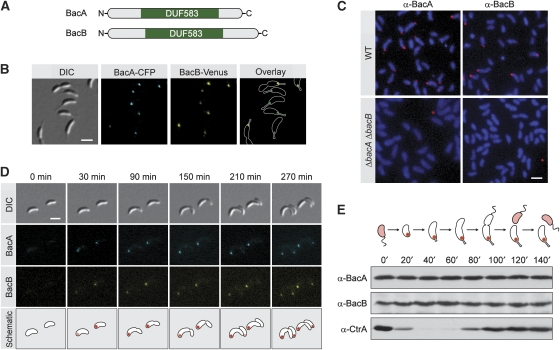
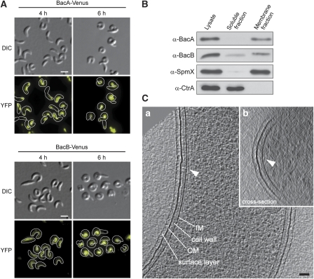
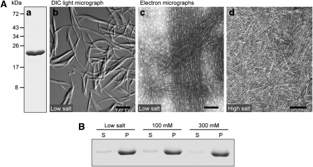
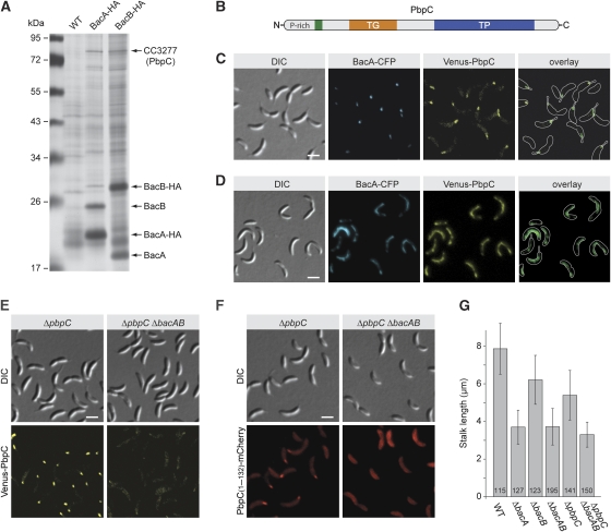
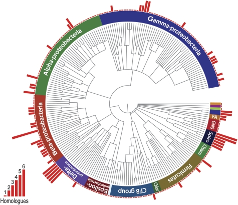
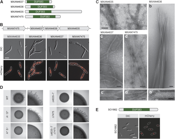
The cytoskeleton has a key function in the temporal and spatial organization of both prokaryotic and eukaryotic cells. Here, we report the identification of a new class of polymer-forming proteins, termed bactofilins, that are widely conserved among bacteria. In Caulobacter crescentus, two bactofilin paralogues cooperate to form a sheet-like structure lining the cytoplasmic membrane in proximity of the stalked cell pole. These assemblies mediate polar localization of a peptidoglycan synthase involved in stalk morphogenesis, thus complementing the function of the actin-like cytoskeleton and the cell division machinery in the regulation of cell wall biogenesis. In other bacteria, bactofilins can establish rod-shaped filaments or associate with the cell division apparatus, indicating considerable structural and functional flexibility. Bactofilins polymerize spontaneously in the absence of additional cofactors in vitro, forming stable ribbon- or rod-like filament bundles. Our results suggest that these structures have evolved as an alternative to intermediate filaments, serving as versatile molecular scaffolds in a variety of cellular pathways.


Conflict of interest statement

The authors declare that they have no conflict of interest.

Figures

Figure 1
Figure 1
Cell-cycle-dependent localization and abundance of BacA and BacB. (A) Schematic representation of BacA and BacB. The position of the conserved DUF583 domain is indicated in green. (B) Localization of BacA and BacB in live cells. Cells of strain JK34 (bacA-ecfp bacB-venus) were grown in PYE-rich medium and visualized by DIC and fluorescence microscopy (bar: 2 μm). (C) Localization of BacA and BacB by immunofluorescence microscopy. Cells of strains CB15N (wild type) and JK5 (ΔbacAB) were probed with anti-BacA and anti-BacB antibodies. Immunocomplexes were detected with a Alexa-Fluor 555-conjugated secondary antibody. To visualize the cells, chromosomal DNA was stained with 4′,6-diamidino-2-phenylindole (DAPI). The micrographs shown were created by overlaying the Alexa-Fluor 555 and DAPI signals (bar: 2 μm). (D) Cell-cycle-dependent subcellular localization of BacA and BacB. Swarmer cells of strain JK34 (bacA-ecfp bacB-venus) were transferred onto an agarose pad (t=0 min) and observed as they progressed through the cell cycle, using DIC and fluorescence microscopy (bar: 2 μm). (E) Cell-cycle-dependent abundance of BacA and BacB. Swarmer cells of wild-type strain CB15N were transferred into M2G minimal medium and incubated for the duration of one cell cycle. At the indicated timepoints, samples were taken from the culture and analysed by immunoblotting using anti-BacA, anti-BacB, and anti-CtrA antiserum. The schematic illustrates the morphology of C. crescentus and the subcellular distribution of BacA and BacB at the different cell-cycle stages.
Figure 2
Figure 2
Assembly of BacA and BacB into membrane-bound polymeric sheets. (A) Filamentous structures and cell shape defects induced by overproduction of BacA and BacB. Cells of wild-type strain CB15N carrying the overexpression plasmid pJK13 (Pxyl-bacA-venus) or pJK14 (Pxyl-bacB-venus), respectively, were grown in PYE-rich medium. Xylose was added to a final concentration of 0.3% to induce synthesis of BacA–Venus or BacB–Venus (t=0 h). At the indicated timepoints, cells were withdrawn from the cultures and visualized by DIC and fluorescence microscopy (bars: 2 μm). The concentrations of BacA–Venus and BacB–Venus were increased about 130- and 360-fold, respectively, over the corresponding wild-type levels (data not shown). (B) Membrane association of BacA and BacB. Whole-cell lysate of wild-type strain CB15N was fractionated by ultracentrifugation. Samples from the lysate, the soluble fraction, and the insoluble membrane fraction were analysed by immunoblotting using anti-BacA, anti-BacB, anti-SpmX, and anti-CtrA antiserum. The intregral membrane protein SpmX (Radhakrishnan et al, 2008) and the cytoplasmic response regulator CtrA (Quon et al, 1996) serve as controls for the fractionation efficiency. (C) Cryo-electron tomography of cells overproducing BacA. Cells of wild-type strain CB15N bearing overexpression plasmid pJK4 (Pxyl-bacA) were grown in PYE medium, induced for 4 h with 0.03% xylose, and analysed by cryo-electron tomography. Shown are a longitudinal section (13-nm slice, panel a) and a cross-section (38-nm slice, panel b) through a reconstructed cell, with the arrow heads pointing to the membrane-associated BacA polymer (bar: 50 nm).
Figure 3
Figure 3
Polymerization of BacA. (A) Polymers formed by BacA. (a) Purified BacA, applied to an 11% SDS–polyacrylamide gel and stained with Coomassie Blue (5 μg of total protein). (b) DIC micrograph of polymers observed after dialysis of BacA (1.6 mg/ml) against 10 mM Tris/HCl (pH 7.5) (bar: 10 μM). (c) Transmission electron micrograph of BacA protofilament bundles formed in a low-salt buffer (buffer LS; see Materials and methods) (bar: 75 nm). (d) Pairs and ribbons of protofilaments obtained by dialysis of BacA against buffer LS containing 300 mM KCl (bar: 50 nm). The same polymerization behaviour was observed in the presence of 100 mM KCl (data not shown). (B) Polymerization efficiency of BacA at different salt concentrations. BacA (0.9 mg/ml) was dialysed against buffer LS containing 0 mM (low salt), 100 mM, or 300 mM KCl. After ultracentrifugation, samples from the supernatant (S) and the pellet (P) were applied to an SDS–gel, and proteins were detected by Coomassie blue staining. Densitometric analysis showed that, in all three conditions, more than 95% of BacA was found in the pellet fraction (data not shown).
Figure 4
Figure 4
Assembly of BacA and BacB into co-polymers. (A) Co-immunoprecipitation analysis. HA-tagged derivatives of BacA and BacB were precipitated from cell lysates of strains KL7 (bacA-HA) and KL8 (bacB-HA), respectively, using anti-HA-affinity beads. Proteins co-precipitating with BacA-HA and BacB-HA were probed with anti-BacB and anti-BacA antibodies, respectively. As a control, the same analyses were performed with lysates of wild-type (WT) strain CB15N. (B) Detection of an interaction between BacA and BacB in vivo. Cells of strain MT260 (bacA-ecfp) bearing overexpression plasmid pJK17 (Pxyl-bacB-venus) were grown in PYE-rich medium, induced for 6 h with 0.3% xylose, and visualized by DIC and fluorescence microscopy (bar: 2 μm).
Figure 5
Figure 5
Interaction of BacA and BacB with the penicillin-binding protein PbpC. (A) Identification of proteins interacting with BacA and BacB. Lysates prepared from formaldehyde-treated cells of strains CB15N (wild type), KL7 (bacA-HA), and KL8 (bacB-HA) were subjected to co-immunoprecipitation analysis using anti-HA-affinity beads. Precipitated proteins were resolved in an 11% SDS–polyacrylamide gel and detected by silver staining. Bands specific for the samples from strains KL7 or KL8 were analysed by mass spectrometry. Owing to the limited amount of starting material, only the five most abundant proteins could be identified. (B) Schematic representation of PbpC. The transmembrane helix (residues 86–108) is indicated in green, the transglycosylase domain (TG; residues 133–300) in orange, and the transpeptidase domain (TP; residues 401–668) in blue. (C) Colocalization of BacA and PbpC. Cells of strain JK271 (bacA-ecfp xylX::Pxyl-venus-pbpC) were grown in PYE-rich medium, induced for 1 h with 0.03% xylose, and visualized by DIC and fluorescence microscopy (bar: 2 μm). (D) Detection of an interaction between BacA and PbpC in vivo. Cells of strain MT279 (xylX::Pxyl-venus-pbpC) carrying overexpression plasmid pJK53 (Pxyl-bacA-ecfp) were grown in PYE-rich medium, induced for 4 h with 0.3% xylose, and visualized by DIC and fluorescence microscopy (bar: 2 μm). Note: For strain MT279 lacking an overexpression plasmid, only polar signals were observed when grown under the same conditions (data not shown). (E) Loss of polar PbpC localization on deletion of bacA and bacB. Strains JK308 (ΔpbpC xylX::Pxyl-venus-pbpC) and JK310 (ΔbacAB ΔpbpC xylX::Pxyl-venus-pbpC) were grown in PYE-rich medium, induced for 1 h with 0.03% xylose, and visualized by DIC and fluorescence microscopy (bar: 2 μm). (F) Determination of the PbpC region responsible for interaction with BacA and BacB. Strains JK308 (ΔpbpC xylX::Pxyl-pbpC[AA 1−132]-mCherry) and JK289 (ΔbacAB ΔpbpC xylX::Pxyl-pbpC[AA 1−132]-mCherry) were grown in PYE-rich medium, induced for 2 h with 0.03% xylose, and visualized by DIC and fluorescence microscopy (bar: 2 μm). (G) Reduced stalk length of strains lacking bactofilin homologues and/or PbpC. Strains CB15N (wild type (WT)), MT257 (ΔbacA), MT259 (ΔbacB), JK5 (ΔbacAB), MT304 (ΔpbpC), and JK281 (ΔbacAB ΔpbpC) were grown in PYE-rich medium, diluted 1:20 in minimal medium lacking phosphate (M2G−P), and cultivated for another 24 h. The graph gives the average stalk length (±s.d.) reached under these conditions and the number of cells analysed.
Figure 6
Figure 6
Conservation of bactofilin among bacteria. The PFAM database (Finn et al, 2008) was used to search for bacterial species possessing DUF583-containing proteins. Where sequence information was available for more than one strain per species, only a single strain was chosen for further analysis. After retrieving the corresponding taxonomy IDs from the National Center for Biotechnology Information (NCBI) website, a phylogenetic tree of the species identified was created using the iTOL server (Letunic and Bork, 2007). For each species shown, the number of bactofilin homologues encoded in the genome is indicated by a red bar. Chlor, green sulphur bacteria; Chlam, chlamydias; Spiro, spirochaetes; GNS, green non-sulphur bacteria; FA, fibrobacteres-acidobacteria group.
Figure 7
Figure 7
Conservation of the filament-forming properties of bactofilin across species. (A) Schematic representation of the bactofilin homologues from Myxococcus xanthus. The DUF583 domain is indicated in green. (B) Subcellular localization of bactofilin homologues in M. xanthus. Cells of strains MT296 (MXAN4635-mCherry), MT297 (MXAN4636-mCherry), MT298 (MXAN4637-mCherry), and MT299 (mCherry-MXAN7475) were grown in CTT-rich medium and visualized by DIC and fluorescence microscopy (bar: 3 μm). The schematic shows the chromosomal organization of the four M. xanthus bactofilin genes investigated. (C) Polymers formed by M. xanthus bactofilin homologues in vitro. Shown are elecron micrographs of polymers formed by purified MXAN4635 (panel a), MXAN4636 (panels b and b′), MXAN4637 (panels c and c′), and MXAN7475 (panels d and d′). Bars: 75 nm (panels a, b, c, d) or 25 nm (panels b′, c′, d′). (D) Involvement of MXAN4635-7 in social gliding motility. Strains DK1622 (WT), DK1217 (aglB1; A S+) (Hodgkin and Kaiser, 1979), DK10416 (pilB; A+ S) (Wu et al, 1997), MT295 (ΔMXAN4635-7), MT300 (ΔMXAN7475), and JK328 (ΔMXAN4635-7 ΔMXAN7475) were grown in CTT medium, spotted on low-percentage agar plates, and analysed for motility. Flare-like structures projecting from the rim of the colony represent rafts of cells moving by means of social motility. (E) Subcellular localization of the S. oneidensis bactofilin homologue. Cells of strain MT288 (SO1662-mCherry) were grown in LB-rich medium and visualized by DIC and fluorescence microscopy (bar: 3 μm). The schematic indicates the position of the DUF583 domain within SO1662.

Similar articles

Cited by

References

    1. Aaron M, Charbon G, Lam H, Schwarz H, Vollmer W, Jacobs-Wagner C (2007) The tubulin homologue FtsZ contributes to cell elongation by guiding cell wall precursor synthesis in Caulobacter crescentus. Mol Microbiol 64: 938–952 - PubMed
    1. Alyahya SA, Alexander R, Costa T, Henriques AO, Emonet T, Jacobs-Wagner C (2009) RodZ, a component of the bacterial core morphogenic apparatus. Proc Natl Acad Sci USA 106: 1239–1244 - PMC - PubMed
    1. Anderson DE, Gueiros-Filho FJ, Erickson HP (2004) Assembly dynamics of FtsZ rings in Bacillus subtilis and Escherichia coli and effects of FtsZ-regulating proteins. J Bacteriol 186: 5775–5781 - PMC - PubMed
    1. Ausmees N, Kuhn JR, Jacobs-Wagner C (2003) The bacterial cytoskeleton: an intermediate filament-like function in cell shape. Cell 115: 705–713 - PubMed
    1. Bagchi S, Tomenius H, Belova LM, Ausmees N (2008) Intermediate filament-like proteins in bacteria and a cytoskeletal function in Streptomyces. Mol Microbiol 70: 1037–1050 - PMC - PubMed

Publication types

MeSH terms