A homozygous p.Glu150Lys mutation in the opsin gene of two Pakistani families with autosomal recessive retinitis pigmentosa
- PMID: 19960070
- PMCID: PMC2787306
A homozygous p.Glu150Lys mutation in the opsin gene of two Pakistani families with autosomal recessive retinitis pigmentosa
Abstract
Purpose: To identify the gene mutations responsible for autosomal recessive retinitis pigmentosa (arRP) in Pakistani families.
Methods: A cohort of consanguineous families with typical RP phenotype in patients was screened by homozygosity mapping using microsatellite markers that mapped close to 21 known arRP genes and five arRP loci. Mutation analysis was performed by direct sequencing of the candidate gene.
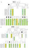
Results: In two families, RP21 and RP53, homozygosity mapping suggested RHO, the gene encoding rhodopsin, as a candidate disease gene on chromosome 3q21. In six out of seven affected members from the two families, direct sequencing of RHO identified a homozygous c.448G>A mutation resulting in the p.Glu150Lys amino acid change. This variant was first reported in PMK197, an Indian arRP family. Single nucleotide polymorphism analysis in RP21, RP53, and PMK197 showed a common disease-associated haplotype in the three families.
Conclusions: In two consanguineous Pakistani families with typical arRP phenotype in the patients, we identified a disease-causing mutation (p.Glu150Lys) in the RHO gene. Single nucleotide polymorphism analysis suggests that the previously reported Indian family (PMK197) and the two Pakistani families studied here share the RHO p.Glu150Lys mutation due to a common ancestry.
Figures


Similar articles
-
A novel nonsense mutation in rhodopsin gene in two Indonesian families with autosomal recessive retinitis pigmentosa.Ophthalmic Genet. 2011 Mar;32(1):57-63. doi: 10.3109/13816810.2010.535892. Epub 2010 Dec 21. Ophthalmic Genet. 2011. PMID: 21174529
-
A novel homozygous R764H mutation in crumbs homolog 1 causes autosomal recessive retinitis pigmentosa.Mol Vis. 2013 Apr 5;19:829-34. Print 2013. Mol Vis. 2013. PMID: 23592920 Free PMC article.
-
Identification of a novel homozygous nonsense mutation in EYS in a Chinese family with autosomal recessive retinitis pigmentosa.BMC Med Genet. 2010 Aug 10;11:121. doi: 10.1186/1471-2350-11-121. BMC Med Genet. 2010. PMID: 20696082 Free PMC article.
-
Genetic analysis of Indian families with autosomal recessive retinitis pigmentosa by homozygosity screening.Invest Ophthalmol Vis Sci. 2009 Sep;50(9):4065-71. doi: 10.1167/iovs.09-3479. Epub 2009 Apr 1. Invest Ophthalmol Vis Sci. 2009. PMID: 19339744 Free PMC article.
-
Retinitis pigmentosa genes implicated in South Asian populations: a systematic review.J Pak Med Assoc. 2017 Nov;67(11):1734-1739. J Pak Med Assoc. 2017. PMID: 29171570
Cited by
-
Unravelling the genetic basis of retinal dystrophies in Pakistani consanguineous families.BMC Ophthalmol. 2023 May 10;23(1):205. doi: 10.1186/s12886-023-02948-8. BMC Ophthalmol. 2023. PMID: 37165311 Free PMC article.
-
Functional characterization of a novel c.614-622del rhodopsin mutation in a French pedigree with retinitis pigmentosa.Mol Vis. 2012;18:581-7. Epub 2012 Mar 2. Mol Vis. 2012. PMID: 22419850 Free PMC article.
-
Electrostatic compensation restores trafficking of the autosomal recessive retinitis pigmentosa E150K opsin mutant to the plasma membrane.J Biol Chem. 2010 Sep 17;285(38):29446-56. doi: 10.1074/jbc.M110.151407. Epub 2010 Jul 13. J Biol Chem. 2010. PMID: 20628051 Free PMC article.
-
In vivo photoreceptor base editing ameliorates rhodopsin-E150K autosomal-recessive retinitis pigmentosa in mice.Proc Natl Acad Sci U S A. 2024 Nov 26;121(48):e2416827121. doi: 10.1073/pnas.2416827121. Epub 2024 Nov 18. Proc Natl Acad Sci U S A. 2024. PMID: 39556729 Free PMC article.
-
Syndromic forms of inherited retinal dystrophies: a comprehensive molecular diagnosis of consanguineous Pakistani families using capture panel sequencing.Mol Vis. 2025 Mar 26;31:69-83. eCollection 2025. Mol Vis. 2025. PMID: 40384762 Free PMC article.
References
-
- Berson EL. Retinitis pigmentosa. The Friedenwald Lecture. Invest Ophthalmol Vis Sci. 1993;34:1659–76. - PubMed
-
- Evans K, Bhattacharya S. Retinal dystrophies: a molecular genetic approach. In: Pawlowitzki IH, Edwards JH, Thompson EA, editors. Genetic Mapping of Disease Genes. San Diego: Academic Press; 1997. p. 247–54.
-
- Bunker CH, Berson EL, Bromley WC, Hayes RP, Roderick TH. Prevalence of retinitis pigmentosa in Maine. Am J Ophthalmol. 1984;97:357–65. - PubMed
-
- Hood DC, Birch DG. Light adaptation of human rod receptors: the leading edge of the human a-wave and models of rod receptor activity. Vision Res. 1993;33:1605–18. - PubMed
Publication types
MeSH terms
Substances
LinkOut - more resources
Full Text Sources
Molecular Biology Databases
