. 2007 Mar 28;20(5):209-220.
doi: 10.1002/cem.993.

Artificial neural networks as supervised techniques for FT-IR microspectroscopic imaging

Affiliations

Artificial neural networks as supervised techniques for FT-IR microspectroscopic imaging

Peter Lasch et al. J Chemom. .

Abstract

In this report the applicability of an improved method of image segmentation of infrared microspectroscopic data from histological specimens is demonstrated. Fourier transform infrared (FT-IR) microspectroscopy was used to record hyperspectral data sets from human colorectal adenocarcinomas and to build up a database of spatially resolved tissue spectra. This database of colon microspectra comprised 4120 high-quality FT-IR point spectra from 28 patient samples and 12 different histological structures. The spectral information contained in the database was employed to teach and validate multilayer perceptron artificial neural network (MLP-ANN) models. These classification models were then employed for database analysis and utilised to produce false colour images from complete tissue maps of FT-IR microspectra. An important aspect of this study was also to demonstrate how the diagnostic sensitivity and specificity can be specifically optimised. An example is given which shows that changes of the number of teaching patterns per class can be used to modify these two interrelated test parameters. The definition of ANN topology turned out to be crucial to achieve a high degree of correspondence between the gold standard of histopathology and IR spectroscopy. Particularly, a hierarchical scheme of ANN classification proved to be superior for the reliable classification of tissue spectra. It was found that unsupervised methods of clustering, specifically agglomerative hierarchical clustering (AHC), were helpful in the initial phases of model generation. Optimal classification results could be achieved if the class definitions for the ANNs were carried out by considering the classification information provided by cluster analysis.


Figures

Figure 1
Figure 1
Dendrogram produced by agglomerative hierarchical clustering (HCA) of representative FT-IR spectra from the colon database. At least two spectra per spectral class and patient were selected for the analysis. The dendrogram illustrates that spectra from fat tissue and the submucosa can be easily differentiated from the majority of the database spectra. HCA classification of the remaining spectra yielded in a number of cases ambiguous results illustrating that unsupervised cluster analysis alone cannot be used to attain consistent classification results (see text for details).
Figure 2
Figure 2
The hierarchical (modular) classification scheme for ANN analysis of IR microspectra from the human colon.
Figure 3
Figure 3
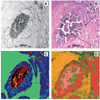
FT-IR microspectroscopic imaging of a cryostat section from a well differentiated (G1) adenocarcinoma of the rectum. (A) Photomicrograph of the unstained cryostat section. Sample area: 1206 × 1231 µm2. (B) Tissue area shown in A after IR microspectroscopy and staining with H&E. 1, necrotic detritus; 2, vital tumour cells; 3, fibrovascular connective tissue and smooth muscle strands; 4, secretion products (mucin); 5, tissue clefts. (C) IR imaging based on 192 × 194 microspectra of the tissue area shown in panel A and hierarchical cluster analysis (five class classification approach). (D) Imaging based on FT-IR microspectroscopy and ANN analysis (‘top-level net’). See text for details.
Figure 4
Figure 4
FT-IR microspectroscopic imaging of a cryostat section from a well differentiated (G1) adenocarcinoma of the rectum. (A) Photomicrograph of the H&E stained cryostat section. Sample area: 1206 × 1231 µm2. (B) Imaging based on FT-IR microspectroscopy and ANN analysis (‘combinet’). See text for details.
Figure 5
Figure 5
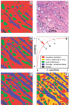
Optimisation of ANNs: illustration of the dependency of sensitivity/specificity on the number of spectra from mucosa structures used for network teaching. (A) ANN image reassembled from FT-IR microspectra of the colon database (four-class classification trial). Very high sensitivity, but low specificity for spectra from the class ‘adenocarcinoma’. (B) Same as A. Moderately improved specificity and high sensitivity for the class ‘adenocarcinoma’ (C) same as A and B. Relatively high specificity and sensitivity for the class ‘adenocarcinoma’ (D) photomicrograph of the adenocarcinoma cryosection after post-staining by H&E. Neoplastic crypts are composed of absorptive epithelia (1) and basal cells (4). The crypts are separated by proliferated fibrovascular connective tissue (2) and filled with detritus and products of secretion (3). (E) Theoretical interrelationship of sensitivity and specificity (receiver operating characteristics, ROC) (F) five-class-classification approach. Sensitivity and specificity can be increased by introducing new spectral classes (see inset).

Similar articles

Cited by

References

    1. Choo LP, Wetzel DL, Halliday WC, Jackson M, LeVine SM, Mantsch HH. In situ characterization of β-amyloid in Alzheimer’s diseased tissue by synchrotron FTIR microspectroscopy. Biophys. J. 1996;71:1672–1679. - PMC - PubMed
    1. Kidder LH, Kalasinsky VF, Luke JL, Levin IW, Lewis EN. Visualization of Silicone Gel in Human Breast Tissue using new Infrared Imaging Spectroscopy. Nat. Med. 1997;3:235–237. - PubMed
    1. Lasch P, Naumann D. FT-IR microspectroscopic imaging of human carcinoma thin sections based on pattern recognition techniques. Cell. Mol. Biol. 1998;44(1):189–202. - PubMed
    1. Chiriboga L, Xie P, Yee H, Zarou D, Zakim W, Diem M. Cell. Mol. Biol. 1998;44(1):219. - PubMed
    1. Lasch P, Haensch W, Kidder L, Lewis EN, Naumann D. Colorectal adenocarcinoma characterization by spatially resolved FT-IR microspectroscopy. Appl. Spectrosc. 2002;56(1):1–9.

LinkOut - more resources