

Autofluorescent proteins as photosensitizer in eukaryontes
- PMID: 19960122
- PMCID: PMC2786992
- DOI: 10.7150/ijms.6.365
Autofluorescent proteins as photosensitizer in eukaryontes
Abstract
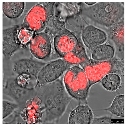
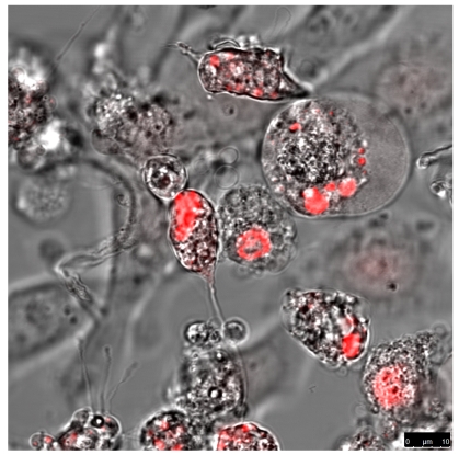
Since the discovery of the green fluorescent green protein (GFP) in 1961 many variants of fluorescent proteins (FP) were detected. The importance was underlined by the Nobel price award in chemistry 2008 for the invention, application, and development of the GFP by Shimomura, Chalfie and Tsien. GFP, first described by Shimomura now is indispensible in the scientific daily life. Since then and also in future fluorescent proteins will lead to new applications as reporters in cell biology. Such FPs can absorb visible day-light and predominantly one variant of the red fluorescent protein, the KillerRed protein (KRED) emits active electrons producing reactive oxygen species (ROS) leading to photokilling processes in eukaryotes. KRED can be activated by daylight as a photosensitizing agent. It is quite obvious that the KRED's expression and localization is critical with respect to damage, mutation and finally killing of eukaryotic cells. We found evidence that the KRED's cytotoxicity is ascendantly location-dependent from the cell membrane over the nuclear lamina to the chromatin in the cell nucleus. Daylight illumination of cells harbouring the KRED protein fused with the histone H2A, a DNA-binding protein which is critical for the formation of the chromatin structure results in cell killing. Therefore the H2A-KRED fusion protein can be considered as an appropriate candidate for the photodynamic therapy (PDT). This finding can be transferred to current photodynamic approaches and can enhance their therapeutic outcome.
Keywords: KillerRed; Melanoma; Photo-Dynamic-Therapy (PDT); ROS; Skin Tumors; fluorescent Proteins; subcellular Localization; topical Application.
Conflict of interest statement
Conflict of Interest: The authors have declared that no conflict of interest exists.
Figures
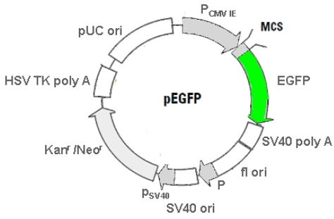
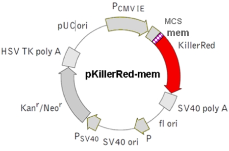
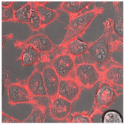
 shows the mem sequence.
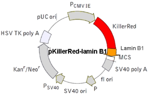
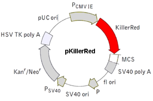
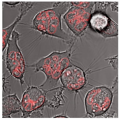
 The Lamin B1 was inserted into the MCS. (The Lamin B1 sequence was kindly provided by Harald Herrmann, this institute)
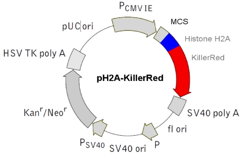
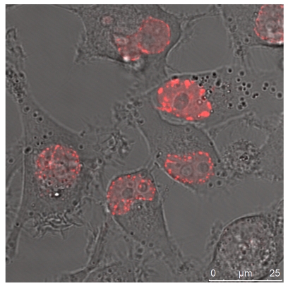
 The histone H2A was inserted into the MCS.
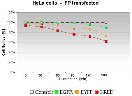
Similar articles
-
Positioning effects of KillerRed inside of cells correlate with DNA strand breaks after activation with visible light.Int J Med Sci. 2011 Jan 21;8(2):97-105. doi: 10.7150/ijms.8.97. Int J Med Sci. 2011. PMID: 21278894 Free PMC article.
-
Towards PDT with Genetically Encoded Photosensitizer KillerRed: A Comparison of Continuous and Pulsed Laser Regimens in an Animal Tumor Model.PLoS One. 2015 Dec 11;10(12):e0144617. doi: 10.1371/journal.pone.0144617. eCollection 2015. PLoS One. 2015. PMID: 26657001 Free PMC article.
-
Advances in the Genetically Engineered KillerRed for Photodynamic Therapy Applications.Int J Mol Sci. 2021 Sep 20;22(18):10130. doi: 10.3390/ijms221810130. Int J Mol Sci. 2021. PMID: 34576293 Free PMC article. Review.
-
A genetically-encoded KillerRed protein as an intrinsically generated photosensitizer for photodynamic therapy.Biomaterials. 2014 Jan;35(1):500-8. doi: 10.1016/j.biomaterials.2013.09.075. Epub 2013 Oct 8. Biomaterials. 2014. PMID: 24112805
-
[The green fluorescent protein that glows in bioscience].Medicina (B Aires). 2009;69(3):370-4. Medicina (B Aires). 2009. PMID: 19622489 Review. Spanish.
Cited by
-
Positioning effects of KillerRed inside of cells correlate with DNA strand breaks after activation with visible light.Int J Med Sci. 2011 Jan 21;8(2):97-105. doi: 10.7150/ijms.8.97. Int J Med Sci. 2011. PMID: 21278894 Free PMC article.
-
Spatial localization of genes determined by intranuclear DNA fragmentation with the fusion proteins lamin KRED and histone KRED und visible light.Int J Med Sci. 2013 Jul 7;10(9):1136-48. doi: 10.7150/ijms.6121. Print 2013. Int J Med Sci. 2013. PMID: 23869190 Free PMC article.
-
Photo-inducible cell ablation in Caenorhabditis elegans using the genetically encoded singlet oxygen generating protein miniSOG.Proc Natl Acad Sci U S A. 2012 May 8;109(19):7499-504. doi: 10.1073/pnas.1204096109. Epub 2012 Apr 24. Proc Natl Acad Sci U S A. 2012. PMID: 22532663 Free PMC article.
-
Optogenetic in vivo cell manipulation in KillerRed-expressing zebrafish transgenics.BMC Dev Biol. 2010 Nov 2;10:110. doi: 10.1186/1471-213X-10-110. BMC Dev Biol. 2010. PMID: 21040591 Free PMC article.
-
Precise spatio-temporal control of rapid optogenetic cell ablation with mem-KillerRed in Zebrafish.Sci Rep. 2017 Jul 11;7(1):5096. doi: 10.1038/s41598-017-05028-2. Sci Rep. 2017. PMID: 28698677 Free PMC article.
References
-
- Szent-Gyorgyi A. The living state and cancer. Physiol Chem. Phys. 1980;12(2):99–110. - PubMed
-
- Velando A, Torres R, Alonso-Alvarez C. Avoiding bad genes: oxidatively damaged DNA in germ line and mate choice. Bioessays. 2008;30(11-12):1212–1219. - PubMed
-
- Mayr M, Sidibe A, Zampetaki A. The paradox of hypoxic and oxidative stress in atherosclerosis. J. Am. Coll. Cardiol. 2008;51(13):1266–1267. - PubMed
-
- Puddu P, Puddu GM, Cravero E, Rosati M, Muscari A. The molecular sources of reactive oxygen species in hypertension. Blood Press. 2008;17(2):70–77. - PubMed
-
- Voeikov VL. Reactive oxygen species--(ROS) pathogens or sources of vital energy? Part 1. ROS in normal and pathologic physiology of living systems. J Altern Complement Med. 2006;12(2):111–118. - PubMed
MeSH terms
Substances
LinkOut - more resources
Full Text Sources
Other Literature Sources
Miscellaneous
