. 2010 Jan 12;49(1):156-65.
doi: 10.1021/bi901550p.

Conformational changes in the nicotinic acetylcholine receptor during gating and desensitization

Affiliations

Conformational changes in the nicotinic acetylcholine receptor during gating and desensitization

Innocent H Yamodo et al. Biochemistry. .

Abstract

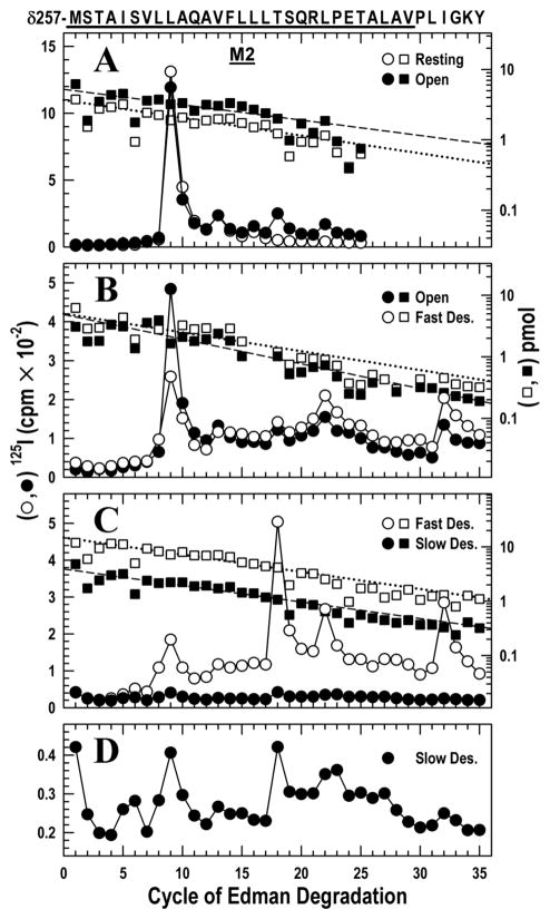
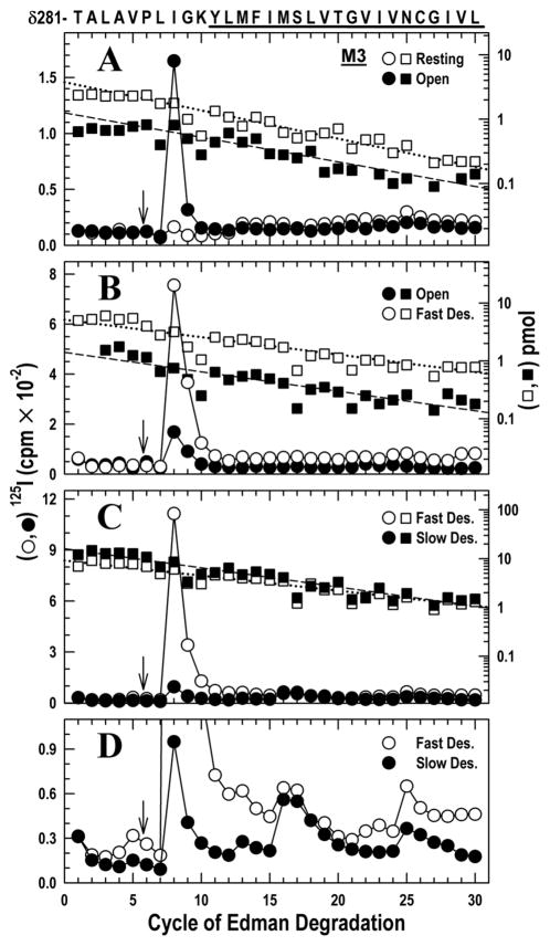
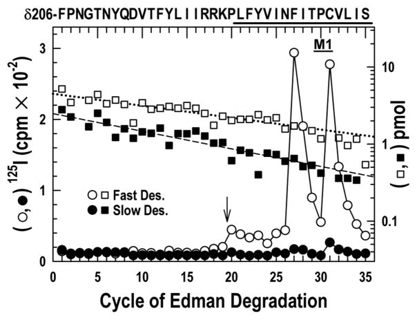
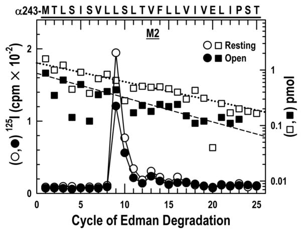
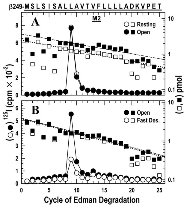
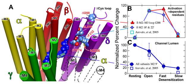
The nicotinic acetylcholine receptor (nAChR) is a member of the important Cys loop ligand-gated ion channel superfamily that modulates neuronal excitability. After they respond to their agonists, their actions are terminated either by removal of ligand or by fast and slow desensitization, processes that play an important role in modulating the duration of conducting states and hence of integrated neuronal behavior. We monitored structural changes occurring during fast and slow desensitization in the transmembrane domain of the Torpedo nAChR using time-resolved photolabeling with the hydrophobic probe 3-(trifluoromethyl)-3-(m-iodophenyl)diazirine (TID). After channel opening, TID photolabels a residue on the delta-subunit's M2-M3 loop and a cluster of four residues on deltaM1 and deltaM2, defining an open state pocket [Arevalo, E., et al. (2005) J. Biol. Chem. 280, 13631-13640]. We now find that photolabeling of this pocket persists during the transition to the fast desensitized state, the extent of photoincorporation decreasing only with the transition to the slow desensitized state. In contrast, the extent of photoincorporation in the channel lumen at the conserved 9'-leucines on the second transmembrane helix (M2-9') decreased successively during the resting to open and open to fast desensitized state transitions, implying that the local conformation is different in each state, a conclusion consistent with the hypothesis that there are separate gates for channel opening and desensitization. Thus, although during fast desensitization there is a conformation change in the channel lumen at the level of M2-9', there is none in the regions of the delta-subunit's M2-M3 loop and the interior of its M1-M4 helix bundle until slow desensitization occurs.


Figures

Figure 1
Figure 1. Photoincorporation of [125I]TID in the δM2 helix and δM2–M3 loop in the resting, open, fast desensitized and slow desensitized states of Torpedo nAChR
nAChR-enriched membranes (4 mg/mL) were equilibrated with 7 mM [125I]TID and then rapidly mixed with an equal volume of buffer (resting), or agonist (Carbachol, 20 mM; 10 mM final concentration) for 15 ms (open state), 1 s (fast desensitized state) or ≥1 hour (slow desensitized state), then frozen in less than 1 ms, and finally photolabeled for 30 min (see Experimental Procedures). In each case, the fragment beginning at δMet-257 was isolated from EndoLys-C digests of δ subunits by SDS-PAGE and reversed phase HPLC as described in the Experimental Procedures (Fig. S1). The panels show 125I (○,●; left axis) and PTH-amino acids (□, ■; right axis) released during the sequencing of δ-subunit peptides. The sequence beginning at the N-terminus of δM2 at Met-257 is shown on the top axis with the M2 transmembrane helix highlighted with a dark bar. A, nAChRs were photolabeled in the resting (○, □) and open (●, ■) states. For sequencing the resting and open state samples, 9,230 and 16,140 cpm of 125I were loaded onto filters respectively. The primary sequence began at δMet-257 (δM2–1′) (□, Io = 3.7 pmol, R = 94%; ■ Io = 5.2 pmol, R = 95%), with a secondary sequence beginning at δAsn-437 in both samples at ~0.3 pmol. Peaks of 125I release (○,●) were detected in the resting state at cycles 9, 13, 16 and in the open state at cycles 9, 13, 16, 18 and 22, corresponding respectively to residues δLeu-265, δVal-269, δLeu-272, δThr-274 and δLeu-278. B, nAChRs were photolabeled in the open (●, ■) and the fast desensitized (○, □) states. For sequencing the open and fast desensitized state samples, 24,021 and 28,221 cpm of 125I were loaded onto filters respectively. The fragment beginning at δMet-257 was present (■: Io = 4.9 pmol, R = 91%; □: Io = 5.0 pmol, R = 93%) along with the fragment beginning at δAsn-437 at ~ 8 pmol in both samples, which contains the M4 helix beginning at cycle 20. Peaks of 125I release were observed in both samples at cycles 9, 13, 18, 22 and 32. Residues in M4 are labeled inefficiently by TID (δSer-457 at δMet-467 at < 1 cpm/pmol) because they are at the lipid interface (37) and they do not contribute to the peaks of 125I release observed here. C, nAChRs were photolabeled in the fast (○,□) and slow desensitized (●, ■) states. For sequencing the fast desensitized and slow desensitized state samples, 15,450 and 2,370 cpm of 125I were loaded onto filters respectively. The primary sequence began at δMet-257 (□, Io = 15 pmol, R = 93%; ■, Io = 3.9 pmol, R = 93%), with the fragment beginning at δAsn-437 also present at ~ 2 pmol in each sample. Peaks of 125I release occurred in the same cycles as in B. D, The data from C for the slow desensitized state are replotted on an expanded scale.
Figure 2
Figure 2. Photoincorporation of [125I]TID in the δM2–M3 loop and δM3 helix in the resting, open, fast and slow desensitized states of Torpedo nAChR
Photolabeled nAChRs were from the experiments described in Fig. 1. The panels show 125I (○, ●; left axis) and PTH-amino acids (□, ■; right axis) released during the sequencing. The sequence of the δ-subunit fragment beginning at δThr-281 (δM2–25′), near the C-terminus of δM2 and continuing through the δM2–δM3 loop into δM3 (highlighted with a dark bar), is shown on the top axis. Aliquots of δ subunits were digested in solution with V8 protease. The digests were fractionated by reversed phase HPLC from which fractions containing the desired peptide were pooled (Fig. S2A–C) for sequencing. To restrict the sequencing to the fragment beginning at δThr-281, each sequencing filter was treated with OPA after 5 cycles of Edman degradation (i.e. when δPro-286 was the N-terminal residue) (↓) to block other peptides that do not contain a proline at that cycle. A, nAChRs were photolabeled in the resting (○, □) and open (●, ■) states. For sequencing the resting and open state samples, 4,650 and 2,750 cpm of 125I were loaded onto filters respectively. The fragment beginning at δThr-281 was the only sequence remaining after the OPA treatment (□, Io = 3.8 pmol, R = 90 %; ■, Io = 1.3 pmol, R = 92 %). A prominent release of 125I was detected in cycle 8 (δIle-288) only in the open state sample. There were minor peaks of 125I release in cycles 13 and 25 (δMet-293 and δAsn-305). B, nAChRs were photolabeled in the open (●, ■) and the fast desensitized (○, □) states. For sequencing the open and fast desensitized state samples, 6,880 and 24,263 cpm of 125I were loaded onto filters respectively. The fragment beginning at δThr-281 was the only sequence remaining after the OPA treatment (■, Io = 1.4, R = 93 %; □, Io = 5.2 pmol, R = 92 %). A prominent release of 125I was detected in cycle 8 (δIleu-288) in the resting and fast desensitized state, with minor release occurring at cycles 13, 22 and 25 (δMet-293, δVal-302 and δAsn-305). C, nAChRs were photolabeled in the fast (○,□) and slow desensitized (●, ■) states. For sequencing the fast desensitized and slow desensitized state samples, 23,900 and 6,850 cpm of 125I were loaded onto filters respectively. The fragment beginning at δThr-281 was the only sequence remaining after the OPA treatment (□, Io = 9.1 pmol, R = 93%; ■, Io = 16 pmol, R = 91%). The prominent 125I releases in cycle 8 (δIle-288) in the fast desensitized state fell dramatically during slow desensitization. D, Because the amount of peptide is higher in C than in A & B, the low photoincorporation in δM3 (cycles 13, 22 and 25; δMet-293, δVal-302 and δAsn-305) is clear when the data is plotted on an expanded axis.
Figure 3
Figure 3. Photoincorporation of [125I]TID in the δM1 helix in the fast desensitized and slow desensitized states of nAChR
125I (○, ●) and PTH-amino acids (□, ■) released during sequencing of the fragment beginning at δPhe-206 and extending through δM1, which was isolated by the reversed phase HPLC fractionation of the EndoLys-C digests of the δ subunit described in Fig. 1 and Fig. S1 from nAChRs photolabeled in the fast desensitized (○, □) and slow desensitized (○, ■) states. For sequencing the fast desensitized and slow desensitized state samples, 7,600 and 840 cpm of 125I were loaded onto filters respectively. To confirm that the 125I release after cycle 20 was attributable to the δM1, both samples were treated with OPA after 19 cycles of Edman degradation (i.e. when δPro-225 was the N-terminal residue) (↓) to prevent further sequencing of any other peptides in the samples not containing an N-terminal proline at that cycle. Even before OPA treatment however, the only sequence detected began at δPhe-206 (□, Io = 4.5 pmol, R = 97 %;■, Io = 2.5 pmol, R = 95 %). Prominent labeling at cycles 27 and 31 was only seen in the fast desensitized state.
Figure 4
Figure 4. Photoincorporation of [125I]TID in the αM2 helix in the resting and open states
125I (○, ●) and PTH-amino acids (□, ■) released while sequencing the fragment beginning at αMet-243 (αM2–1′), which was isolated from the α-subunits photolabeled in the experiment described in Fig. 1A by HPLC purification of an EndoLys-C digest (Fig. S3) of a 20 kDa fragment produced by digestion with V8 protease. For sequencing the resting and open state samples, 2,217 and 1,874 cpm of 125I were loaded onto filters respectively. Sequencing revealed a single peptide (resting, □, Io = 1.3 pmol, R = 92%; open state, ■, Io = 0.9 pmol, R = 90%). A major peak of 125I release was observed in cycle 9 (αLeu-251), with minor peaks at cycles 13 (αVal-255) and 16 (αLeu-258).
Figure 5
Figure 5. Photoincorporation of [125I]TID in the βM2 helix in the resting, open and fast desensitized states
125I (○, ●) and PTH-amino acids (□, ■) released while sequencing the fragment beginning at βMet-249 (βM2–1′), which was isolated from the β-subunits photolabeled in the experiments described in Figs. 1A & B by HPLC purification (Fig. S4) of an ~10 kDa fragment isolated by Tricine SDS-PAGE from trypsin digests. A, nAChRs were photolabeled in the resting (○, □) and open (●, ■) states. For sequencing the resting and open state samples, 4252 and 4523 cpm of 125I were loaded onto filters respectively. Sequencing revealed a single peptide beginning at βMet-249 (□: Io = 2.5 pmol, R= 93%; ■: Io = 3.6 pmol, R= 94%). There was a major peak of 125I release in both states at cycle 9, corresponding to βLeu-257. B, nAChRs were photolabeled in the open (●, ■) and the fast desensitized (○, □) states. For sequencing the open and fast desensitized state samples, 3166 and 1060 cpm of 125I were loaded onto filters respectively. Sequencing revealed a single peptide beginning at βMet-249 (■: Io = 3.9 pmol, R= 92%; □: Io = 3.8 pmol, R= 92%). The major peak of 125I release in both states was at cycle 9 (βLeu-257), with minor release at cycles 13 (βVal-261) and 16 (βLeu-264).
Figure 6
Figure 6. Location (Panel A) and contrasting state–dependence of the activation–dependent (Panel B) and the channel lumen (Panel C) groups of residues
Panel A: A representation of the transmembrane region of the Torpedo nAChR based on the 2005 structure (2gb9.pdb: (5)). The receptor is viewed from the extracellular, N-terminal side and the structure is sliced to show the transmembrane domain (TMD) only. The subunits are color coded as follows (Alpha, yellow; Beta, red; Gamma, green; Delta, purple; the TMD of the second α–subunit that is situated between the γ– and δ–subunits is shown only in outline for clarity). The channel lumen residues are shown in dark blue; the M2–9′ residues are shown for each subunit and the M2–13′ & 16′ residues for the δ-subunit. The δM2–9′ Leu-265 is identified in green lettering. For the activation dependent–residues on the δ–subunit the carbon atoms are shown in corn blue, oxygen in red and nitrogen in mid blue; residues are identified in red lettering. In the top right near δIle-288 a portion of the δ–subunit’s Cys–loop from the LBD is shown in ribbon representation. Molecular graphics images were produced using the UCSF Chimera package from the Resource for Biocomputing, Visualization, and Informatics at the University of California, San Francisco (supported by NIH P41 RR-01081) (48). Panels B & C: The normalized efficiency of photoincorporation of [125I]TID as the nAChR passes sequentially from the resting through the open and fast desensitized states to the slow desensitized state. Panel B: The biphasic state dependence of photoincorporation into the activation–dependent residus in the δ–subunit; the residues are identified in the key. The percent change in each pair of experiments was calculated. The data for δM2–18′ & 22′ (Thr-274 and Leu-278) were grouped and averaged (2 replicates per point). For the δM2–M3 loop residue, Ile-288, there were two replicates for each state transition (Fig. 1 and Fig 2.). The data were then normalized with error propagation to the open state, which was set to 100. There was no photolabeling in the resting state, equal degrees of photoincorporation in the open and fast desensitized states and only modest photolabeling in the slow desensitized state. Panel C: The state dependence of the efficiency of photoincorporation into the channel lumen M2–9′ residues on the α–, β–& δ–subunits (αM2 Leu-251; βM2 Leu-257 & δM2 Leu-265). The data were analyzed as in Panel B. The percent change in each pair of experiments was calculated. We assumed 9′ leucine photoincorporation was equal in all subunits and averaged the data, which were then normalized with error propagation to the resting state, which was set to 100. For comparison the squares show data from our previous study (28) for the mean of δM2–18 & 22′ normalized to the open state (B) and for δM2 Leu-265 normalized to the resting state (C).

Similar articles

Cited by

References

    1. Cederholm JM, Schofield PR, Lewis TM. Gating mechanisms in Cys-loop receptors. Eur Biophys J. 2009 in press. - PubMed
    1. Sine SM, Engel AG. Recent advances in Cys-loop receptor structure and function. Nature. 2006;440:448–455. - PubMed
    1. Giniatullin R, Nistri A, Yakel JL. Desensitization of nicotinic ACh receptors: shaping cholinergic signaling. Trends Neurosci. 2005;28:371–378. - PubMed
    1. Wells GB. Structural answers and persistent questions about how nicotinic receptors work. Front Biosci. 2008;13:5479–5510. - PMC - PubMed
    1. Unwin N. Refined structure of the nicotinic acetylcholine receptor at 4A resolution. Journal of molecular biology. 2005;346:967–989. - PubMed

Publication types