Review
. 2009;114(4):193-205.
doi: 10.3109/03009730903366075.

Pulsatility of insulin release--a clinically important phenomenon

Affiliations
Review

Pulsatility of insulin release--a clinically important phenomenon

Bo Hellman. Ups J Med Sci. 2009.

Abstract

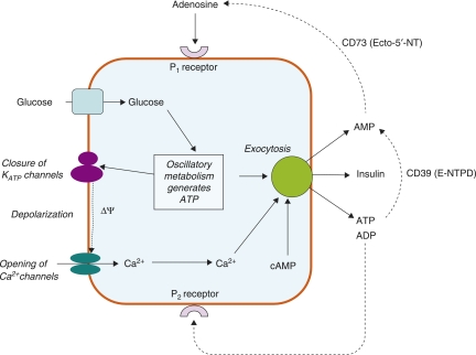
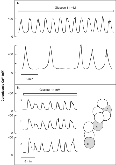
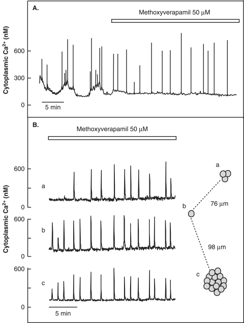
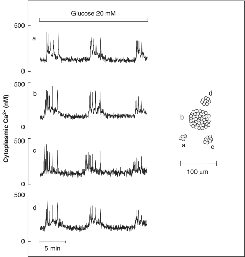
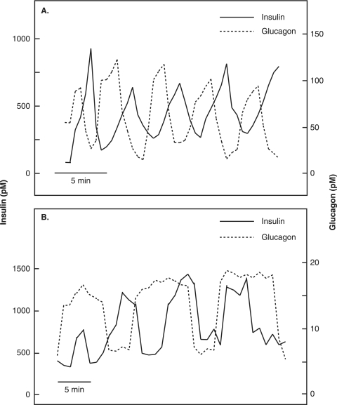
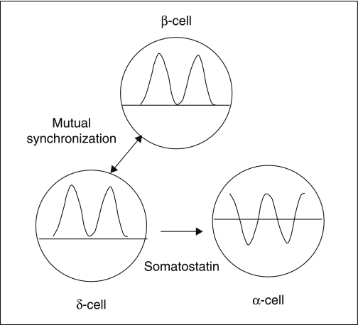
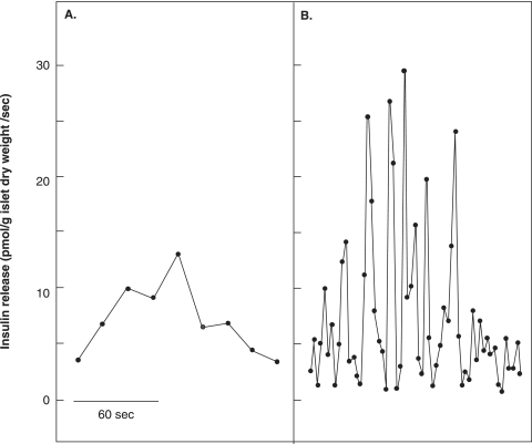
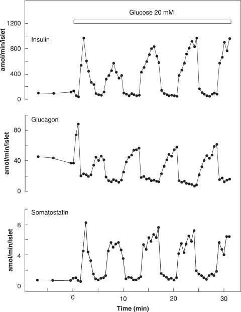
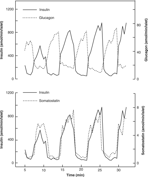
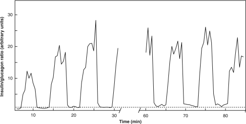
The mechanisms and clinical importance of pulsatile insulin release are presented against the background of more than half a century of companionship with the islets of Langerhans. The insulin-secreting beta-cells are oscillators with intrinsic variations of cytoplasmic ATP and Ca(2+). Within the islets the beta-cells are mutually entrained into a common rhythm by gap junctions and diffusible factors (ATP). Synchronization of the different islets in the pancreas is supposed to be due to adjustment of the oscillations to the same phase by neural output of acetylcholine and ATP. Studies of hormone secretion from the perfused pancreas of rats and mice revealed that glucose induces pulses of glucagon anti-synchronous with pulses of insulin and somatostatin. The anti-synchrony may result from a paracrine action of somatostatin on the glucagon-producing alpha-cells. Purinoceptors have a key function for pulsatile release of islet hormones. It was possible to remove the glucagon and somatostatin pulses with maintenance of those of insulin with an inhibitor of the P2Y(1) receptors. Knock-out of the adenosine A(1) receptor prolonged the pulses of glucagon and somatostatin without affecting the duration of the insulin pulses. Studies of isolated human islets indicate similar relations between pulses of insulin, glucagon, and somatostatin as found during perfusion of the rodent pancreas. The observation of reversed cycles of insulin and glucagon adds to the understanding how the islets regulate hepatic glucose production. Current protocols for pulsatile intravenous infusion therapy (PIVIT) should be modified to mimic the anti-synchrony between insulin and glucagon normally seen in the portal blood.


Figures

Figure 1.
Figure 1.
Model showing how glucose-induced oscillations of cytoplasmic ATP generate pulsatile release of insulin from a β-cell. Rhythmic glycolysis triggers periodic rises of cytoplasmic ATP that inhibits a specific K+ channel. Resulting depolarization evokes oscillations of cytoplasmic Ca2+ due to entry of the ion via voltage-dependent channels. The right part of the Figure shows that oscillatory rises of cytoplasmic Ca2+, ATP, and cAMP evoke exocytosis of secretory granules containing insulin together with ATP, ADP, and AMP. After release from the β-cell these nucleotides, degraded or not to adenosine by ectonucleotidases (CD39 and CD73), serve as regulators of pulsatile release of insulin by binding to P1 and P2 receptors.
Figure 2.
Figure 2.
Oscillations of cytoplasmic Ca2+ in two mouse β-cells lacking contact (A) and in three cells situated in an aggregate (B). Contacts between the cells result in synchronization of the Ca2+ oscillations. The traces refer to the cells shown to the right.
Figure 3.
Figure 3.
Transients of cytoplasmic Ca2+ in ob/ob mouse β-cells superfused with a medium containing 20 mM glucose and 20 nM glucagon. A: Suppression of the Ca2+ entry with methoxyverapamil removes the Ca2+ oscillations, allowing the transients to start from the basal level. B: Synchronized cytoplasmic Ca2+ transients in single cell/aggregates (shown to the right) exposed to methoxyverapamil.
Figure 4.
Figure 4.
Co-ordination of [Ca2+]i oscillations in the four aggregates shown to the right. Most of the superimposed transients appear in synchrony not only within but also among the aggregates. Modified from Grapengiesser et al. 2003 (8) with permission.
Figure 5.
Figure 5.
Relation between repetitive insulin and glucagon pulses during perfusion of rodent pancreas with 20 mM glucose. A: The pulses of insulin are anti-synchronous to those of glucagon in rat pancreas. From Grapengiesser et al. 2006 (49) with permission. B: The pulses of glucagon are prolonged compared with insulin in mice with knock-out of the adenosine A1 receptor. From Salehi et al. 2009 (31) with permission.
Figure 6.
Figure 6.
Model of cell interactions important for glucose generation of pulsatile hormone release from an islet. Dotted lines indicate level of basal release before the rise of glucose. Glucose generates simultaneous pulses of insulin and somatostatin release by mutual synchronization of β- and δ-cells. The oscillations of glucagon appear in anti-synchrony and have nadirs below the basal level. Paracrine release of somatostatin from δ-cells accounts for the appearance of glucagon pulses 180° out of phase.
Figure 7.
Figure 7.
Insulin release from a human islet exposed to 11 mM glucose. A: Pulse observed with a sampling time of 17.5 seconds. B: The same pulse analysed with a sampling time of 2.5 seconds.
Figure 8.
Figure 8.
Effects of raising glucose from 3 to 20 mM on the release of insulin, glucagon, and somatostatin from a batch of 15 human islets. The hormones were measured in 30-second samples of the perifusate.
Figure 9.
Figure 9.
Relation between the repetitive release pulses of insulin and glucagon in the experiment shown in Figure 8. The insulin pulses are anti-synchronous to the glucagon pulses (upper panel) and coincide with the somatostatin pulses (lower panel).
Figure 10.
Figure 10.
Variations of the insulin/glucagon ratio during superfusion of 15 human islets with 20 mM glucose. Sampling was interrupted for 28.5 min in the middle of the experiment. The insulin/glucagon ratio is given in arbitrary units with the average of nadir values set to 1.0 (dotted line).

References

    1. Hellman B, Diderholm H. The diabetogenic effects of alloxan after elimination of extra-pancreatic factors. Acta Endocrinol (Copenh) 1955;20:81–7. - PubMed
    1. Hellman B. Actual distribution of the number and volume of the islets of Langerhans in different size classes in non-diabetic humans of varying ages. Nature. 1959;184:1498–9. - PubMed
    1. Hellman B, Sehlin J, Täljedal IB. Calcium and secretion: Distinction between two pools of glucose-sensitive calcium in pancreatic islets. Science. 1976;194:1421–3. - PubMed
    1. Hellman B. β-cell cytoplasmic Ca2+ balance as a determinant for glucose-stimulated insulin release. Diabetologia. 1985;28:494–501. - PubMed
    1. Grapengiesser E, Gylfe E, Hellman B. Three types of cytoplasmic Ca2+ oscillations in stimulated pancreatic β-cells. Arch Biochem Biophys. 1989;268:404–7. - PubMed