

Profound human/mouse differences in alpha-dystrobrevin isoforms: a novel syntrophin-binding site and promoter missing in mouse and rat
- PMID: 19961569
- PMCID: PMC2796648
- DOI: 10.1186/1741-7007-7-85
Profound human/mouse differences in alpha-dystrobrevin isoforms: a novel syntrophin-binding site and promoter missing in mouse and rat
Abstract
Background: The dystrophin glycoprotein complex is disrupted in Duchenne muscular dystrophy and many other neuromuscular diseases. The principal heterodimeric partner of dystrophin at the heart of the dystrophin glycoprotein complex in the main clinically affected tissues (skeletal muscle, heart and brain) is its distant relative, alpha-dystrobrevin. The alpha-dystrobrevin gene is subject to complex transcriptional and post-transcriptional regulation, generating a substantial range of isoforms by alternative promoter use, alternative polyadenylation and alternative splicing. The choice of isoform is understood, amongst other things, to determine the stoichiometry of syntrophins (and their ligands) in the dystrophin glycoprotein complex.
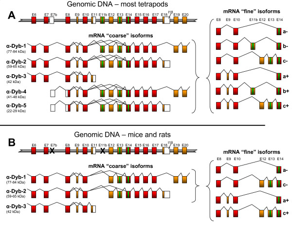
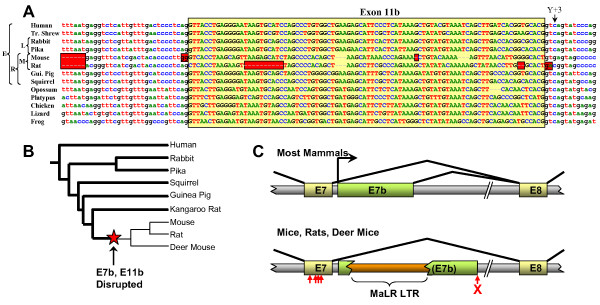
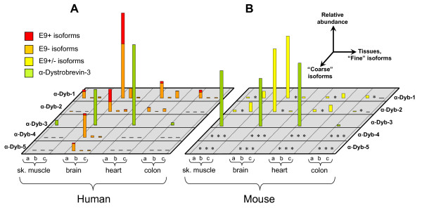
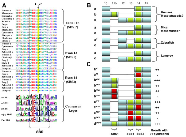
Results: We show here that, contrary to the literature, most alpha-dystrobrevin genes, including that of humans, encode three distinct syntrophin-binding sites, rather than two, resulting in a greatly enhanced isoform repertoire. We compare in detail the quantitative tissue-specific expression pattern of human and mouse alpha-dystrobrevin isoforms, and show that two major gene features (the novel syntrophin-binding site-encoding exon and the internal promoter and first exon of brain-specific isoforms alpha-dystrobrevin-4 and -5) are present in most mammals but specifically ablated in mouse and rat.
Conclusion: Lineage-specific mutations in the murids mean that the mouse brain has fewer than half of the alpha-dystrobrevin isoforms found in the human brain. Our finding that there are likely to be fundamental functional differences between the alpha-dystrobrevins (and therefore the dystrophin glycoprotein complexes) of mice and humans raises questions about the current use of the mouse as the principal model animal for studying Duchenne muscular dystrophy and other related disorders, especially the neurological aspects thereof.
Figures
Similar articles
-
Biophysical characterization of the dystrophin C-terminal domain: Dystrophin interacts differentially with dystrobrevin isoforms.J Biol Chem. 2024 Dec;300(12):108002. doi: 10.1016/j.jbc.2024.108002. Epub 2024 Nov 17. J Biol Chem. 2024. PMID: 39551137 Free PMC article.
-
The syntrophin-dystrobrevin subcomplex in human neuromuscular disorders.J Neuropathol Exp Neurol. 2005 Apr;64(4):350-61. doi: 10.1093/jnen/64.4.350. J Neuropathol Exp Neurol. 2005. PMID: 15835271
-
Differential expression and developmental regulation of a novel alpha-dystrobrevin isoform in muscle.Gene. 1999 Oct 1;238(2):479-88. doi: 10.1016/s0378-1119(99)00358-3. Gene. 1999. PMID: 10570976
-
Dystrobrevins in muscle and non-muscle tissues.Neuromuscul Disord. 2007 Feb;17(2):123-34. doi: 10.1016/j.nmd.2006.11.003. Epub 2007 Jan 23. Neuromuscul Disord. 2007. PMID: 17251025 Review.
-
Dystrobrevin dynamics in muscle-cell signalling: a possible target for therapeutic intervention in Duchenne muscular dystrophy?Neuromuscul Disord. 2002 Oct;12 Suppl 1:S110-7. doi: 10.1016/s0960-8966(02)00091-3. Neuromuscul Disord. 2002. PMID: 12206805 Review.
Cited by
-
Dystrobrevin increases dystrophin's binding to the dystrophin-glycoprotein complex and provides protection during cardiac stress.J Mol Cell Cardiol. 2014 Nov;76:106-15. doi: 10.1016/j.yjmcc.2014.08.013. Epub 2014 Aug 24. J Mol Cell Cardiol. 2014. PMID: 25158611 Free PMC article.
-
Evolutionary profiling reveals the heterogeneous origins of classes of human disease genes: implications for modeling disease genetics in animals.BMC Evol Biol. 2014 Oct 4;14:212. doi: 10.1186/s12862-014-0212-1. BMC Evol Biol. 2014. PMID: 25281000 Free PMC article.
-
Evolution and developmental functions of the dystrophin-associated protein complex: beyond the idea of a muscle-specific cell adhesion complex.Front Cell Dev Biol. 2023 Jun 13;11:1182524. doi: 10.3389/fcell.2023.1182524. eCollection 2023. Front Cell Dev Biol. 2023. PMID: 37384252 Free PMC article. Review.
-
The role of dystrophin isoforms and interactors in the brain.Brain. 2025 Apr 3;148(4):1081-1098. doi: 10.1093/brain/awae384. Brain. 2025. PMID: 39673425 Free PMC article. Review.
-
Absence of glial α-dystrobrevin causes abnormalities of the blood-brain barrier and progressive brain edema.J Biol Chem. 2012 Nov 30;287(49):41374-85. doi: 10.1074/jbc.M112.400044. Epub 2012 Oct 5. J Biol Chem. 2012. PMID: 23043099 Free PMC article.
References
Publication types
MeSH terms
Substances
Grants and funding
LinkOut - more resources
Full Text Sources
Molecular Biology Databases
