. 2009 Dec 4:4:43.
doi: 10.1186/1749-8104-4-43.

Multiple non-cell-autonomous defects underlie neocortical callosal dysgenesis in Nfib-deficient mice

Affiliations

Multiple non-cell-autonomous defects underlie neocortical callosal dysgenesis in Nfib-deficient mice

Michael Piper et al. Neural Dev. .

Abstract

Background: Agenesis of the corpus callosum is associated with many human developmental syndromes. Key mechanisms regulating callosal formation include the guidance of axons arising from pioneering neurons in the cingulate cortex and the development of cortical midline glial populations, but their molecular regulation remains poorly characterised. Recent data have shown that mice lacking the transcription factor Nfib exhibit callosal agenesis, yet neocortical callosal neurons express only low levels of Nfib. Therefore, we investigate here how Nfib functions to regulate non-cell-autonomous mechanisms of callosal formation.

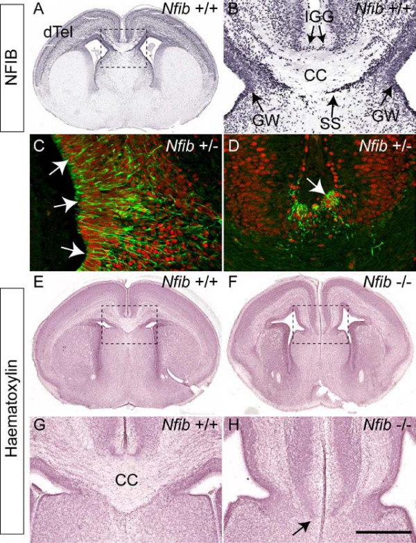
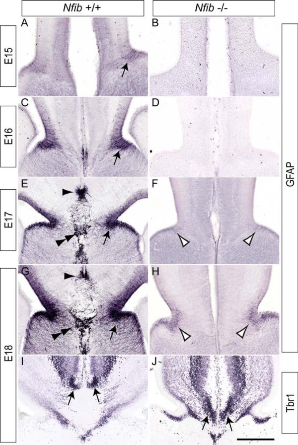
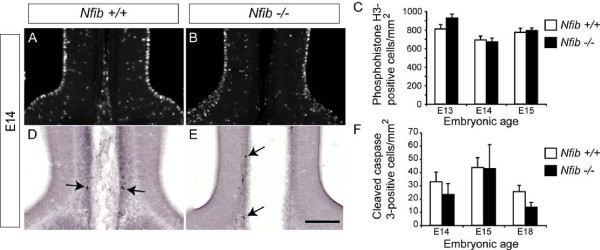
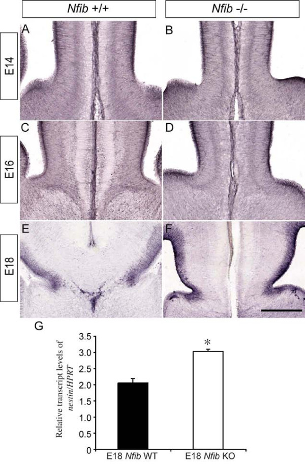
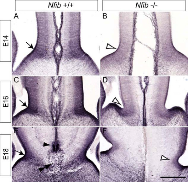
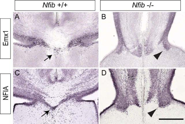
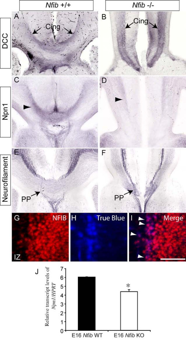
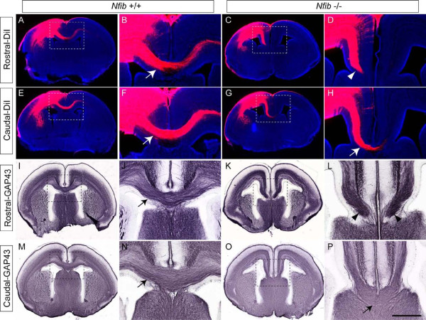
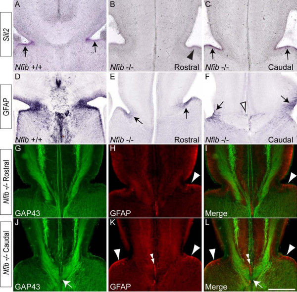
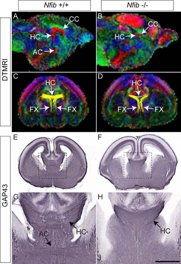
Results: Our investigations confirmed a reduction in glial cells at the midline in Nfib-/- mice. To determine how this occurs, we examined radial progenitors at the cortical midline and found that they were specified correctly in Nfib mutant mice, but did not differentiate into mature glia. Cellular proliferation and apoptosis occurred normally at the midline of Nfib mutant mice, indicating that the decrease in midline glia observed was due to deficits in differentiation rather than proliferation or apoptosis. Next we investigated the development of callosal pioneering axons in Nfib-/- mice. Using retrograde tracer labelling, we found that Nfib is expressed in cingulate neurons and hence may regulate their development. In Nfib-/- mice, neuropilin 1-positive axons fail to cross the midline and expression of neuropilin 1 is diminished. Tract tracing and immunohistochemistry further revealed that, in late gestation, a minor population of neocortical axons does cross the midline in Nfib mutants on a C57Bl/6J background, forming a rudimentary corpus callosum. Finally, the development of other forebrain commissures in Nfib-deficient mice is also aberrant.

Conclusion: The formation of the corpus callosum is severely delayed in the absence of Nfib, despite Nfib not being highly expressed in neocortical callosal neurons. Our results indicate that in addition to regulating the development of midline glial populations, Nfib also regulates the expression of neuropilin 1 within the cingulate cortex. Collectively, these data indicate that defects in midline glia and cingulate cortex neurons are associated with the callosal dysgenesis seen in Nfib-deficient mice, and provide insight into how the development of these cellular populations is controlled at a molecular level.


Figures

Figure 1
Figure 1
Expression of NFIB in midline glial populations. (A, B) Coronal section of an E18 wildtype brain stained with NFIB. (A) NFIB was expressed broadly throughout the dorsal telencephalon (dTel). (B) Higher magnification view of the boxed region in (A) showing NFIB expression at the cortical midline. NFIB was expressed in the glial wedge (GW), indusium griseum glia (IGG) and subcallosal sling (SS). (C, D) Confocal sections of E18 Nfib heterozygote brains, demonstrating the co-expression of the β-galactosidase (β-gal) reporter (red) and glial fibrillary acidic protein (GFAP; green) within the glial wedge (C) and the indusium griseum glia (D). β-gal-positive nuclei were often surrounded by GFAP-positive fibres (arrows in C, D), indicating that GFAP-expressing glia likely express Nfib. (E-H) Coronal sections of wildtype (E, G) and Nfib knockout (F, H) brains stained with haematoxylin. The corpus callosum (CC) does not form rostrally in mice lacking Nfib (arrow in (H)). Panels (G) and (H) are higher magnifications of the boxed regions in (E) and (F), respectively. Scale bars: 500 μm (A, E, F); 200 μm (B, G, H); 100 μm (C, D).
Figure 2
Figure 2
Reduced expression of GFAP in Nfib knockout mice. (A-H) Expression of GFAP at the cortical midline of wildtype (A, C, E, G) and Nfib-deficient (B, D, F, H) mice. In the wildtype, expression of GFAP in the glial wedge was initiated at E15, and became progressively stronger as development proceeded (arrows in (A, C, E, G)). GFAP expression in the indusium griseum glia (arrowhead) and midline zipper glia (double arrowhead) of the wildtype was also evident from E17 onwards (E, G). However, in the mutant, low levels of GFAP expression in the glial wedge were only observed from E17 onwards (open arrowheads in (F, H)), whereas expression in the indusium griseum glia and midline zipper glia was absent. Neurons in the indusium griseum of the wildtype expressed Tbr1 (arrows in (I)). Neurons expressing Tbr1 in the indusium griseum were also observed in the mutant (arrows in (J)). Scale bars: 280 μm (A, B); 250 μm (C, D); 225 μm (E, F); 200 μm (G-J).
Figure 3
Figure 3
Normal proliferation and cell death at the cortical midline of mice lacking Nfib. (A, B) Proliferation at the cortical midline in wildtype (A) and Nfib-deficient (B) mice was assessed with immunohistochemistry against the mitotic marker phosphohistone H3. (C) Counts of phosphohistone H3-positive cells at the cortical midline demonstrated that there was no significant difference in proliferation between Nfib null mutants and controls at E13, E14 or E15. (D, E) Apoptosis at the midline in wildtype (D) and Nfib-deficient (E) brains was assessed via expression of the marker for cell death, cleaved caspase 3. There were few apoptotic cells observed in either wildtype or knockout samples (arrows in (D, E)), and these were predominantly observed around the area where fusion between the cerebral hemispheres occurs. (F) We did not observe any significant differences in the numbers of apoptotic cells in mice lacking Nfib compared to wildtype controls at E14, E15 or E18. n = 3 independent replicates for both wildtype and Nfib mutants. Error bars indicate standard error of the mean. Scale bar: 300 μm.
Figure 4
Figure 4
Radial progenitor cells express nestin at higher levels at E18 in Nfib mutant mice. (A-F) Coronal sections of wildtype (A, C, E) and Nfib-deficient brains (B, D, F) demonstrating expression of nestin. At E14 (A, B), E16 (C, D) and E18 (E, F), expression of nestin in the mutant was comparable to that in the control. (G) At E18, levels of nestin mRNA were significantly higher in Nfib mutants than in littermate controls (*P < 0.05; t-test). RNA from three independent replicates for both wildtype (WT) and Nfib mutants (Nfib knockout (KO)) was analysed. Error bars indicate standard error of the mean. Scale bar: 300 μm (A, B); 250 μm (C, D); 200 μm (E, F).
Figure 5
Figure 5
Diminished expression of GLAST at the cortical midline of Nfib-deficient mice. (A-F) Expression of GLAST at E14 (A, B), E16 (C, D) and E18 (E, F) in coronal sections of wildtype (A, C, E) and Nfib knockout (B, D, F) brains. At E14 in the wildtype (A), GLAST was expressed in the glial wedge (arrow), and this expression intensified as development proceeded (arrows in (C, E)). Furthermore, GLAST expression was observed in the indusium griseum glia (arrowhead) and midline zipper glia (double arrowhead) at E18 in the wildtype (E). In the mutant, however, GLAST expression in the glial wedge was reduced in comparison to controls (open arrowheads; compare (B) to (A), and (D) to (C)). Moreover, the indusium griseum glia and midline zipper glia in the Nfib null mutant were not apparent in the mutant via GLAST immunohistochemistry at E18 (F). Scale bars: 300 μm (A, B); 250 μm (C, D); 200 μm (E, F).
Figure 6
Figure 6
The subcallosal sling fails to form correctly in Nfib knockout mice. (A-D) Coronal sections of E18 brains showing expression of Emx1 (A, B) and NFIA (C, D). In the wildtype, cells of the subcallosal sling were seen crossing the midline immediately ventral to the CC (arrows in (A, C)). In the mutant, however, cells of the sling did not cross the midline, and instead remained ipsilateral (arrowheads in (B, D)). Scale bar: 200 μm.
Figure 7
Figure 7
Expression of guidance receptors on the axons of cingulate pioneering neurons. (A-F) Expression of DCC (A, B), Npn1 (C, D) and neurofilament (E, F) in coronal sections of E18 wildtype (A, C, E) and Nfib-deficient (B, D, F) brains. Axons from neurons in the cingulate cortex initiate callosal tract formation, and express the guidance receptor DCC. Expression was seen on axons from the cingulate cortex (Cing) in both the wildtype and Nfib null mutant (arrows in (A, B)). However, expression of Npn1, another guidance receptor localised to cingulate pioneering axons, was diminished in the knockout in comparison to littermate controls (arrowheads in (C, D)). The perforating pathway (PP), shown via expression of the axonal marker neurofilament in wildtype sections (arrow in (E)), appeared relatively normal in mice lacking Nfib (arrow in (F)). (G-I) The retrograde tracer True Blue was injected into the cingulate cortex of E17 wildtype embryos in utero. At E18, immunohistochemistry on coronal sections against NFIB (G) demonstrated that the retrograde tracer (H) and NFIB were co-localised in a population of callosally projecting neurons in the cingulate cortex (arrowheads in (I)). (J) At E16, levels of Npn1 mRNA were significantly lower in Nfib mutants than in littermate controls (*P < 0.05; t-test). RNA from three independent replicates for both wild type (WT) and Nfib mutants (Nfib knockout (KO)) was quantified. Error bars indicate standard error of the mean. IZ, intermediate zone. Scale bar: A-F 200 μm; G-I 30 μm.
Figure 8
Figure 8
Nfib-deficient mice exhibit callosal dysgenesis. (A-H) Carbocyanine tract tracing in wildtype (A, B, E, F) and Nfib mutant (C, D, G, H) brains at E18. DiI was injected into the neocortex of wildtype and knockout brains, thereby labelling all neocortical projections, including the CC. In the wildtype at rostral (A, B) and caudal (E, F) levels, callosal axons were seen projecting across the midline (arrows in (B, F)). In the knockout at rostral levels, no axons were observed crossing the midline (arrowhead in (D)). However, at more caudal levels, a small number of axons were seen crossing into the contralateral cortex (arrow in (H)). (I-P) Immunohistochemistry against the axonal marker GAP43 in wildtype (I, J, M, N) and Nfib mutant (K, L, O, P) brains at E18. The CC was clearly observed in the wildtype at rostral and caudal levels (arrows in (J, N)). In the mutant at rostral levels, no GAP43-positive axons were seen crossing the midline. Instead, axons stopped adjacent to the midline (arrowheads in (L)). More caudally, however, a small CC was evident in the mutant (arrow in (P)). Panels (B, D, F, H, J, L, N, P) are higher magnifications of the boxed regions in (A, C, E, G, I, K, M, O), respectively. Scale bars: 500 μm (A, C, E, G, I, K, M, O); 200 μm (B, D, F, H, J, L, N, P).
Figure 9
Figure 9
Slit2 expression at the cortical midline. (A) In the wildtype at E18, expression of the axon guidance cue Slit2 was observed within the glial wedge region (arrows in (A)). (B) In the Nfib null mutant at rostral levels, expression of Slit2 was diminished (arrowhead). (C) At more caudal levels in the mutant, expression of Slit2 in the glial wedge region was more noticeable (arrows). (D) Expression of GFAP in the wildtype at E18. (E) In Nfib knockout sections at rostral levels, GFAP was observed in the glial wedge (arrows). (F) Further caudally, GFAP was observed in both the glial wedge (arrows) and indusium griseum glia (open arrowhead). (G-L) Co-immunofluorescent labelling of Nfib knockout sections at rostral (G-I) and caudal (J-L) levels with the axonal marker GAP43 (green) and the astrocytic marker GFAP (red). At rostral levels, no callosal axons could be seen crossing the midline, and few GFAP-positive glia were observed within the glial wedge (arrowheads in (H, I)). At caudal levels, however, more GFAP-positive glia were detected within the glial wedge (arrowheads in (K, L)) and GFAP-positive glia were also seen within the indusium griseum (double arrowheads in (K, L)). Callosal axons are also seen crossing the midline at this level (arrows in (J, L)). Scale bar: 200 μm.
Figure 10
Figure 10
Hippocampal commissure formation in Nfib null mutants. (A-D) Colour-coded anisotropy maps of E18 wildtype (A, C) and Nfib-deficient (B, D) brains. The colour code indicates the direction of axon fibre tracts (blue, dorso-ventrally projecting tracts; red, medio-laterally projecting tracts; green, rostro-caudally projecting tracts). Sections in (A, B) are mid-sagittal views. In the wildtype (A), the three major forebrain commissures were evident; the corpus callosum (CC), the hippocampal commissure (HC) and the anterior commissure (AC). In the Nfib mutant, the anterior commissure was absent, but the corpus callosum and the hippocampal commissure were evident, although much reduced in size. (C, D) Coronal views of the brains scanned in (A, B), in which tractography (yellow lines) was performed on the hippocampal fimbria. The tracts to the hippocampal commissure and the fornix (FX) could be seen at this rostro-caudal position. In the Nfib-deficient brain (D), the size of the hippocampal commissure was reduced in comparison to that of the wildtype control (C). (E-H) The brains represented in (A, B) were cut coronally and axon tracts were revealed via expression of the axonal marker GAP43. In the wildtype (E, G), the hippocampal commissure and anterior commissure were seen crossing the midline. In the Nfib mutant (F, H), a reduced hippocampal commissure was revealed by GAP43 immunohistochemistry, and the anterior commissure was absent. Panels (G, H) are higher magnifications of the boxed regions in (E, F), respectively. Scale bars: 800 μm (A, B); 500 μm (C-F); 200 μm (G, H).

Similar articles

Cited by

References

    1. Lindwall C, Fothergill T, Richards LJ. Commissure formation in the mammalian forebrain. Curr Opin Neurobiol. 2007;17:3–14. doi: 10.1016/j.conb.2007.01.008. - DOI - PubMed
    1. Yorke CH Jr, Caviness VS Jr. Interhemispheric neocortical connections of the corpus callosum in the normal mouse: a study based on anterograde and retrograde methods. J Comp Neurol. 1975;164:233–245. doi: 10.1002/cne.901640206. - DOI - PubMed
    1. Paul LK, Brown WS, Adolphs R, Tyszka JM, Richards LJ, Mukherjee P, Sherr EH. Agenesis of the corpus callosum: genetic, developmental and functional aspects of connectivity. Nat Rev Neurosci. 2007;8:287–299. doi: 10.1038/nrn2107. - DOI - PubMed
    1. LaMantia AS, Rakic P. Axon overproduction and elimination in the corpus callosum of the developing rhesus monkey. J Neurosci. 1990;10:2156–2175. - PMC - PubMed
    1. Alcamo EA, Chirivella L, Dautzenberg M, Dobreva G, Farinas I, Grosschedl R, McConnell SK. Satb2 regulates callosal projection neuron identity in the developing cerebral cortex. Neuron. 2008;57:364–377. doi: 10.1016/j.neuron.2007.12.012. - DOI - PubMed

Publication types

MeSH terms