A huge intraductal papillary mucinous carcinoma of the bile duct treated by right trisectionectomy with caudate lobectomy
- PMID: 19961613
- PMCID: PMC2797779
- DOI: 10.1186/1477-7819-7-93
A huge intraductal papillary mucinous carcinoma of the bile duct treated by right trisectionectomy with caudate lobectomy
Abstract
Background: Because intraductal papillary mucinous neoplasm of the bile duct (IPMN-B) is believed to show a better clinical course than non-papillary biliary neoplasms, it is important to make a precise diagnosis and to perform complete surgical resection.
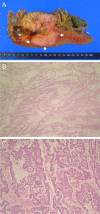
Case presentation: We herein report a case of malignant IPMN-B treated by right trisectionectomy with caudate lobectomy and extrahepatic bile duct resection. Radiologic images showed marked dilatation of the left medial sectional bile duct (B4) resulting in a bulky cystic mass with multiple internal papillary projections. Duodenal endoscopic examination demonstrated very patulous ampullary orifice with mucin expulsion and endoscopic retrograde cholangiogram confirmed marked cystic dilatation of B4 with luminal filling defects. These findings suggested IPMN-B with malignancy potential. The functional volume of the left lateral section was estimated to be 45%. A planned extensive surgery was successfully performed. The remnant bile ducts were also dilated but had no macroscopic intraluminal tumorous lesion. The histopathological examination yielded the diagnosis of mucin-producing oncocytic intraductal papillary carcinoma of the bile duct with poorly differentiated carcinomas showing neuroendocrine differentiation. The tumor was 14.0 x 13.0 cm-sized and revealed no stromal invasiveness. Resection margins of the proximal bile duct and hepatic parenchyma were free of tumor cell. The patient showed no postoperative complication and was discharged on 10th postoperative date. He has been regularly followed at outpatient department with no evidence of recurrence.
Conclusion: Considering a favorable prognosis of IPMN-B compared to non-papillary biliary neoplasms, this tumor can be a good indication for aggressive surgical resection regardless of its tumor size.
Figures


Similar articles
-
Mucin-hypersecreting bile duct neoplasm characterized by clinicopathological resemblance to intraductal papillary mucinous neoplasm (IPMN) of the pancreas.World J Surg Oncol. 2007 Aug 28;5:98. doi: 10.1186/1477-7819-5-98. World J Surg Oncol. 2007. PMID: 17725824 Free PMC article.
-
Cyst-forming intraductal papillary neoplasm of the bile ducts: description of imaging and pathologic aspects.AJR Am J Roentgenol. 2011 Nov;197(5):1111-20. doi: 10.2214/AJR.10.6363. AJR Am J Roentgenol. 2011. PMID: 22021503
-
Simultaneous liver mucinous cystic and intraductal papillary mucinous neoplasms of the bile duct: a case report.World J Gastroenterol. 2014 Apr 14;20(14):4102-5. doi: 10.3748/wjg.v20.i14.4102. World J Gastroenterol. 2014. PMID: 24744602 Free PMC article.
-
Mucinous Cystic Neoplasm of the Liver or Intraductal Papillary Mucinous Neoplasm of the Bile Duct? A Case Report and a Review of Literature.Ann Hepatol. 2018 May-June;17(3):519-524. doi: 10.5604/01.3001.0011.7397. Epub 2018 Apr 9. Ann Hepatol. 2018. PMID: 29735801 Review.
-
Current status of diagnosis and therapy for intraductal papillary neoplasm of the bile duct.World J Gastroenterol. 2021 Apr 21;27(15):1569-1577. doi: 10.3748/wjg.v27.i15.1569. World J Gastroenterol. 2021. PMID: 33958844 Free PMC article. Review.
Cited by
-
Intraductal papillary neoplasm of the bile ducts: A case report and literature review.World J Gastroenterol. 2015 Nov 21;21(43):12498-504. doi: 10.3748/wjg.v21.i43.12498. World J Gastroenterol. 2015. PMID: 26604656 Free PMC article. Review.
-
A rare case of symptomatic grossly-visible biliary intraepithelial neoplasia mimicking cholangiocarcinoma.World J Surg Oncol. 2019 Nov 11;17(1):191. doi: 10.1186/s12957-019-1737-y. World J Surg Oncol. 2019. PMID: 31711502 Free PMC article.
-
Gd-EOB-DTPA-enhanced magnetic resonance imaging for bile duct intraductal papillary mucinous neoplasms.World J Gastroenterol. 2015 Jul 7;21(25):7824-33. doi: 10.3748/wjg.v21.i25.7824. World J Gastroenterol. 2015. PMID: 26167082 Free PMC article.
-
Cystic tumor of the liver without ovarian-like stroma or bile duct communication: two case reports and a review of the literature.World J Surg Oncol. 2014 Jul 21;12:229. doi: 10.1186/1477-7819-12-229. World J Surg Oncol. 2014. PMID: 25047921 Free PMC article. Review.
References
-
- Zen Y, Sasaki M, Fujii T, Chen TC, Chen MF, Yeh TS, Jan YY, Huang SF, Nimura Y, Nakanuma Y. Different expression patterns of mucin core proteins and cytokeratins during intrahepatic cholangiocarcinogenesis from biliary intraepithelial neoplasia and intraductal papillary neoplasm of the bile duct-an immunohistochemical study of 110 cases of hepatolithiasis. J Hepatol. 2006;44:350–358. - PubMed
-
- Kloppel G, Kosmahl M. Is the intraductal papillary mucinous neoplasia of the biliary tract a counterpart of pancreatic papillary mucinous neoplasm? J Hepatol. 2006;44:249–250. - PubMed
-
- Isogai M, Nimura Y, Hayakawa N. A case of mucus producing cholangiocarcinoma with superficial spread. Jpn J Gastroenteroi Surg. 1986;19:710–713.
-
- Suh KS, Roh HR, Koh YT, Lee KU, Park YH, Kim SW. Clinicopathologic features of the intraductal growth type of peripheral cholangiocarcinoma. Hepatology. 2000;31:12–17. - PubMed
-
- Paik KY, Heo JS, Choi SH, Choi DW. Intraductal papillary neoplasm of the bile ducts: the clinical features and surgical outcome of 25 cases. J Surg Oncol. 2008;97:508–512. - PubMed
Publication types
MeSH terms
LinkOut - more resources
Full Text Sources
Medical
Molecular Biology Databases

