. 2010 Feb 15;338(2):127-35.
doi: 10.1016/j.ydbio.2009.11.030. Epub 2009 Dec 1.

Wnt signaling in heart valve development and osteogenic gene induction

Affiliations

Wnt signaling in heart valve development and osteogenic gene induction

Christina M Alfieri et al. Dev Biol. .

Abstract

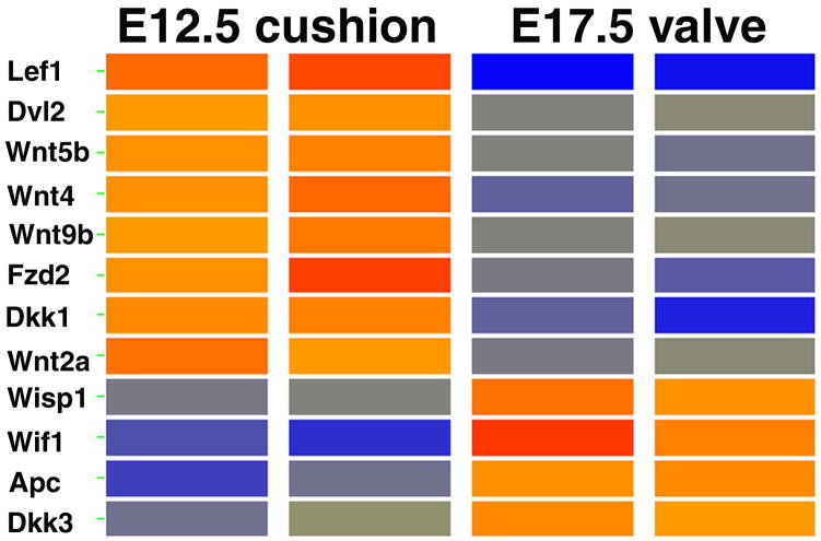
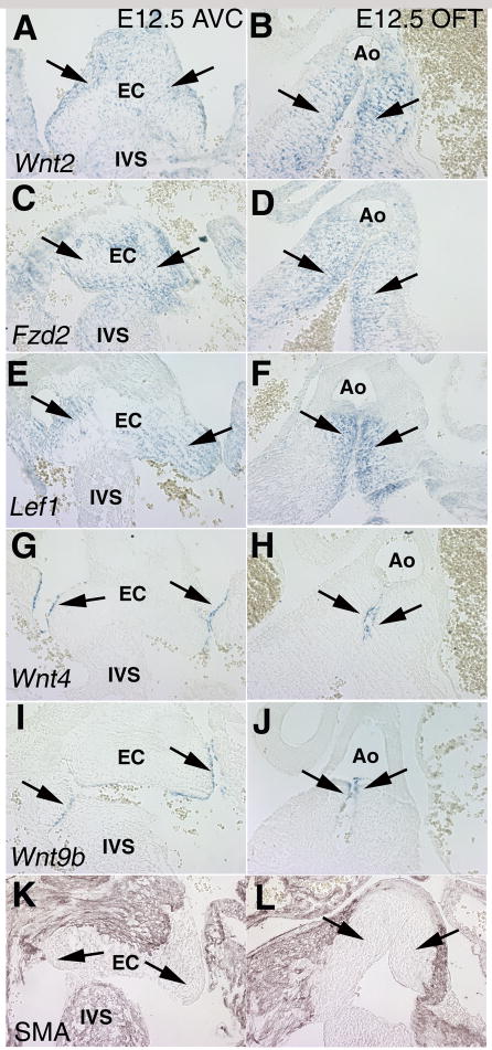
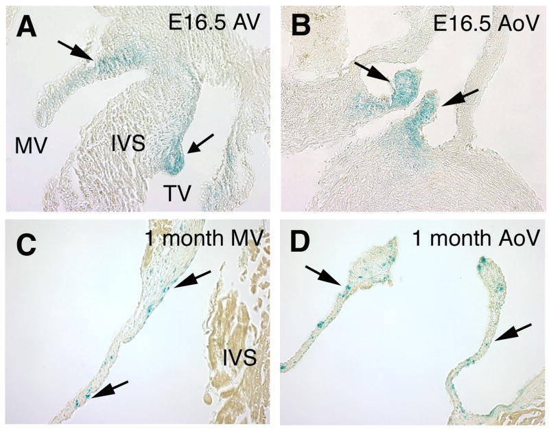
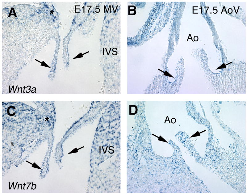
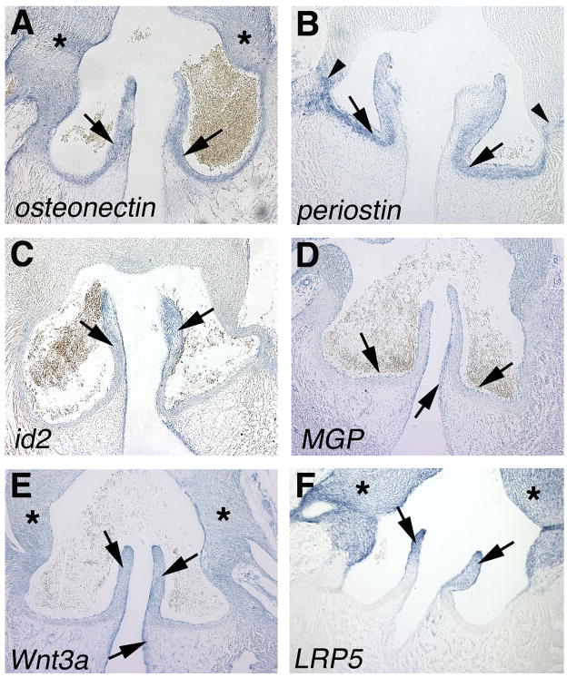
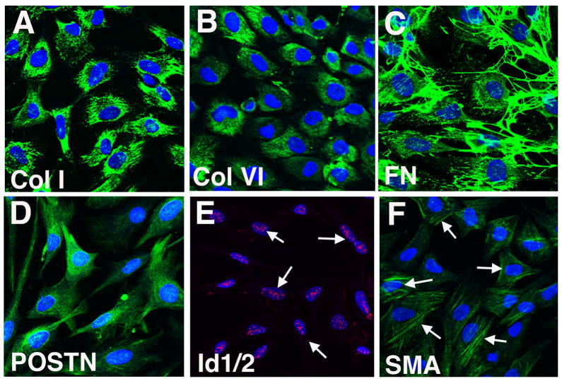
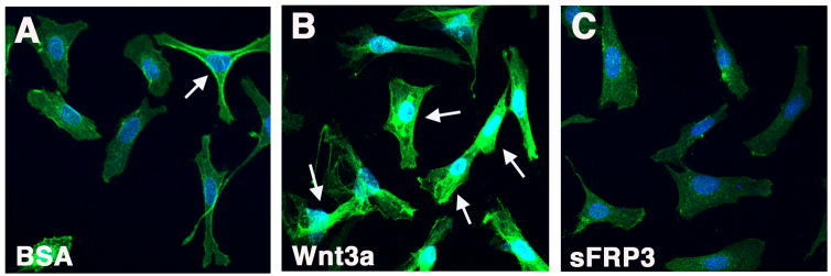
Wnt signaling mediated by beta-catenin has been implicated in early endocardial cushion development, but its roles in later stages of heart valve maturation and homeostasis have not been identified. Multiple Wnt ligands and pathway genes are differentially expressed during heart valve development. At E12.5, Wnt2 is expressed in cushion mesenchyme, whereas Wnt4 and Wnt9b are predominant in overlying endothelial cells. At E17.5, both Wnt3a and Wnt7b are expressed in the remodeling atrioventricular (AV) and semilunar valves. In addition, the TOPGAL Wnt reporter transgene is active throughout the developing AV and semilunar valves at E16.5, with more localized expression in the stratified valve leaflets after birth. In chicken embryo aortic valves, genes characteristic of osteogenic cell lineages including periostin, osteonectin, and Id2 are expressed specifically in the collagen-rich fibrosa layer at E14. Treatment of E14 aortic valve interstitial cells (VICs) in culture with osteogenic media results in increased expression of multiple genes associated with bone formation. Treatment of VIC with Wnt3a leads to nuclear localization of beta-catenin and induction of periostin and matrix gla protein but does not induce genes associated with later stages of osteogenesis. Together, these studies provide evidence for Wnt signaling as a regulator of endocardial cushion maturation as well as valve leaflet stratification, homeostasis, and pathogenesis.


Figures

Figure 1
Figure 1. Wnt pathway genes are differentially expressed in the developing AV valves at E12.5 and E17.5
An Affymetrix microarray gene expression analysis was performed on duplicate samples of RNA isolated from E12.5 AV cushions or E17.5 AV valves. The presence of Wnt signaling pathway genes was determined among genes with >2.0 fold statistically significant change at E12.5 versus E17.5 (Chakraborty et al., 2008). A heat map was generated of relative gene expression levels based on raw intensity values. Red indicates increased expression, blue indicates decreased expression, and yellow indicates no change in expression.
Figure 2
Figure 2. Wnt pathway genes are differentially expressed in mesenchymal and endothelial cells of mouse E12.5 endocardial cushions
In situ hybridizations of sectioned hearts show Wnt pathway gene expression in both atrioventricular canal (AVC; left panels) and outflow tract (OFT; right panels) cushions at E12.5. The ligand Wnt2 (A, B), receptor frizzled2 (Fzd2; C,D) and downstream transcription factor LEF1 (E,F) are expressed in cushion mesenchyme (indicated by arrows). In contrast, the ligands Wnt4 (G,H) and Wnt9b (I,J) are expressed specifically in the cushion endothelial layer (indicated by arrows). Antibody staining for smooth muscle α-actin (SMA) indicates muscle cells (K,L). Endocardial cushions (EC), interventricular septum (IVS) and Aorta (Ao) are indicated.
Figure 3
Figure 3. The Wnt pathway is active in mouse mitral and aortic valves as indicated by TOPGAL reporter expression
(A–D) Analysis of TOPGAL Wnt pathway reporter mice demonstrates pathway activation in the mitral valve (MV) and tricuspid valve (TV) primordia as well as the aortic valve (AoV) at E16.5 (A, B), as indicated by X-gal staining. Localized TOPGAL activity also is apparent in MV and AoV leaflets 1 month after birth (C,D), predominantly on the fibrosa side (indicated by arrows). The interventricular septum (IVS) is indicated.
Figure 4
Figure 4. Wnt3a and Wnt7b are expressed in mouse mitral and aortic valves at E17.5
(A–D) In situ hybridization of sectioned E17.5 mouse hearts demonstrates that Wnt3a (A,B) and Wnt7b (C,D) are expressed in the remodeling mitral (MV) and aortic (AoV) valves. Wnt3a and Wnt7b expression in the AV valves is apparent at the tips of the leaflets (indicated by arrows) and in the AV canal (indicated by asterisks). Wnt3a is expressed preferentially on the fibrosa side the aortic valve leaflets (arrows, B). Wnt7b expression is apparent throughout the AoV leaflets (arrows, D). The interventricular septum (IVS) and aorta (Ao) are indicated.
Figure 5
Figure 5. ECM, osteogenic and Wnt pathway genes are locally expressed during aortic valve stratification in chicken embryos
In situ hybridization of sectioned E14 chicken embryo hearts shows expression of (A) osteonectin, (B) periostin, (C) id2, (D) matrix gla protein (MGP), (E) Wnt3a and (F) LRP5 in aortic valve cusps (arrows). Expression also is apparent in the aorta (asterisks). The aortic valve annulus is indicated by arrowheads in B.
Figure 6
Figure 6. Cultured E14 chicken aortic valve cells have a fibrosa-like phenotype
Immunostaining and confocal analysis demonstrates expression of (A) Collagen I, (B) Collagen VI, (C) fibronectin (FN), (D) periostin (POSTN), (E) Id1/2 and (F) smooth muscle α-actin (SMA) in aortic valve cells cultured for 5d. Indicated proteins are shown in green with the exception of Id1/2, which is red. Nuclear staining of Id1/2 and distribution of SMA are indicated by arrows. Nuclei are stained blue with ToPro3.
Figure 7
Figure 7. β-catenin protein expression is increased in E14 Aortic VIC treated with Wnt3a but not sFRP3
Primary E14 aortic VIC cultures were treated with (A) control vehicle BSA, (B) Wnt3a or (C) sFRP3 for 1hr. Increased nuclear and cytoplasmic β-catenin protein (green) was detected by immunofluorescence and confocal microscopy (arrows) in Wnt3a-treated relative to control or sFRP3-treated cells. Nuclei (blue) were visualized using ToPro3.
Figure 8
Figure 8. Induction of osteoblast-associated genes in avian E14 aortic VIC cultures treated with β-glycerophosphate/ascorbic acid is enhanced by increased Wnt signaling
(A) E14 embryonic aortic valve cells were cultured in the presence or absence of osteogenic (osteo) media containing β-glycerophosphate (10mM) and ascorbic acid (50μg/ml). Expression levels of periostin (POSTN), matrix-gla protein (MGP), Runx2, osteocalcin (OCN) and alkaline phosphatase (ALP) transcripts were determined by qRT-PCR in untreated (white bars) and treated (black bars) VIC cultures. Control values were set to 1 and fold differences are shown with error bars representing s.e.m. Statistically significant increases in gene expression in induced cells (p<0.05) are indicated by asterisks. Baseline levels of expression are POSTN (19.26), MGP (3710.23), Runx2 (957.22), OCN (10.90), ALP (100.37) relative fluorescence units. (B) Primary chicken embryo (E14) aortic valve cells were cultured as in (A) with the addition of groups treated with Wnt3a or the Wnt antagonist sFRP3. Expression levels of POSTN, MGP, Runx2, OCN and ALP transcripts were determined by qRT-PCR for all groups. Control values were set to 1, and fold differences are shown for a representative experiment. Note induction of MGP and POSTN in the presence of osteogenic media is enhanced by Wnt3a and is inhibited by sFRP3 treatments.

Similar articles

Cited by

References

    1. Aikawa E, Nahrendorf M, Sosnovik D, Lok VM, Jaffer FA, Aikawa M, Weissleder R. Multimodality molecular imaging identifies proteolytic and osteogenic activities in early aortic valve disease. Circulation. 2007;115:377–386. - PubMed
    1. Aikawa E, Whittaker P, Farber M, Mendelson K, Padera RF, Aikawa M, Schoen FJ. Human semilunar cardiac valve remodeling by activated cells from fetus to adult. Circulation. 2006;113:1344–1352. - PubMed
    1. Armstrong EJ, Bischoff J. Heart valve development: Endothelial cell signaling and differentiation. Circ Res. 2004;95:459–470. - PMC - PubMed
    1. Barone LM, Owen TA, Tassinari MS, Bortell R, Stein GS, Lian JB. Developmental expression and hormonal regulation of the rat matrix Gla protein (MGP) gene in chondrogenesis and osteogenesis. J Cell Biochem. 1991;46:351–365. - PubMed
    1. Caira FC, Stock SR, Gleason TG, McGee EC, Huang J, Bonow RO, Spelsberg TC, McCarthy PM, Rahimtoola SH, Rajamannan NM. Human degenerative valve disease is associated with up-regulation of low-density lipoprotein-related protein 5 receptor-mediated bone formation. J Am Coll Cardiol. 2006;47:1707–1712. - PMC - PubMed

Publication types