. 2010 Apr;48(4):713-24.
doi: 10.1016/j.yjmcc.2009.11.015. Epub 2009 Dec 1.

Cyclic GMP/PKG-dependent inhibition of TRPC6 channel activity and expression negatively regulates cardiomyocyte NFAT activation Novel mechanism of cardiac stress modulation by PDE5 inhibition

Affiliations

Cyclic GMP/PKG-dependent inhibition of TRPC6 channel activity and expression negatively regulates cardiomyocyte NFAT activation Novel mechanism of cardiac stress modulation by PDE5 inhibition

Norimichi Koitabashi et al. J Mol Cell Cardiol. 2010 Apr.

Abstract

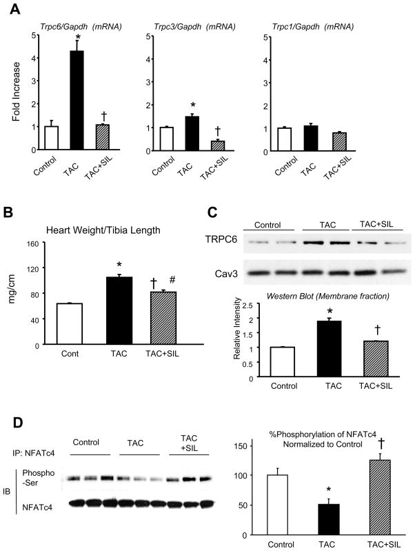
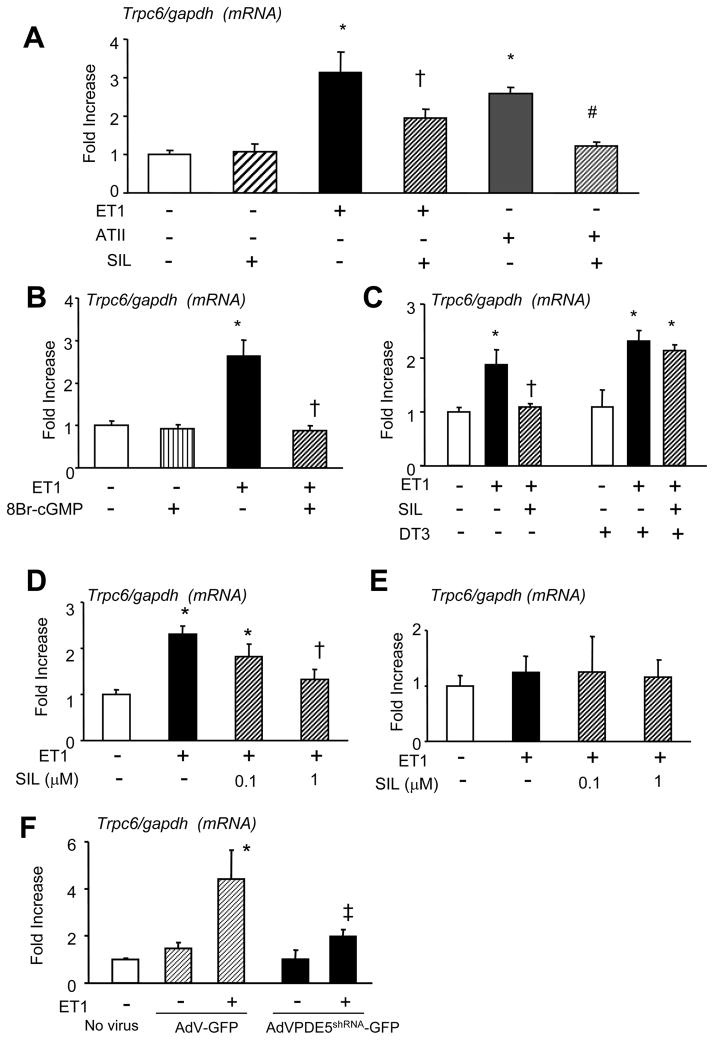
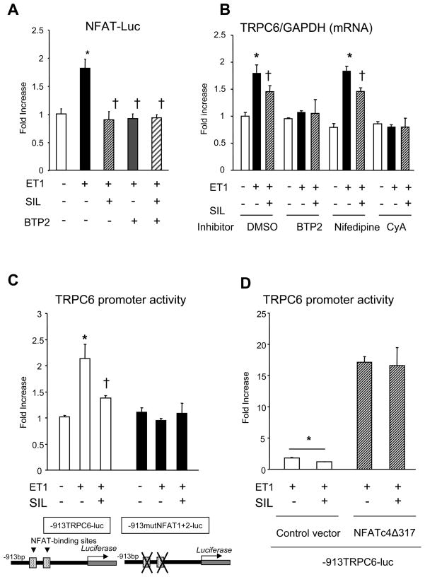
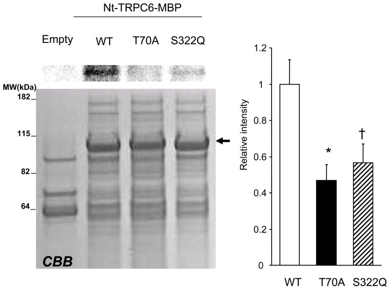
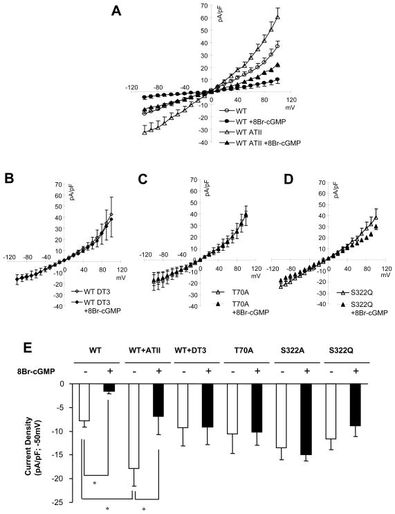
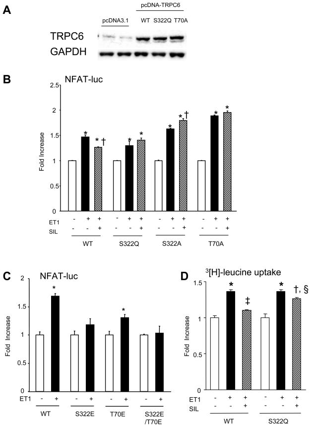
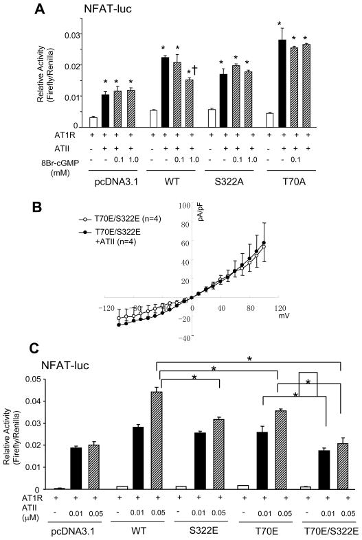
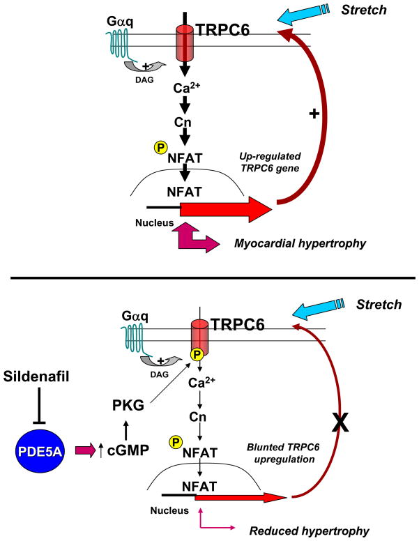
Increased cyclic GMP from enhanced synthesis or suppressed catabolism (e.g. PDE5 inhibition by sildenafil, SIL) activates protein kinase G (PKG) and blunts cardiac pathological hypertrophy. Suppressed calcineurin (Cn)-NFAT (nuclear factor of activated T-cells) signaling appears to be involved, though it remains unclear how this is achieved. One potential mechanism involves activation of Cn/NFAT by calcium entering via transient receptor potential canonical (TRPC) channels (notably TRPC6). Here, we tested the hypothesis that PKG blocks Cn/NFAT activation by modifying and thus inhibiting TRPC6 current to break the positive feedback loop involving NFAT and NFAT-dependent TRPC6 upregulation. TRPC6 expression rose with pressure-overload in vivo, and angiotensin (ATII) or endothelin (ET1) stimulation in neonatal and adult cardiomyocytes in vitro. 8Br-cGMP and SIL reduced ET1-stimulated TRPC6 expression and NFAT dephosphorylation (activity). TRPC6 upregulation was absent if its promoter was mutated with non-functional NFAT binding sites, whereas constitutively active NFAT triggered TRPC6 expression that was not inhibited by SIL. PKG phosphorylated TRPC6, and both T70 and S322 were targeted. Both sites were functionally relevant, as 8Br-cGMP strongly suppressed current in wild-type TRPC6 channels, but not in those with phospho-silencing mutations (T70A, S322A or S322Q). NFAT activation and increased protein synthesis stimulated by ATII or ET1 was blocked by 8Br-cGMP or SIL. However, transfection with T70A or S322Q TRPC6 mutants blocked this inhibitory effect, whereas phospho-mimetic mutants (T70E, S322E, and both combined) suppressed NFAT activation. Thus PDE5-inhibition blocks TRPC6 channel activation and associated Cn/NFAT activation signaling by PKG-dependent channel phosphorylation.


Figures

Figure 1
Figure 1
Influence of pressure-overload and PDE5 inhibition (sildenafil, SIL) on TRPC6 expression. A) Tprc6, 3, and 1, mRNA normalized to Gapdh were assessed in mice subjected to 7-days of transverse aortic constriction (TAC). Results are shown normalized to normal controls (n=4 for each group, * p<0.01 versus control and TAC1w; † p<0.001 vs TAC1w; ‡ p<0.05 vs TAC1w). B) SIL effect on cardiac hypertrophy (heart weight/tibia length) after TAC; n=4–5/group; * p<0.01 versus control; †-p<0.01 versus TAC; # p=0.028 versus control). C) Western blot of TRPC6 in membrane fraction from whole heart tissue isolate. Caveolin3 (Cav3) was used as a protein loading control; (n=4/group; *-p<0.05 versus control; †-p<0.01 versus 1-wk TAC hearts) D) SIL blunts NFAT dephosphorylation induced by TAC. NFATc4 was immune-precipitated and isolated then proved with a phospho-Ser specific antibody. Summary data for ratio of phosphorylated/total NFATc4 is shown at the right (n=4/group; *-p<0.05 versus control; †-p<0.01 versus 1-wk TAC hearts).
Figure 2
Figure 2
Effect of SIL or 8Br-cGMP on Trpc6 mRNA expression in neonatal rat (AC) and adult mouse (D and E) cultured cardiomyocytes. A) Trpc6 mRNA expression normalized to Gapdh rose with 0.1 μM endothelin-1(ET1) or 1 μM angiotensin II (ATII), and both changes were suppressed by co-incubation with 1 μM SIL for 24hrs. (n=5–12/group; * p<0.01 vs vehicle (Veh), †p<0.05 vs ET1; ‡p<0.01 vs ATII). B) ET1 stimulated Trpc6 mRNA expression is suppressed by co-treatment of 8Br-GMP (1 mM, n=5–6/group, *p<0.002 vs other groups) for 24hrs. C) SIL-mediated suppression of ET1 activated Trpc6 expression is blocked by inhibiting PKG activity with DT3 (n=4/group; * p<0.05 vs Veh; †p<0.05 vs ET1). D) SIL (0.1, 1 μM) suppresses ET-1 triggered Trpc6 gene expression in adult mouse cardiac myocytes. (24 hr incubation study, n=9/group, * p<0.01 versus vehicle control, † p<0.05 versus ET1 stimulation. E) In contrast, ET-1 did not stimulate Trpc3 expression in these cells. F) PDE5 gene-silencing blocks ET1-stimulated Trpc6 gene expression. Cells were transfected with (AdVPDE5shRNA GFP) or control (AdV-GFP) [13] were stimulated by 0.1 μM ET1 for 24hrs (n=3/group, * p<0.001 for ET1 effect, ‡ p<0.05 for effect from PDE5 gene silencing on ET1 response, 2-way ANOVA).
Figure 3
Figure 3
Effect of SIL on cardiomyocyte NFAT activation and TRPC6 expression. A) Activation of NFAT (luciferase reporter assay, NFAT-Luc) by ET1 (0.1 μM) in NRVM is prevented by SIL (1 μM) or by the TRP-channel blocker BTP2 (1 μM), with no additive effects from their combination (4-hr incubation, n=3/group, *P<0.01 versus other groups). B) ET-1 stimulated Trpc6 expression is prevented by the Cn inhibitor cyclosporin A (1 μM) and TRP channel antagonist BTP2, but not LTCC blocker nifedipine (1 μM). SIL still suppress expression in the presence of nifedipine; studies performed in NRVM; DMSO, dimetyl sulfoxide (n=3/group, *P<0.01 versus vehicle, †p<0.01 versus 24hr ET1 stimulation.) C) TRPC6 promoter activity in constructs with (−913TRPC6-luc) or without (−913 mutNFAT1+2TRPC6-luc) NFAT binding sites, generated as described(18). N=6/group, *P<0.01 versus vehicle, †p<0.01 versus ET1 stimulation. 4hour stimulation D) SIL suppression of TRPC6 promoter activity with or without constitutive active form of NFATc4: NFATc4Δ317. *P<0.05. 4hr stimulation.
Figure 4
Figure 4
PKG phosphorylation of N-terminus human TRPC6 protein. Left panel shows representative in vitro PKG phosphorylation of TRPC6 fused with maltose binding protein (Nt-TRPC6-MBP) with wild type (WT) or phospho-silenced mutants. CBB: Coomassie Brilliant Blue staining of the synthetic peptides was used for the phosphorylation assay, MW: molecular weight. ; top panel shows autoradiogram data, and right panel summary of radiogram for each condition. (n=4/group, *P<0.01 versus WT, † p<0.05 versus WT).
Figure 5
Figure 5
cGMP-mediated TRPC6 current inhibition in HEK293T cells expressing wild-type (WT) or mutant TRPC6. A) Averaged current-voltage (I–V) relationship in cells expressing wild-type TRPC6 channels in absence (open circle; N=6) or presence (closed circle; N=5) of 8Br-cGMP (1mM) and in ATII (200nM) stimulated cells with (open triangle; N=5) or without (closed triangle; N=5) 8Br-cGMP. ATII type I receptor (AT1R) was co-transfected into HEK cells for ATII experiments. In both conditions, cGMP depressed TRPC6 current. B) Inhibition of PKG with DT3 (0.2 μM) prevents suppression of TRPC6 current by cGMP. C,D) I-V relationships in HEK cells expressing T70A or S322Q mutant TRPC6 channels (phospho-silenced) with and without co-treatment by 8Br-cGMP. (n=5–6/group). In both instances, the inhibition of channel current by cGMP was absent. E) Summary data for the preceding studies (also for S322A mutant) displaying averaged density of TRPC6 current measured at −50mV from a holding potential −60mV. * p<0.05 versus comparison as depicted.
Figure 6
Figure 6
Effect of TRPC6 phospho-mutants on the efficacy of SIL to block NFAT activation by ET1. A) Example Western blot showing robust expression of various TRPC6 forms after transient transfection of TRPC6 plasmids in NRVM. B) NFAT luciferase assay in NRVM transfected with WT of phospho-silenced TRPC6 mutants (S322Q, S322A, or T70A). SIL suppression of NFAT activation by ET1 was absent with each mutant. (n=3/group for both, * p<0.01 versus vehicle, †-p<0.01 vs ET1). C) In contrast, expression of phospho-mimetic mutants of either S322E or T70E reduced ET1-induced NFAT activation, and the double mutant (S322E/T70E) fully prevented activation. (n=6/group for both, * p<0.01 versus vehicle). D) NRVM expressing WT-TRPC6 undergo enhanced protein synthesis (leucine incorporation) with ET1 that is blunted by SIL. In contrast, cells expressing the S322Q-TRPC6 have less impact from SIL. (n=5–6/group, *P<0.01 versus vehicle, † p<0.01 vs vehicle, p<0.01 vs ET1; ‡p<0.005 vs vehicle, p<0.01 vs ET1; § p<0.02 for interaction of TRPC6 mutation and ET1 effect (2-way ANOVA). 4hr stimulation.
Figure 7
Figure 7
Effect of TRPC mutants on NFAT activation in HEK293T cells. A) NFAT activity in cells transfected with WT or phospho-silenced TRPC6 mutants. Cells were co-transfected with ATII type 1a receptor (AT1R) and stimulated with 0.01 μM ATII in the presence or absence of 8Br-cGMP (0.1–1mM, 4–6 hr incubation). Without TRPC6 co-transfection, 8Br-cGMP did not suppress NFAT activation induced by ATII. This suppression was also absent in cells expressing S322A or T70A mutants. B) Averaged current-voltage (I-V) relationship in cells expressing a phospho-mimetic mutant of TRPC6 channel (S322E/T70E double mutant) and AT1-R in the absence (open circle; N=4) or presence (closed circle; N=4) of ATII (200nM). C) Effect of TRPC6 phospho-mimetic mutations on NFAT activation from AII in HEK cells co-transfected with AT1-R. Co-transfection with WT-TRPC6 resulted in an augmented NFAT activation and ATII dose response. This was blunted with S322E and T70E mutants, and completely suppressed (to empty vector control levels) with the double mutant. N=6 of each group († p<0.01 versus WT-0.05 μM ATII stimulation; # P<0.05 versus S322E or T70E with 0.05 μM ATII; ¶ P<0.05 versus WT-0.01 μM ATII stimulation). 4hr stimulation.
Figure 8
Figure 8
Schematic summarizing the signaling involving TRPC6-Cn-NFAT in the absence (top) and presence (bottom) of activated PKG (due to enhanced cGMP generation or blocked catabolism). See text for details.

Similar articles

Cited by

References

    1. Mudd JO, Kass DA. Tackling heart failure in the twenty-first century. Nature. 2008;451:919–928. - PubMed
    1. Molkentin JD, Lu JR, Antos CL, Markham B, Richardson J, Robbins J, Grant SR, Olson EN. A calcineurin-dependent transcriptional pathway for cardiac hypertrophy. Cell. 1998;93:215–228. - PMC - PubMed
    1. Bueno OF, Wilkins BJ, Tymitz KM, Glascock BJ, Kimball TF, Lorenz JN, Molkentin JD. Impaired cardiac hypertrophic response in Calcineurin Abeta -deficient mice. Proc Natl Acad Sci U S A. 2002;99:4586–4591. - PMC - PubMed
    1. Hsu S, Nagayama T, Koitabashi N, Zhang M, Zhou L, Bedja D, Gabrielson KL, Molkentin JD, Kass DA, Takimoto E. Phosphodiesterase 5 inhibition blocks pressure overload-induced cardiac hypertrophy independent of the calcineurin pathway. Cardiovasc Res. 2009;81:301–309. - PMC - PubMed
    1. Hogan PG, Chen L, Nardone J, Rao A. Transcriptional regulation by calcium, calcineurin, and NFAT. Genes Dev. 2003;17:2205–2232. - PubMed

Publication types

MeSH terms