Angiotensin II-activated protein kinase D mediates acute aldosterone secretion
- PMID: 19961896
- PMCID: PMC2814994
- DOI: 10.1016/j.mce.2009.11.017
Angiotensin II-activated protein kinase D mediates acute aldosterone secretion
Abstract
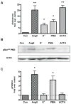
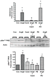
Dysregulation of the renin-angiotensin II (AngII)-aldosterone system can contribute to cardiovascular disease, such that an understanding of this system is critical. Diacylglycerol-sensitive serine/threonine protein kinase D (PKD) is activated by AngII in several systems, including the human adrenocortical carcinoma cell line NCI H295R, where this enzyme enhances chronic (24h) AngII-evoked aldosterone secretion. However, the role of PKD in acute AngII-elicited aldosterone secretion has not been previously examined. In primary cultures of bovine adrenal glomerulosa cells, which secrete detectable quantities of aldosterone in response to secretagogues within minutes, PKD was activated in response to AngII, but not an elevated potassium concentration or adrenocorticotrophic hormone. This activation was time- and dose-dependent and occurred through the AT1, but not the AT2, receptor. Adenovirus-mediated overexpression of constitutively active PKD resulted in enhanced AngII-induced aldosterone secretion; whereas overexpression of a dominant-negative PKD construct decreased AngII-stimulated aldosterone secretion. Thus, we demonstrate for the first time that PKD mediates acute AngII-induced aldosterone secretion.
Figures


Similar articles
-
A role for phospholipase D in angiotensin II-induced protein kinase D activation in adrenal glomerulosa cell models.Mol Cell Endocrinol. 2013 Feb 5;366(1):31-7. doi: 10.1016/j.mce.2012.11.008. Epub 2012 Nov 20. Mol Cell Endocrinol. 2013. PMID: 23178798 Free PMC article.
-
Protein kinase C and Src family kinases mediate angiotensin II-induced protein kinase D activation and acute aldosterone production.Mol Cell Endocrinol. 2014 Jul 5;392(1-2):173-81. doi: 10.1016/j.mce.2014.05.015. Epub 2014 May 22. Mol Cell Endocrinol. 2014. PMID: 24859649 Free PMC article.
-
Angiotensin II-induced protein kinase D activates the ATF/CREB family of transcription factors and promotes StAR mRNA expression.Endocrinology. 2014 Jul;155(7):2524-33. doi: 10.1210/en.2013-1485. Epub 2014 Apr 7. Endocrinology. 2014. PMID: 24708239 Free PMC article.
-
Angiotensin-responsive adrenal glomerulosa cell proteins: characterization by protease mapping, species comparison, and specific angiotensin receptor antagonists.Endocrinology. 1997 Jun;138(6):2530-6. doi: 10.1210/endo.138.6.5207. Endocrinology. 1997. PMID: 9165045
-
AngII induces transient phospholipase D activity in the H295R glomerulosa cell model.Mol Cell Endocrinol. 2003 Aug 29;206(1-2):113-22. doi: 10.1016/s0303-7207(03)00211-9. Mol Cell Endocrinol. 2003. PMID: 12943994
Cited by
-
Protein kinase D1 deficiency promotes differentiation in epidermal keratinocytes.J Dermatol Sci. 2014 Dec;76(3):186-95. doi: 10.1016/j.jdermsci.2014.09.007. Epub 2014 Oct 2. J Dermatol Sci. 2014. PMID: 25450094 Free PMC article.
-
A role for phospholipase D in angiotensin II-induced protein kinase D activation in adrenal glomerulosa cell models.Mol Cell Endocrinol. 2013 Feb 5;366(1):31-7. doi: 10.1016/j.mce.2012.11.008. Epub 2012 Nov 20. Mol Cell Endocrinol. 2013. PMID: 23178798 Free PMC article.
-
VLDL-activated cell signaling pathways that stimulate adrenal cell aldosterone production.Mol Cell Endocrinol. 2016 Sep 15;433:138-46. doi: 10.1016/j.mce.2016.05.018. Epub 2016 May 21. Mol Cell Endocrinol. 2016. PMID: 27222295 Free PMC article.
-
Protein kinase C and Src family kinases mediate angiotensin II-induced protein kinase D activation and acute aldosterone production.Mol Cell Endocrinol. 2014 Jul 5;392(1-2):173-81. doi: 10.1016/j.mce.2014.05.015. Epub 2014 May 22. Mol Cell Endocrinol. 2014. PMID: 24859649 Free PMC article.
-
Aquaporin-3 re-expression induces differentiation in a phospholipase D2-dependent manner in aquaporin-3-knockout mouse keratinocytes.J Invest Dermatol. 2015 Feb;135(2):499-507. doi: 10.1038/jid.2014.412. Epub 2014 Sep 18. J Invest Dermatol. 2015. PMID: 25233074 Free PMC article.
References
-
- Abedi H, Rozengurt E, Zachary I. Rapid activation of the novel serine/threonine protein kinase, protein kinase D by phorbol esters, angiotensin II and PDGF-BB in vascular smooth muscle cells. FEBS Lett. 1998;427:209–212. - PubMed
-
- Bassett MH, Zhang Y, White PC, Rainey WE. Regulation of human CYP11B2 and CYP11B1: comparing the role of the common CRE/Ad1 element. Endocr Res. 2000;26:941–951. - PubMed
-
- Betancourt-Calle S, Bollag WB, Jung EM, Calle RA, Rasmussen H. Effects of angiotensin II and adrenocorticotropic hormone on myristoylated alanine-rich C-kinase substrate phosphorylation in glomerulosa cells. Mol Cell Endocrinol. 1999;154:1–9. - PubMed
-
- Betancourt-Calle S, Jung EM, White S, Ray S, Zheng X, Calle RA, Bollag WB. Elevated K(+) induces myristoylated alanine-rich C-kinase substrate phosphorylation and phospholipase D activation in glomerulosa cells. Mol Cell Endocrinol. 2001;184:65–76. - PubMed
-
- Bird IM, Hanley NA, Word RA, Mathis JM, McCarthy JL, Mason JI, Rainey WE. Human NCI-H295 adrenocortical carcinoma cells: a model for angiotensin-II-responsive aldosterone secretion. Endocrinology. 1993;133:1555–1561. - PubMed
Publication types
MeSH terms
Substances
Grants and funding
LinkOut - more resources
Full Text Sources
Research Materials

