Review
. 2010 Feb;1800(2):49-56.
doi: 10.1016/j.bbagen.2009.11.016. Epub 2009 Dec 2.

O-GlcNAc protein modification in plants: Evolution and function

Affiliations
Review

O-GlcNAc protein modification in plants: Evolution and function

Neil E Olszewski et al. Biochim Biophys Acta. 2010 Feb.

Abstract

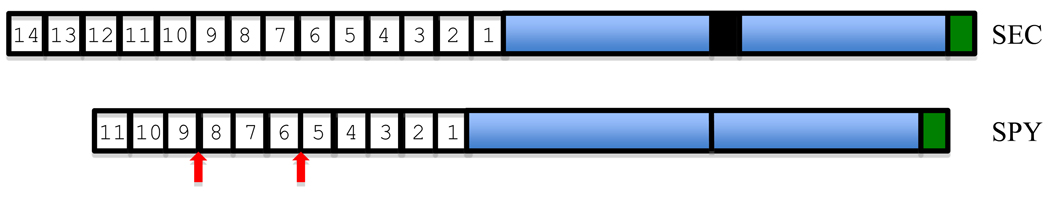
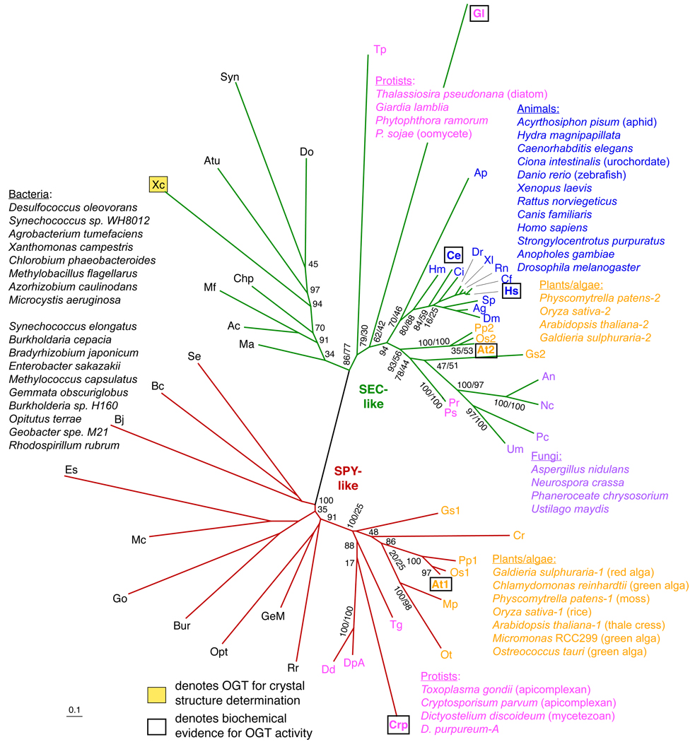
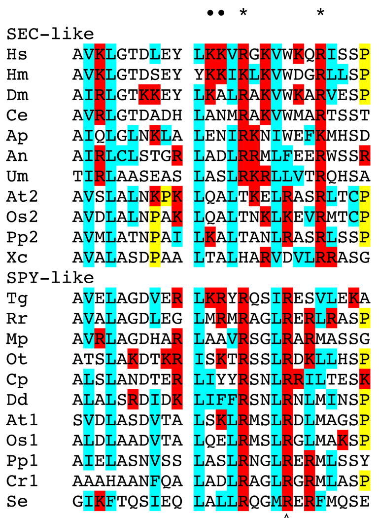
The role in plants of posttranslational modification of proteins with O-linked N-acetylglucosamine and the evolution and function of O-GlcNAc transferases responsible for this modification are reviewed. Phylogenetic analysis of eukaryotic O-GlcNAc transferases (OGTs) leads us to propose that plants have two distinct OGTs, SEC- and SPY-like, that originated in prokaryotes. Animals and some fungi have a SEC-like enzyme while plants have both. Green algae and some members of the Apicomplexa and amoebozoa have the SPY-like enzyme. Interestingly the progenitor of the Apicomplexa lineage likely had a photosynthetic plastid that persists in a degenerated form in some species, raising the possibility that plant SPY-like OGTs are derived from a photosynthetic endosymbiont. OGTs have multiple tetratricopeptide repeats (TPRs) that within the SEC- and SPY-like classes exhibit evidence of strong selective pressure on specific repeats, suggesting that the function of these repeats is conserved. SPY-like and SEC-like OGTs have both unique and overlapping roles in the plant. The phenotypes of sec and spy single and double mutants indicate that O-GlcNAc modification is essential and that it affects diverse plant processes including response to hormones and environmental signals, circadian rhythms, development, intercellular transport and virus infection. The mechanistic details of how O-GlcNAc modification affects these processes are largely unknown. A major impediment to understanding this is the lack of knowledge of the identities of the modified proteins.


Figures

Fig. 1
Fig. 1. Structure of SEC and SPY from Arabidopsis
The numbered boxes indicate individual tetratricopeptide repeats (34 amino acids in length). The blue boxes indicate the two regions that together form the catalytic domain. The black box in SEC indicates the location of a variable length insertion between the catalytic domains. This insertion also occurs in animal and fungal OGTs. The green box indicates the phospholipid-binding region. The vertical arrows indicate the most common locations of amino acid insertions between TPR repeats of SPY and SPY-like enzymes.
Fig. 2
Fig. 2. Phylogenetic relationship of OGT-like catalytic domain sequences
For the phylogenetic analysis of the aligned sequences (see Supplemental Table 1 and Supplemental Fig. 1), ProtTest 2.2 [70] was used to find the best fitting amino acid substitution model. A LG+G+I+F Le & Gascuel (LG) substitution model [71] was selected with a gamma rate (G) distribution (4 categories, alpha=1.515), a proportion of invariable (I) sites (0.006), and empirically determined amino acid frequencies (F). This model was implemented in a maximum likelihood framework, executed using the program PhyML [72] with 9,766 replicates to search for the best tree. A likelihood ratio test (LRT), as implemented in PhyML, and bootstrapping, were used to calculate support for the tree, and are listed as percent values at the nodes (LRT,bootstrap; or LRT alone). Sequences with experimentally verified OGT activity are bolded and boxed. The shaded box denotes the X. campestris sequence that has been analyzed crystallographically.
Fig. 3
Fig. 3. Alignment of predicted phospholipid binding domain sequences
Alignment of the phospholipid binding domains of: Hs, Homo sapiens; Hm, Hydra magnipapillata; Dm, Drosophila melanogaster, Ce, Caenorhabditis elegans; Ap, Acyrthosiphon pisum; An, Aspergillus niger; Um, Ustilago maydis; At2, Arabidopsis thaliana SEC; Os2, Oryza sativa SEC; Pp2, Physcomitrella patens SEC; Xc, Xanthomonas campestris; Tg, Toxoplasma gondii; Rr, Rhodospirillum rubrum; Mp, Micromonas RCC299; Ot, Ostreococcus tauri; Cp, Cryptosporidium parvum; Dd, Dictyostelium discoideum; At1, Arabidopsis thalaian SPY, Os1, Oryza sativa SPY; Pp1, Physcomitrella patens SPY; Cr1, Chlamydomonas reinhardtii; Se, Synechococcus elongatus PCC 6301. Amino acids demonstrated by mutagenesis of human OGT (•) or predicted from the Xanthomonas campestris crystal structure (*) to be important for PIP3 binding are indicated above the alignment. The amino acid affected in the spy-16 and -17 alleles is indicated below the alignment (◊). Hydrophobic amino acids are highlighted in blue, basic amino acids are highlighted in red, and Pro in yellow.

Similar articles

Cited by

References

    1. Schwechheimer C. Understanding gibberellic acid signaling--are we there yet? Curr. Opin. Plant Biol. 2008;11:9–15. - PubMed
    1. Hartweck LM. Gibberellin signaling. Planta. 2008;229:1–13. - PubMed
    1. Jacobsen SE, Olszewski NE. Mutations at the SPINDLY locus of Arabidopsis alter gibberellin signal transduction. Plant Cell. 1993;5:887–896. - PMC - PubMed
    1. Silverstone AL, Tseng TS, Swain SM, Dill A, Jeong SY, Olszewski NE, Sun TP. Functional analysis of SPINDLY in gibberellin signaling in Arabidopsis. Plant Physiol. 2007;143:987–1000. - PMC - PubMed
    1. Wilson RN, Somerville CR. Phenotypic suppression of the gibberellin-insensitive mutant (gai) of Arabidopsis. Plant Physiol. 1995;108:495–502. - PMC - PubMed

Publication types

MeSH terms

LinkOut - more resources