

doi: 10.1016/j.mric.2009.09.001.
Origins of intraoperative MRI
Affiliations
- PMID: 19962089
- PMCID: PMC4120097
- DOI: 10.1016/j.mric.2009.09.001
Item in Clipboard
Origins of intraoperative MRI
Magn Reson Imaging Clin N Am.
2010 Feb.
Abstract
Neurosurgical diagnosis and intervention has evolved through improved neuroimaging, allowing better visualization of anatomy and pathology. This article discusses the various systems that have been designed over the last decade to meet the requirements of neurosurgical patients and opines on the potential future developments in the technology and application of intraoperative MRI. Because the greatest amount of experience with intraoperative MRI comes from its use in brain tumor resection, this article focuses on the origins of intraoperative MRI in relation to this field.
Figures

Brain shift. (A) T1-weighted axial MRI before craniotomy.
(B) Same axial plane MRI after craniotomy performed.
(C) MRI after lesion resection. Note the significant shift
of intracranial contents after craniotomy, cerebrospinal fluid drainage, and
lesion resection.
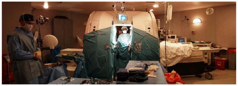
The MRT unit at BWH. The General Electric Signa 0.5T iMRI is an
open-configuration “double donut” system that allows the surgeon
to operate between each superconductive magnet coil (pictured).

Intraoperative colocalization using iMRI images and 3-D Slicer software.
(A) Tumor, functional MRI, and diffusion tensor imaging are
colocalized with standard MRI. (B) After craniotomy is
performed, 3-D Slicer compensates for brain shift, allowing surgeons to
visualize not only shift of gross neuroanatomy but also functional regions and
white matter tracts.

The PoleStar iMRI. The open-bore configuration PoleStar N-20, despite its
low-field 0.15T magnet, has allowed many institutions to take advantage of iMRI
without completely remodeling their operative suite to accommodate a larger,
high-field stationary iMRI. The Polestar is compact enough to be stored in a
shielded room (pictured on the right side of the figure) when not in use. If a
smaller room cannot be dedicated to storage, an in-suite
“hangar” can be set up to shield the magnet when not in use.
(Courtesy of Medtronic Navigation, Louisville, CO; with
permission.)

The IMRIS iMRI. The IMRIS iMRI suite features a high-field, closed-bore magnet.
(A) The IMRIS magnet in its storage room, shielding doors
opened to show its relation to the operative suite when not in use.
(B) The magnet has moved on its ceiling-mounted rails to
its position over the region of the patient's head. The 1.5-T magnet can
be brought from its park position in the storage room to a fully operational
position within the operating room in less than 90 seconds, allowing for
efficient scanning while still remaining unobtrusive. (C) An
example of the IMRIS magnet serving two separate operating rooms. (The magnet
can swivel 180° in the storage room to orient the working end toward the
appropriate operating room.) (Courtesy of IMRIS, Inc.,
Winninpeg, Manitoba, Canada.)

The NeuroArm MRI-compatible neurosurgical robot. (A) Detailed
picture of one of the NeuroArm's two operative limbs, with bipolar
cautery attachment. (B) Command center for NeuroArm robot,
where surgeon is seated and driving the movements of the robot through
haptic-feedback controllers. (C) Real-time virtual reality
display of the robot's position is delivered to the surgeon. Other
virtual reality displays feature colocalization of MRI, functional MRI, and
diffusion tensor imaging. (Courtesy of NeuroArm, Calgary,
Alberta, Canada.)

The Advanced Multimodality Image Guided Operative (AMIGO) suite at the National
Center for Image Guided Therapy, BWH and the Harvard Medical School. In AMIGO,
real-time anatomic imaging modalities such as radiography and ultrasound are
combined with the cross-sectional digital imaging systems of PET-CT and MRI.
(A, B) Cross-sectional and oblique views of the three-room
suite composed of a PET-CT (left room), state-of-the-art
operating room, and 3.0T MRI (right room). (Courtesy
of GE Healthcare, Wauwatosa, WI; with permission.)
Republished from
-
Origins of intraoperative MRI.Neurosurg Clin N Am. 2009 Apr;20(2):137-46. doi: 10.1016/j.nec.2009.04.002. Neurosurg Clin N Am. 2009. PMID: 19555875 Free PMC article.
References
-
- Tronnier VM, Wirtz CR, Knauth M, et al. Intraoperative diagnostic and interventional magnetic resonance imaging in neurosurgery. Neurosurgery. 1997;40(5):891–900. - PubMed
-
- Wirtz CR, Tronnier VM, Bonsanto MM, et al. Image-guided neurosurgery with intraoperative MRI: update of frameless stereotaxy and radicality control. Stereotact Funct Neurosurg. 1997;68(1–4 Pt 1):39–43. - PubMed
-
- Maurer CR, Jr, Hill DL, Martin AJ, et al. Investigation of intraoperative brain deformation using a 1.5-T interventional MR system: preliminary results. IEEE Trans Med Imaging. 1998;17(5):817–25. - PubMed
-
- Nimsky C, Ganslandt O, Hastreiter P, et al. Intraoperative compensation for brain shift. Surg Neurol. 2001;56(6):357–64. discussion: 64–5. - PubMed
-
- Ferrant M, Nabavi A, Macq B, et al. Serial registration of intraoperative MR images of the brain. Med Image Anal. 2002;6(4):337–59. - PubMed
Grants and funding
LinkOut - more resources
Full Text Sources
