. 2010 Mar;31(7):1626-35.
doi: 10.1016/j.biomaterials.2009.11.035. Epub 2009 Dec 3.

Physiologic compliance in engineered small-diameter arterial constructs based on an elastomeric substrate

Affiliations

Physiologic compliance in engineered small-diameter arterial constructs based on an elastomeric substrate

Peter M Crapo et al. Biomaterials. 2010 Mar.

Abstract

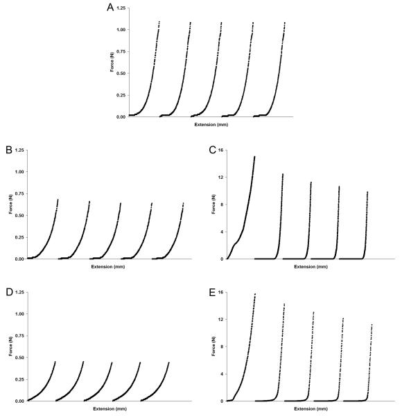
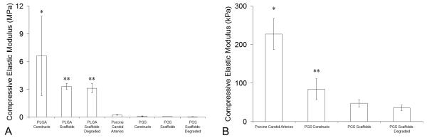
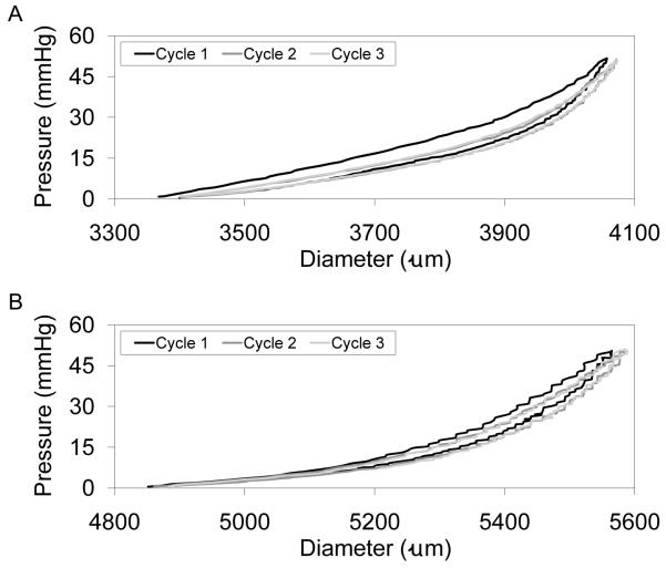
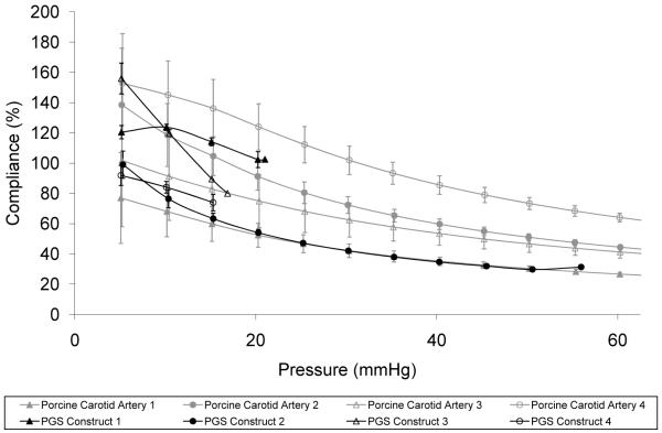
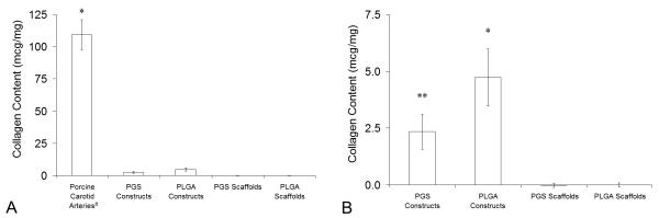
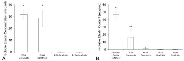
Compliance mismatch is a significant challenge to long-term patency in small-diameter bypass grafts because it causes intimal hyperplasia and ultimately graft occlusion. Current engineered grafts are typically stiff with high burst pressure but low compliance and low elastin expression. We postulated that engineering small arteries on elastomeric scaffolds under dynamic mechanical stimulation would result in strong and compliant arterial constructs. This study compares properties of engineered arterial constructs based on biodegradable polyester scaffolds composed of either rigid poly(lactide-co-glycolide) (PLGA) or elastomeric poly(glycerol sebacate) (PGS). Adult baboon arterial smooth muscle cells (SMCs) were cultured in vitro for 10 days in tubular, porous scaffolds. Scaffolds were significantly stronger after culture regardless of material, but the elastic modulus of PLGA constructs was an order of magnitude greater than that of porcine carotid arteries and PGS constructs. Deformation was elastic in PGS constructs and carotid arteries but plastic in PLGA constructs. Compliance of arteries and PGS constructs were equivalent at pressures tested. Altering scaffold material from PLGA to PGS significantly decreased collagen content and significantly increased insoluble elastin content in constructs without affecting soluble elastin concentration in the culture medium. PLGA constructs contained no appreciable insoluble elastin. This research demonstrates that: (1) substrate stiffness directly affects in vitro tissue development and mechanical properties; (2) rigid materials likely inhibit elastin incorporation into the extracellular matrix of engineered arterial tissues; and (3) grafts with physiologic compliance and significant elastin content can be engineered in vitro after only days of cell culture.


Figures

Figure 1
Figure 1. Electron Micrographs of Scaffold Luminal Microstructure and Cellular Luminal Confluence
Luminal surfaces appeared similar in micrographs of (A) PGS and (B) PLGA scaffolds (scale bars = 100 μm). Luminal microstructure also appeared similar in high-magnification micrographs of (C) PGS and (D) PLGA scaffolds (scale bars = 10 μm). Cellular confluence appeared equivalent on the luminal surfaces of (E) PGS and (F) PLGA arterial constructs cultured with adult baboon arterial SMCs for 10 days. Topography varied due to scaffold compaction and individual SMCs were visible on their luminal surfaces in some locations. Dark spots (E) are charging artifacts.
Figure 2
Figure 2. Macroscopic and Histological Appearance of Arteries and Engineered Arterial Constructs
Macroscopic appearance of (A) the tunicae media and intima of a porcine carotid artery, a PGS construct, and a PLGA construct (shown left to right). Staining with H&E to qualitatively assess cell and protein distribution showed that (B) porcine carotid arteries were dense and highly organized while (C) PGS constructs and (D) PLGA constructs had concentrations of cells and proteins at the luminal and abluminal surfaces and cells distributed throughout the scaffold walls. Ruler divisions and scale bars = 1.0 mm. For images of complete histological cross-sections see Figure S6.
Figure 3
Figure 3. Deformation and Elastic Recovery of Arteries and Engineered Arterial Constructs in Transverse Compression
Segments of porcine carotid arteries, PGS or PLGA constructs, and PGS or PLGA scaffolds were compressed perpendicular to their axis. Five force-extension cycles are shown chronologically for each tested segment. Elastic recovery was observed in (A) arteries and (B) PGS constructs, while plastic deformation was observed in (C) PLGA constructs. Scaffolds composed of (D) PGS or (E) PLGA showed similar behavior to their construct counterparts with or without degradation (degraded scaffold data not shown). Increases in moduli caused by ECM production were evident from steeper regions near the end of the force-extension curves of (B) PGS constructs compared to (D) PGS scaffolds and a notable difference in the toe regions and near the end of the force-extension curves of (B) PLGA constructs compared to (D) PLGA scaffolds (see also Fig. 4). The tunica adventitia of arteries was removed prior to testing.
Figure 4
Figure 4. Elastic Moduli of Arteries and Engineered Arterial Constructs
Segments of porcine carotid arteries, PGS or PLGA constructs, and PGS or PLGA scaffolds with or without degradation were compressed perpendicular to their axis. Transverse compressive elastic moduli (Ec) were determined from the second, third, and fourth force-extension cycles to 50% strain. (A) Comparison of all experimental groups and controls (n = 12, 12, 12, 8, 10, 12, and 12 from left to right). *Ec of PLGA constructs was significantly higher than all other groups. **Ec of undegraded and degraded PLGA scaffolds were significantly higher than Ec of porcine carotid arteries and all PGS groups. (B) Comparison of positive control and PGS groups only. *Ec of arteries was significantly higher than all PGS groups. **Ec of PGS constructs was significantly higher than Ec of undegraded and degraded PGS scaffolds. #The tunica adventitia of arteries was removed prior to testing.
Figure 5
Figure 5. Elastic Recovery of Arteries and Engineered Arterial Constructs
Segments of porcine carotid arteries and whole PGS constructs were pressure-diameter tested for comparison. Constructs were cycled to each target pressure three times, with hysteresis observed only during the first cycle. Complete elastic recovery was observed after the initial cycle to each target pressure in (A) porcine carotid arteries up to and including the highest pressure tested (240 mmHg) and in (B) PGS constructs up to pressures near their burst pressure (cycles to 50 mmHg are shown for a construct with a burst pressure of 56 mmHg). The tunica adventitia of porcine carotid arteries was removed prior to testing.
Figure 6
Figure 6. Physiologic Compliance of Arteries and Engineered Arterial Constructs
The compliance of porcine carotid arteries (gray, n = 4) and PGS constructs (black, n = 4) was remarkably similar at the pressures tested. There was no statistical difference between the two groups. PGS scaffolds hold no pressure due to their high porosity. #The tunica adventitia of porcine carotid arteries was removed prior to testing.
Figure 7
Figure 7. Total Collagen Content in Arteries and Engineered Arterial Constructs
Total collagen contents in porcine carotid arteries, PGS or PLGA constructs, and PGS or PLGA scaffolds were measured per unit wet weight by quantifying 4-hydroxyproline content. (A) Comparison of experimental groups and controls (n = 4, 4, 4, 8, and 8 from left to right). *Porcine carotid arteries contained significantly more collagen than all other groups. (B) Comparison of experimental groups and negative controls only. *PLGA constructs contained significantly more collagen than PGS constructs and uncultured scaffolds. **PGS constructs contained significantly more collagen than uncultured scaffolds. #The tunica adventitia of porcine carotid arteries was removed prior to testing.
Figure 8
Figure 8. Elastin in Arteries and Engineered Arterial Constructs
Elastin production was quantified using an elastin-binding dye. (A) Soluble elastin concentrations in culture medium collected at the termination of culture of PGS or PLGA constructs were measured after centrifugation to remove any insoluble elastin (n = 4, 4, 10, and 10 from left to right). *PGS and PLGA constructs released significant amounts of soluble elastin into culture medium. (B) Insoluble elastin contents in porcine carotid arteries, PGS or PLGA constructs, and PGS or PLGA scaffolds were measured per unit wet weight after acid hydrolysis of each construct, which destroyed all proteins except elastin, and centrifugation to pellet insolubles (n = 4, 4, 4, 9, and 9 from left to right). *Arteries contained significantly more insoluble elastin than all other groups. ** PGS constructs contained significantly more insoluble elastin than PLGA constructs and uncultured scaffolds, but PLGA constructs did not contain significantly more insoluble elastin compared to uncultured scaffolds (even if the positive control was excluded). #The tunica adventitia of porcine carotid arteries was removed prior to testing.

Similar articles

Cited by

References

    1. Rosamond W, Flegal K, Furie K, Go A, Greenlund K, Haase N, et al. Heart disease and stroke statistics--2008 update: A report from the American Heart Association Statistics Committee and Stroke Statistics Subcommittee. Circulation. 2008;117:e25–146. - PubMed
    1. Daemen J, Serruys PW. Optimal revascularization strategies for multivessel coronary artery disease. Curr Opin Cardiol. 2006;21:595–601. - PubMed
    1. Desai ND, Fremes SE. Radial artery conduit for coronary revascularization: as good as an internal thoracic artery. Curr Opin Cardiol. 2007;22:534–40. - PubMed
    1. Suma H. Arterial grafts in coronary bypass surgery. Ann Thorac Cardiovasc Surg. 1999;5:141–5. - PubMed
    1. Kassab GS, Navia JA. Biomechanical considerations in the design of graft: the homeostasis hypothesis. Annu Rev Biomed Eng. 2006;8:499–535. - PubMed

Publication types

MeSH terms