. 2009 Dec 18;31(6):986-98.
doi: 10.1016/j.immuni.2009.09.020. Epub 2009 Dec 3.

Differential contribution of chemotaxis and substrate restriction to segregation of immature and mature thymocytes

Affiliations

Differential contribution of chemotaxis and substrate restriction to segregation of immature and mature thymocytes

Lauren I Richie Ehrlich et al. Immunity. .

Abstract

T cell development requires sequential localization of thymocyte subsets to distinct thymic microenvironments. To address mechanisms governing this segregation, we used two-photon microscopy to visualize migration of purified thymocyte subsets in defined microenvironments within thymic slices. Double-negative (CD4(-)8(-)) and double-positive (CD4(+)8(+)) thymocytes were confined to cortex where they moved slowly without directional bias. DP cells accumulated and migrated more rapidly in a specialized inner-cortical microenvironment, but were unable to migrate on medullary substrates. In contrast, CD4 single positive (SP) thymocytes migrated directionally toward the medulla, where they accumulated and moved very rapidly. Our results revealed a requisite two-step process governing CD4 SP cell medullary localization: the chemokine receptor CCR7 mediated chemotaxis of CD4 SP cells towards medulla, whereas a distinct pertussis-toxin sensitive pathway was required for medullary entry. These findings suggest that developmentally regulated responses to both chemotactic signals and specific migratory substrates guide thymocytes to specific locations in the thymus.


Figures

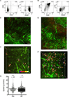
Figure 1
Figure 1. EGFP slices are structurally intact and support thymocyte migration in all regions
(A,B) Individual images from cortex (A) or medulla (B) of EGFP slices illustrate the distinct stromal morphology in each region. Medullary stroma are broader with sheet-like morphology, in contrast to the reticular projections of cortical stroma. Scale bars, 20 µm. (C) Flow cytometric analysis of input thymocytes from B6-Ka mice. Left, CD4 versus CD8 staining gated on live events. Right, c-Kit versus CD25 staining gated on lin CD4CD8 cells. (D) Thymocytes migrate into both cortex and medulla several hours after incubation atop EGFP slices. CMTPX-labeled thymocytes are shown in red, and EGFP-labeled stroma is green. Dotted line approximates the CMJ. Scale bar, 50 µm. Image is a maximum intensity projection (MIP) of individual z-sections.
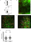
Figure 2
Figure 2. Double-negative (DN) and double-positive (DP) thymocytes localize exclusively to cortex and migrate slowly and tortuously
(A,D) Flow cytometric analysis of input DN cells from Rag2−/− mice (A) or DP cells from LN3βx Rag2−/− mice (D). (B,E) CMTPX-labeled DN (B) or DP cells (E) (red) preferentially localize to cortex in EGFP slices (green). Image properties as in Figure 1D. Scale bar, 50 µm. A duplicate image of (B) with white circles placed over DN cells to aid in visualization can be found in Figure S3. The apparent lower density of DN relative to DP cells in these fields likely reflects addition of only 1/10 the number of thymocytes to the DN versus DP slice. (C,F) Trajectories of individual DN (C) or DP cells (F) at higher magnification in cortex throughout a ~17 min imaging session are displayed as color-coded tracks running from start (blue) to end (white) of the timecourse, as indicated by the timebar in (C). Major tics = 10 µm. (G) Cortical DN and DP speeds. Each point represents the average speed for a single tracked cell. Mean speeds ± s.e.m. for each population are given above each column and represented by a bar. n=261 for DN from 10 imaging fields in 4 slices over 3 experiments; n= 899 for DP from 10 imaging fields in 3 slices over 3 experiments.
Figure 3
Figure 3. CD4SP cells accumulate in medulla, and migrate rapidly in all regions of thymus
(A) Flow cytometric analysis of CD4SP cells obtained by depletion. (B) CMTPX-labeled CD4SP cells (red) are localized to both cortex and medulla in EGFP slices (green), but accumulate preferentially in medulla. A duplicate of this image with white circles placed over CD4SP cells to aid in visualization can be found in Figure S3. Image properties as in Figure 1D. Scale bar, 50 µm. (C,D) Time-encoded tracks of CD4SP cells from high-magnification cortical (C) or medullary (D) fields imaged for ~17 min. Each major tic represents 10 µm. (E) Average track speeds (µm/min) ± s.e.m. of cortical and medullary CD4SP cells. Medullary CD4SP cells migrate faster than cortical CD4SP cells (**, P<0.01 using the two-tailed Mann-Whitney U test). n=340 cells for cortex from 8 imaging fields in 2 slices over 2 experiments; n=289 for medulla from 3 imaging fields in 2 slices over 2 experiments.
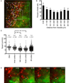
Figure 4
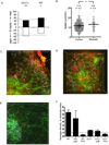
Figure 4. The CMJ presents a functional barrier for pre-selection DP cells to enter medulla
(A) Time-encoded tracks of individual DP cells from a high-magnification field at the CMJ imaged for ~17 min. Note that all DP cells are found in cortex (left side of field) and not in medulla (right side), and that tracks do not cross CMJ (representative of 3 slices containing 141 tracked DP cells). Occasional red fluorescence in medulla can be distinguished from live cells by its punctate morphology and lack of movement (Movie S6). Each major tic represents 10 µm. The area within the white box is displayed in (D) at increased magnification. (B) DP cells accumulate near medulla. The normalized percentage ± s.e.m. of cells found within 50-µm increments from the medulla is displayed. n=876 cells analyzed from 10 series (see Methods) taken from 5 separate slices over 2 experiments. (C) Average track speeds (µm/min) ± s.e.m. of DP cells found in cortical subregions. Velocity increases significantly from outer or mid to inner cortex (**, P<0.01). CMJ, n=141 cells from 3 imaging fields in 3 slices over 3 experiments; inner cortex, n= 244 cells from 4 imaging fields in 2 slices over 2 experiments; mid cortex, n=264 cells from 4 imaging fields in 2 slices over 2 experiments; outer cortex, n=250 cells from 4 imaging fields in 2 slices over 2 experiments; all cortex, n=899 cells from 10 imaging fields in 3 slices over 3 experiments. (D) Example of a DP cell (►) that closely approaches but fails to cross into the medulla. Shown are individual z-planes taken at indicated times. Scale bar, 10 µm.
Figure 5
Figure 5. Cortical CD4SP cells demonstrate directional bias towards medulla and cross the CMJ preferentially from cortex into medulla
(A) The graph represents the percentage of cortical tracks for each thymocyte subset whose net displacement after 13–17 min is towards medulla (positive values) or towards capsule (negative values). Dashed lines are at the 50%, which would be expected from a population with no directional bias. CD4SP cells display significant bias towards medulla, while DN cells (from Rag2−/−) and DP cells are not biased in either direction (*, P<0.05, chi-squared test;). n=131 Rag2/−, 465 DP, and 122 CD4SP tracks each from 2 imaging fields in 2 slices over 2 experiments. (B) Time-encoded tracks of individual CD4SP cells from a high-magnification field clearly cross the CMJ (dashed line) in both directions over the 17 min imaging period. Major tics = 10 µm. (C) The graph represents the percentage of CD4SP crossing events at the CMJ over ~17 min timecourses that are towards medulla (positive values) or towards cortex (negative values). Dashed lines are at 50%, which would be expected if there were no retention or preferential attraction in either region. WT CD4SP cells cross preferentially from cortex to medulla, indicating attraction towards and/or retention within the medulla (*, P<0.05, chi-squared test;). n=151 WT CD4SP crossing events from 6 imaging fields over 2 experiments.
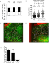
Figure 6
Figure 6. PTX treatment of CD4SP cells reveals a separable GPCR-mediated medullary entry step
(A) PTX treatment eliminates CD4SP medullary bias (**, P<0.01; *, P<0.05). Analysis as in Figure 5A. Time courses ranged from 8–17 min for PTX and 11–16 min for suB. PTX, n=572 tracks from 3 imaging fields in 2 slices over 2 experiments; suB, n= 508 tracks from 3 imaging fields in 3 slices over 2 experiments. For comparison, untreated data are duplicated from Figure 5A. (B) PTX treatment reduces the speed (± s.e.m.) of cortical CD4SP cells relative to untreated and suB-treated CD4SP cells (**, P<0.01, non-parametric one-way ANOVA, followed by Dunn’s post test). PTX, n=691 from 5 imaging fields in 4 slices over 2 experiments; suB, n= 508 from 3 imaging fields in 3 slices over 2 experiments. For comparison, untreated data are duplicated from Figure 3. (C,D) CMTPX-labeled CD4SP cells (red) are rarely found in medulla following PTX treatment (C) but readily enter medulla following suB control treatment (D). Time-encoded tracks of cell trajectories from a low-magnification field of slices containing CD4SP cells pre-treated with 100 ng/ml PTX (C) or suB (D) and imaged for ~8 min (C) or 11 min (D). Major tics = 20 µm. (E) PTX treatment inhibits CD4SP cells from entering medulla. The density of CD4SP cells in medulla relative to cortex was determined in multiple low magnification fields, and then ratios were averaged for different fields, indicating the average fold enrichment of cells in medulla relative to cortex (± s.e.m.). While untreated CD4SP cells are enriched 6.5-fold in the medulla relative to the cortex, PTX treatment results in a medullary: cortical density ratio of 0.4, indicating that GPCR blockade actively prevents cells from entering medulla. Untreated, n=1593 cells from 9 imaging fields in 6 slices from 4 experiments; suB, n=821 cells from 5 imaging fields in 4 slices in 3 experiments; PTX, n = 685 cells from 10 imaging fields in 5 slices from 3 experiments.
Figure 7
Figure 7. CCR7 is responsible for CD4SP chemotaxis towards medulla, but is not necessary for medullary entry
(A) Ccr7−/− CD4SP cells no longer display directional bias towards medulla, but instead migrate towards capsule. Data were analyzed over ~17 min period. Analysis as in Figure 5A (**, P<0.01; *, P<0.05). Ccr7−/−, n=602 cells from 2 fields in 2 slices over 2 experiments. For comparison, WT CD4SP data is duplicated from Figure 5A. (B) Ccr7−/− CD4SP cells migrate rapidly (± s.e.m.) in both cortex and medulla, though medullary migration is faster (*, P<0.05). Cortex, n=602 cells from 2 imaging fields over 2 experiments; medulla, n=110 cells from 2 imaging fields in 2 slices over 2 experiments. (C) CMTPX-labeled Ccr7−/− CD4SP cells (red) are found in cortex, but are also present in medulla in EGFP slices (green). Data collected over ~17 min. Major tics = 20 µm. (D) Ccr7−/− CD4SP cells migrate productively in medulla, as shown by time-encoded tracks from high-magnification medullary fields imaged for ~15 min. Major tics = 10 µm. (E) Ccr7−/− CD4SP cells do not accumulate in medulla to the same extent as WT CD4SP cells. CMTPX-labeled Ccr7−/− CD4SP cells (red) and indo-PE3-labeled WT CD4SP cells (blue) were added to the same EGFP thymic slice. Whereas WT cells clearly accumulate in medulla, Ccr7−/− CD4SP cells are more evenly distributed across the cortex and medulla. Major tics = 20 µm. (F) Ccr7−/− CD4SP cells fail to accumulate in medulla, but are not blocked from medullary entry. Analysis as in Figure 6E; for comparison, all WT data are duplicated from Figure 6E. In contrast to the 6.5-fold accumulation of WT CD4SP cells in medulla, Ccr7−/− CD4SP cells accumulate only 1.6-fold in medulla relative to cortex. Furthermore, Ccr7−/− CD4SP cells are sensitive to PTX, which reduces the medullary: cortical density ratio to 0.6. For Ccr7−/− CD4SP cells: untreated, n=1361 from 11 imaging fields in 8 slices over 4 experiments; suB, n=476 from 4 imaging fields in 2 slices over 2 experiments; PTX, n=185 from 4 imaging fields in 2 slices over 2 experiments. These analyses include data from slices in which two populations were differentially labeled (CMTPX versus Indo-PE3) and added simultaneously to the slice as follows: 3 imaging fields with PTX-treated and suB-treated Ccr7−/− CD4SP cells; 2 imaging fields with PTX-treated and suB-treated WT CD4SP cells; and 6 imaging fields with untreated WT and untreated Ccr7−/− CD4SP cells, as in (D). The relative medullary: cortical enrichments we observed were not significantly different between the slices containing one of the above populations or two differentially labeled populations, so the two data sets were combined.

Comment in

  • Everything in its place.
    Swann JB, Boehm T. Swann JB, et al. Immunity. 2009 Dec 18;31(6):856-8. doi: 10.1016/j.immuni.2009.11.005. Immunity. 2009. PMID: 20064445

Similar articles

Cited by

References

    1. Alfano M, Schmidtmayerova H, Amella CA, Pushkarsky T, Bukrinsky M. The B-oligomer of pertussis toxin deactivates CC chemokine receptor 5 and blocks entry of M-tropic HIV-1 strains. J Exp Med. 1999;190:597–605. - PMC - PubMed
    1. Anderson MS, Venanzi ES, Klein L, Chen Z, Berzins SP, Turley SJ, von Boehmer H, Bronson R, Dierich A, Benoist C, Mathis D. Projection of an immunological self shadow within the thymus by the aire protein. Science. 2002;298:1395–1401. - PubMed
    1. Asperti-Boursin F, Real E, Bismuth G, Trautmann A, Donnadieu E. CCR7 ligands control basal T cell motility within lymph node slices in a phosphoinositide 3-kinase-independent manner. J Exp Med. 2007;204:1167–1179. - PMC - PubMed
    1. Bajenoff M, Egen JG, Koo LY, Laugier JP, Brau F, Glaichenhaus N, Germain RN. Stromal cell networks regulate lymphocyte entry, migration, and territoriality in lymph nodes. Immunity. 2006;25:989–1001. - PMC - PubMed
    1. Bajenoff M, Glaichenhaus N, Germain RN. Fibroblastic reticular cells guide T lymphocyte entry into and migration within the splenic T cell zone. J Immunol. 2008;181:3947–3954. - PMC - PubMed

Publication types