. 2010 Feb;221(2):335-45.
doi: 10.1016/j.expneurol.2009.11.020. Epub 2009 Dec 3.

State-dependent control of lumbar motoneurons by the hypocretinergic system

Affiliations

State-dependent control of lumbar motoneurons by the hypocretinergic system

Jack Yamuy et al. Exp Neurol. 2010 Feb.

Abstract

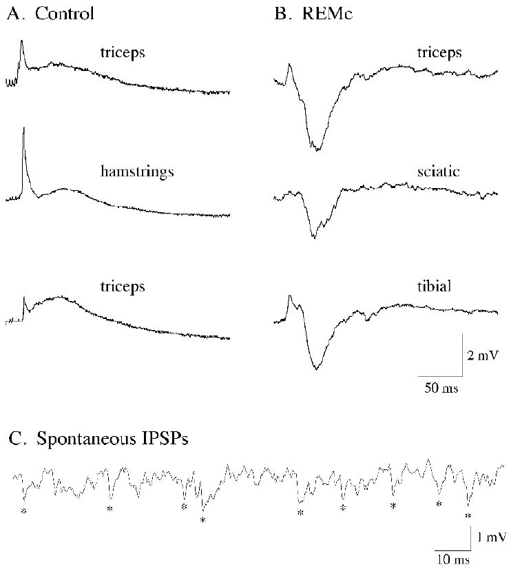
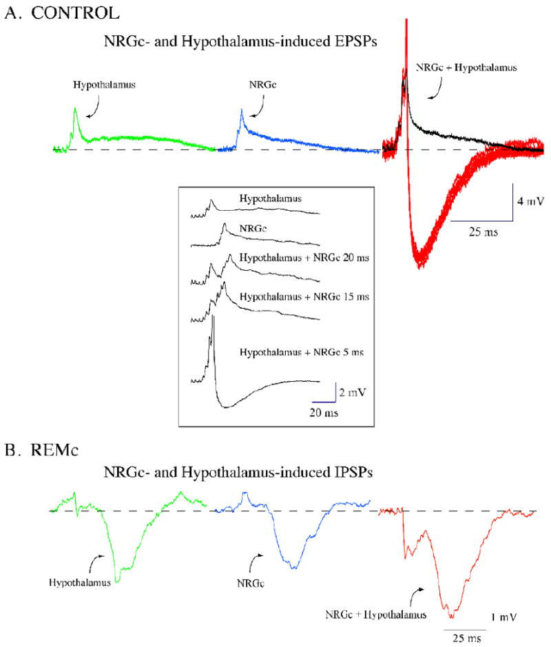
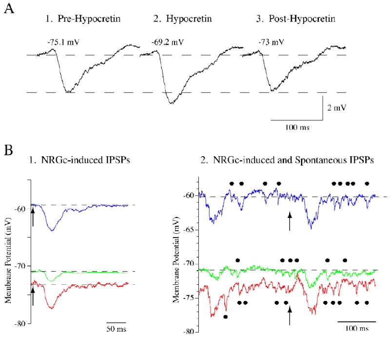
Neurons in the lateral hypothalamus (LH) that synthesize hypocretins (Hcrt-1 and Hcrt-2) are active during wakefulness and excite lumbar motoneurons. Because hypocretinergic cells also discharge during phasic periods of rapid eye movement (REM) sleep, we sought to examine their action on the activity of motoneurons during this state. Accordingly, cat lumbar motoneurons were intracellularly recorded, under alpha-chloralose anesthesia, prior to (control) and during the carbachol-induced REM sleep-like atonia (REMc). During control conditions, LH stimulation induced excitatory postsynaptic potentials (composite EPSP) in motoneurons. In contrast, during REMc, identical LH stimulation induced inhibitory PSPs in motoneurons. We then tested the effects of LH stimulation on motoneuron responses following the stimulation of the nucleus reticularis gigantocellularis (NRGc) which is part of a brainstem-spinal cord system that controls motoneuron excitability in a state-dependent manner. LH stimulation facilitated NRGc stimulation-induced composite EPSP during control conditions whereas it enhanced NRGc stimulation-induced IPSPs during REMc. These intriguing data indicate that the LH exerts a state-dependent control of motor activity. As a first step to understand these results, we examined whether hypocretinergic synaptic mechanisms in the spinal cord were state dependent. We found that the juxtacellular application of Hcrt-1 induced motoneuron excitation during control conditions whereas motoneuron inhibition was enhanced during REMc. These data indicate that the hypocretinergic system acts on motoneurons in a state-dependent manner via spinal synaptic mechanisms. Thus, deficits in Hcrt-1 may cause the coexistence of incongruous motor signs in cataplectic patients, such as motor suppression during wakefulness and movement disorders during REM sleep.


Figures

Figure 1
Figure 1
Stimulation of the lateral hypothalamus (LH) promotes motoneuron responses that depend on the ongoing behavioral state. During control conditions, the electrical stimulation of the LH induced excitatory responses in three representative lumbar motoneurons (A; the motoneuron type is indicated in each trace). During the REM sleeplike state of carbachol-induced REM sleep, identical hypothalamic stimulation induced predominantly inhibitory responses in another three representative lumbar motoneurons in the same cats (B; the motoneuron type is indicated in each trace). The appearance of large-amplitude, spontaneous IPSPs in motoneurons was typical during carbachol-induced REM sleep atonia (C; recording obtained from a sciatic motoneuron). The traces in A and B are averages of 15 to 20 sweeps. In each row for panels A and B are responses from lumbar motoneurons that were recorded in the same animal. Hypothalamic stimulation consisted of a 10-ms train (delivered at 0.5 Hz) of four pulses of 0.8 ms duration, at intensities of 0.5 to 1.0 mA.
Figure 2
Figure 2
Stimulation of the lateral hypothalamus (LH) facilitates the response of lumbar motoneurons to the stimulation of the nucleus reticularis gigantocellularis (NRGc). During control conditions, the motoneuron responses to the separate stimulation of the lateral hypothalamus and NRGc consisted of a sequence of EPSPs (green and blue traces, respectively, in A). The stimulation of the NRGc, with a 5-ms delay after that of the LH, promoted a larger motoneuron excitatory response (black trace in A) that included several spikes (red traces in A). During carbachol-induced REM sleep atonia, a representative motoneuron responded with a large-amplitude IPSP to the stimulation of the LH and NRGc (green and blue traces, respectively, in B). The stimulation of the NRGc with a 5-ms delay after that of the LH promoted a larger motoneuron inhibitory response (red trace in B). Facilitation of excitatory and inhibitory responses occurred at delays that ranged from 0 to 30 ms between hypothalamic and NRGc stimulation. Thus, both excitatory and inhibitory motoneuron responses to the NRGc are expected to be facilitated if neurons in the LH discharge concurrently. The traces are averages of 15 to 20 sweeps except for the spikes in the right-hand traces in A. Recordings in A and B were obtained from two different motoneurons (both responsive to the stimulation of the triceps nerve) in the same animal. Hypothalamic and NRGc stimulation consisted of a 10-ms train (delivered at 0.5 Hz) of four pulses of 0.8 ms duration, at intensities of 0.5 mA and 80 μA, respectively.
Figure 3
Figure 3
The juxtacellular application of hypocretin-1 (Hcrt-1) induces motoneuron depolarization and facilitates the response to subthreshold stimulation of the nucleus reticularis gigantocellularis (NRGc). A hamstrings motoneuron responded with a composite EPSP to the stimulation of the nucleus reticularis gigantocellularis (NRGc; green trace in A). Following the juxtacellular pressure ejection of Hcrt-1 while stimulating the NRGc, this motoneuron depolarized approximately 14 mV and discharged action potentials to NRGc stimulation (black trace in A; note that the spikes are truncated). After a period of 25 s, the motoneuron ceased to discharge and, while still depolarized, showed a response to NRGc stimulation that was reduced in amplitude (blue trace in A). Compared to control conditions, the amplitude and duration of the NRGc-induced excitatory response decreased following Hcrt-1 application (traces in B). In another lumbar motoneuron that responded to sciatic nerve stimulation, the application of Hcrt-1 promoted a 16.5 mV depolarization (green and blue traces in C). The membrane potential was then clamped to a level slightly more hyperpolarized than control (red trace in C); note that the amplitude of the second peak of the NRGc-induced response was still reduced (the averaged responses are aligned in the inset in C). The traces in A are individual sweeps whereas in B and C they are averages of 15 to 20 sweeps. NRGc stimulation consisted of a 10-ms train (delivered at 0.5 Hz) of four pulses of 0.8 ms duration, at intensities of 60-100 μA. Hcrt-1 was ejected at 5 PSI during 5 s
Figure 4
Figure 4
Hypocretin-1 (Hcrt-1) enhances the amplitude of spontaneous and nucleus reticularis gigantocellularis (NRGc)-induced IPSPs and depolarizes lumbar motoneurons during REMc. The NRGc stimulation-induced IPSP in a representative lumbar motoneuron prior to Hcrt-1 application (A1) increased in amplitude following the pressure ejection of Hcrt-1 (A2). After approximately 35 s, the IPSP amplitude returned to control levels (A3). The change in IPSP amplitude occurred in conjunction with membrane depolarization. Note, however, that the amount of depolarization observed in this motoneuron during REMc was considerably smaller than that observed in motoneurons during control conditions (see Fig. 3 and Table 1). NRGc stimulation consisted of a 10-ms train (delivered at 0.5 Hz) of four pulses of 0.8 ms duration, at intensities of 60 μA. Hcrt-1 was ejected at 5 PSI during 5 s. Another triceps motoneuron responded with an IPSP of approximately 1.5 mV to the stimulation of the NRGc (arrows in B1); its membrane potential was -71 mV (green trace in B1). Following the juxtacellular application of Hcrt-1, the cell depolarized approximately 11 mV and the NRGc-induced IPSP increased its amplitude to more than 4 mV (blue trace in B1). Hyperpolarizing current was then passed through the recording electrode to clamp the membrane potential to a level similar to that present prior to Hcrt-1 application. At a membrane potential of approximately −73 mV, the NRGc-induced IPSP was similar to that recorded at depolarized levels (4 mV in amplitude; red trace in B1). This triceps motoneuron showed spontaneous IPSPs during REMc (indicated by filled circles in the green trace in B2). These IPSPs, which also occur typically during natural REM sleep, increased their number and amplitude following the application of Hcrt-1 (indicated by filled circles in blue trace in B2). It can be seen that spontaneous IPSPs also maintained their increased amplitude when the membrane potential was clamped at hyperpolarized levels (indicated by filled circles in red trace in B2). The traces in B1 are averages of 10-20 sweeps whereas in B2 they are individual sweeps. The arrows in B1 and B2 indicate the onset of NRGc stimulation which consisted of a 10-ms train (delivered at 0.5 Hz) of four pulses of 0.8 ms duration, at intensities of 80 μA. Hcrt-1 was ejected at 12 PSI during 20 s.
Figure 5
Figure 5
Hypocretin-1 antagonizes the postsynaptic inhibitory action of glycine on motoneurons. The subthreshold stimulation of the nucleus reticularis gigantocellularis (NRGc) elicited a depolarizing synaptic response in a lumbar motoneuron (green trace in A); following the application of Hcrt-1, the motoneuron depolarized and responded with a train of action potentials to NRGc stimulation (red trace in A). In contrast, the NRGc-induced depolarizing potential in the same motoneuron (green trace in B) decreased three-fold in amplitude following the juxtacellular iontophoretic application of glycine (red trace in B). The iontophoresis of glycine onto the same lumbar motoneuron produced a hyperpolarization of approximately 3 mV (C, glycine application is indicated by the black-filled bar). After 21 s of glycine iontophoresis, Hcrt-1 was pressure-ejected (red bar in C); subsequently, the motoneuron depolarized, discharged action potentials, and an increase in synaptic noise appeared. Approximately 45 s after the application of Hcrt-1, while glycine was still being iontophorezed, the motoneuron began to hyperpolarize. The membrane potential returned to baseline levels after 8 s of discontinuing the application of glycine. The traces in A and B are averages of 10-20 sweeps except for the upper trace in A which is an individual sweep. All recordings in A and B were obtained from the same motoneuron which discharged antidromic spikes following the stimulation of the ventral root. NRGc stimulation consisted of a 10-ms train (delivered at 0.5 Hz) of four pulses of 0.8 ms duration, at an intensity of 100 μA. Hcrt-1 was ejected at 15 PSI during 20 s; glycine was iontophorezed from two barrels using a current of 400 nA for each barrel.

Similar articles

Cited by

References

    1. Acuna-Goycolea C, van den Pol AN. Neuroendocrine proopiomelanocortin neurons are excited by hypocretin/orexin. J Neurosci. 2009;29:1503–1513. - PMC - PubMed
    1. Amatruda TT, III, Black DA, McKenna TM, McCarley RW, Hobson JA. Sleep cycle control and cholinergic mechanisms: differential effects of carbachol injections at pontine brain stem sites. Brain Res. 1975;98:501–515. - PubMed
    1. Anand BK, Chhina GS, Singh B. Effect of glucose on the activity of hypothalamic “feeding centers”. Science. 1962;138:597–598. - PubMed
    1. Baghdoyan HA, Rodrigo-Angulo ML, McCarley RW, Hobson JA. Site-specific enhancement and suppression of desynchronized sleep signs following cholinergic stimulation of three brainstem regions. Brain Res. 1984;306:39–52. - PubMed
    1. Baghdoyan HA, Rodrigo-Angulo ML, McCarley RW, Hobson JA. A neuroanatomical gradient in the pontine tegmentum for the cholinoceptive induction of desynchronized sleep signs. Brain Res. 1987;414:245–261. - PubMed

Publication types

MeSH terms