

Cardiac arrhythmias induced by glutathione oxidation can be inhibited by preventing mitochondrial depolarization
- PMID: 19962380
- PMCID: PMC2837795
- DOI: 10.1016/j.yjmcc.2009.11.011
Cardiac arrhythmias induced by glutathione oxidation can be inhibited by preventing mitochondrial depolarization
Abstract
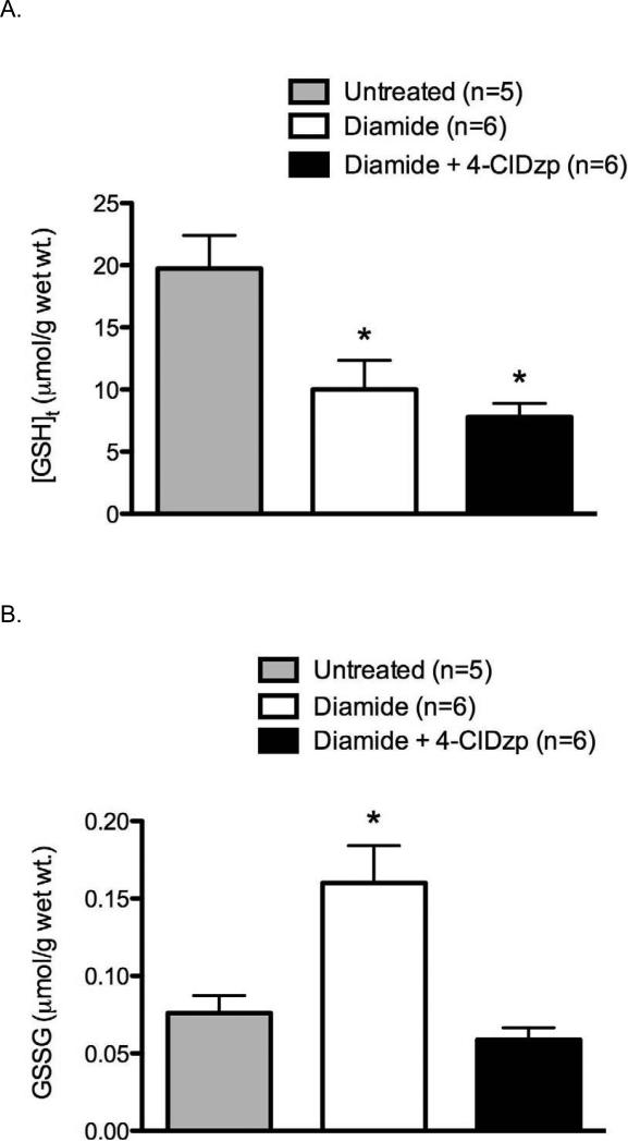
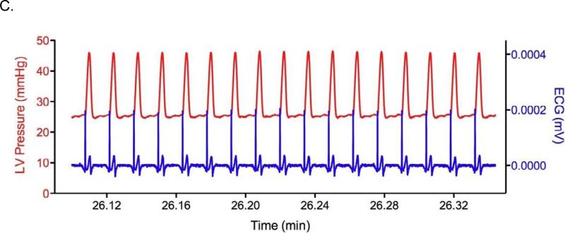
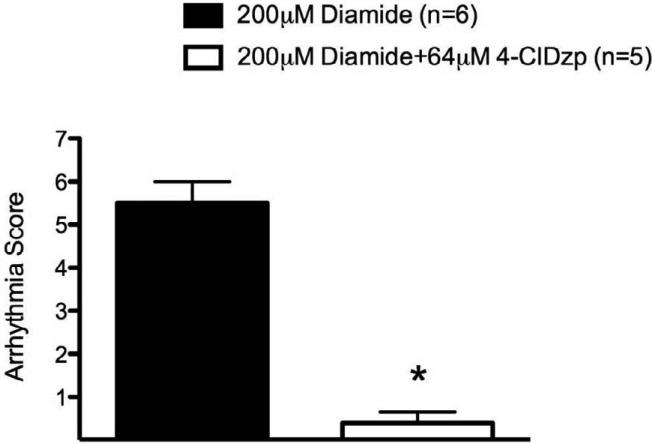
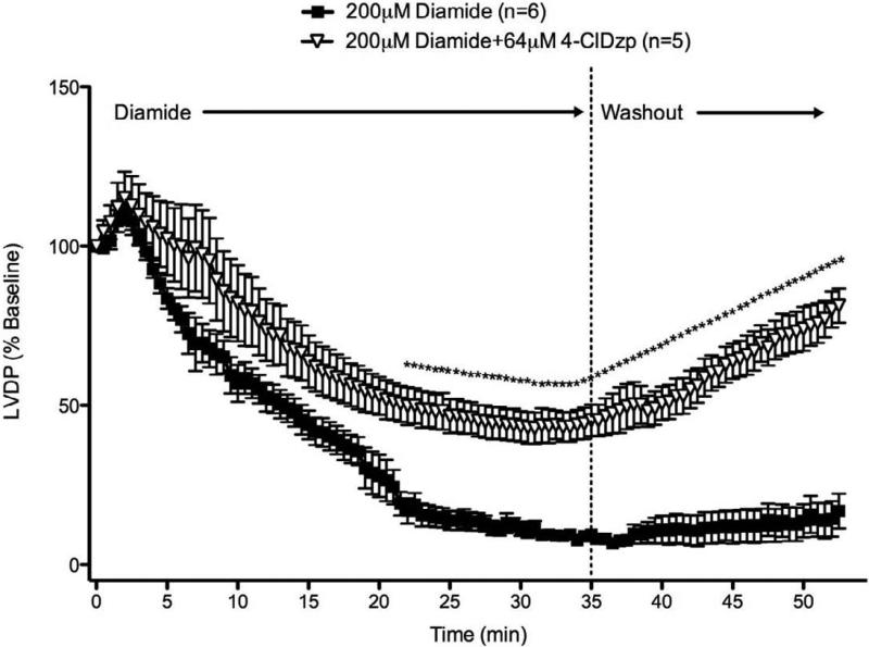
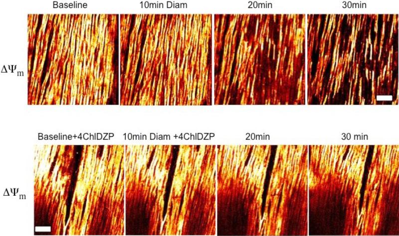
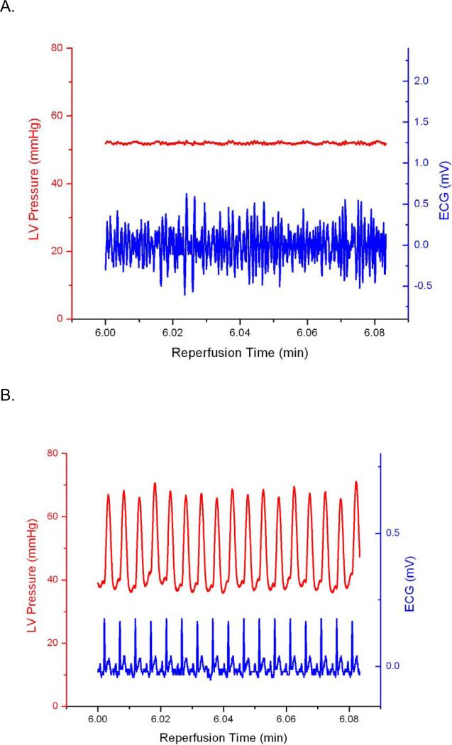
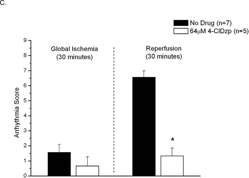
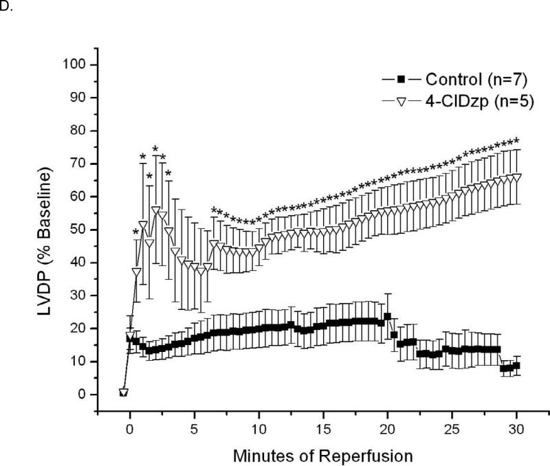
We have previously proposed that the heterogeneous collapse of mitochondrial inner membrane potential (DeltaPsi(m)) during ischemia and reperfusion contributes to arrhythmogenesis through the formation of metabolic sinks in the myocardium, wherein clusters of myocytes with uncoupled mitochondria and high K(ATP) current levels alter electrical propagation to promote reentry. Single myocyte studies have also shown that cell-wide DeltaPsi(m) depolarization, through a reactive oxygen species (ROS)-induced ROS release mechanism, can be triggered by global depletion of the antioxidant pool with diamide, a glutathione oxidant. Here we examine whether diamide causes mitochondrial depolarization and promotes arrhythmias in normoxic isolated perfused guinea pig hearts. We also investigate whether stabilization of DeltaPsi(m) with a ligand of the mitochondrial benzodiazepine receptor (4'-chlorodiazepam; 4-ClDzp) prevents the formation of metabolic sinks and, consequently, precludes arrhythmias. Oxidation of the GSH pool was initiated by treatment with 200 microM diamide for 35 min, followed by washout. This treatment increased GSSG and decreased both total GSH and the GSH/GSSG ratio. All hearts receiving diamide transitioned from sinus rhythm into ventricular tachycardia and/or ventricular fibrillation during the diamide exposure: arrhythmia scores were 5.5+/-0.5; n=6 hearts. These arrhythmias and impaired LV function were significantly inhibited by co-administration of 4-ClDzp (64 microM): arrhythmia scores with diamide+4-ClDzp were 0.4+/-0.2 (n=5; P<0.05 vs. diamide alone). Imaging DeltaPsi(m) in intact hearts revealed the heterogeneous collapse of DeltaPsi(m) beginning 20 min into diamide, paralleling the timeframe for the onset of arrhythmias. Loss of DeltaPsi(m) was prevented by 4-ClDzp treatment, as was the increase in myocardial GSSG. These findings show that oxidative stress induced by oxidation of GSH with diamide can cause electromechanical dysfunction under normoxic conditions. Analogous to ischemia-reperfusion injury, the dysfunction depends on the mitochondrial energy state. Targeting the mitochondrial benzodiazepine receptor can prevent electrical and mechanical dysfunction in both models of oxidative stress.
Copyright (c) 2009 Elsevier Ltd. All rights reserved.
Figures
References
-
- Woodward B, Zakaria MN. Effect of some free radical scavengers on reperfusion induced arrhythmias in the isolated rat heart. J Mol Cell Cardiol. 1985 May;17(5):485–93. - PubMed
-
- Aon MA, Cortassa S, Marban E, O'Rourke B. Synchronized whole cell oscillations in mitochondrial metabolism triggered by a local release of reactive oxygen species in cardiac myocytes. The Journal of biological chemistry. 2003 Nov 7;278(45):44735–44. - PubMed
-
- O'Rourke B, Ramza BM, Marban E. Oscillations of membrane current and excitability driven by metabolic oscillations in heart cells. Science. 1994 Aug 12;265(5174):962–6. - PubMed
-
- Gavish M, Bachman I, Shoukrun R, Katz Y, Veenman L, Weisinger G, et al. Enigma of the peripheral benzodiazepine receptor. Pharmacol Rev. 1999 Dec;51(4):629–50. - PubMed
MeSH terms
Substances
Grants and funding
LinkOut - more resources
Full Text Sources
Medical
