Review
. 2010 May;48(5):866-75.
doi: 10.1016/j.yjmcc.2009.11.014. Epub 2009 Dec 3.

Phosphorylation and function of cardiac myosin binding protein-C in health and disease

Affiliations
Review

Phosphorylation and function of cardiac myosin binding protein-C in health and disease

David Barefield et al. J Mol Cell Cardiol. 2010 May.

Abstract

During the past 5 years there has been an increasing body of literature describing the roles cardiac myosin binding protein C (cMyBP-C) phosphorylation play in regulating cardiac function and heart failure. cMyBP-C is a sarcomeric thick filament protein that interacts with titin, myosin and actin to regulate sarcomeric assembly, structure and function. Elucidating the function of cMyBP-C is clinically important because mutations in this protein have been linked to cardiomyopathy in more than sixty million people worldwide. One function of cMyBP-C is to regulate cross-bridge formation through dynamic phosphorylation by protein kinase A, protein kinase C and Ca(2+)-calmodulin-activated kinase II, suggesting that cMyBP-C phosphorylation serves as a highly coordinated point of contractile regulation. Moreover, dephosphorylation of cMyBP-C, which accelerates its degradation, has been shown to associate with the development of heart failure in mouse models and in humans. Strikingly, cMyBP-C phosphorylation presents a potential target for therapeutic development as protection against ischemic-reperfusion injury, which has been demonstrated in mouse hearts. Also, emerging evidence suggests that cMyBP-C has the potential to be used as a biomarker for diagnosing myocardial infarction. Although many aspects of cMyBP-C phosphorylation and function remain poorly understood, cMyBP-C and its phosphorylation states have significant promise as a target for therapy and for providing a better understanding of the mechanics of heart function during health and disease. In this review we discuss the most recent findings with respect to cMyBP-C phosphorylation and function and determine potential future directions to better understand the functional role of cMyBP-C and phosphorylation in sarcomeric structure, myocardial contractility and cardioprotection.


Conflict of interest statement

Disclosure statement

There are no unlabeled/unapproved uses of drugs or products, and no real or apparent conflicts of interest to report.

Figures

Fig. 1.
Fig. 1.
Schematic diagram of the cardiac sarcomere and arrangement of cMyBP-C. The sarcomere is the basic functional unit of a cardiac muscle’s cross-striated myofibril, which is defined as the boundary between two neighboring Z-lines. The I-band, A-band, H-zone and M-lines are shown. The thick filament protein is composed of myosin, cMyBP-C and titin. cMyBP-C is localized in the inner two-thirds of the A-band, i.e., the C-zone. cMyBP-C is oriented perpendicularly to the long axis of the myosin filaments. The two groups of 7–9 strips of cMyBP-C bands on either side of the H-zone give a characteristic doublet appearance within the A-band. The thin filament is composed of actin monomers, troponins (cTnT and cTnI) and α-TM, which is connected to nebulin in the I-bands.
Fig. 2.
Fig. 2.
General domain structure of mouse cMyBP-C and identified phosphorylated serines. There are eight Ig (oval) and three fibronectin type-3 domains (hexagonal) numbered C0 to C10. The Pro-Ala–rich region (Pro-Ala), phosphorylated M domain, a 28-amino acid cardiac-specific insertion within the C5 domain and regions that interact with actin, myosin and titin sites are shown. The M domain contains multiple phosphorylation sites that can be phosphorylated by PKA, CaMKII and PKC. The amino acid sequences are conserved between species in the M domain. Domains 7–10 participate in binding of myosin and titin, which are necessary for cMyBP-C stability and sarcomere organization.
Fig. 3.
Fig. 3.
Dynamic phosphorylation of cMyBP-C’s M domain influences myosin–actin interactions. cMyBP-C facilitates myosin–actin interactions by tethering the fibers together and regulating the ability of the myosin head to bind to the actin filament during cross-bridge cycling. cMyBP-C interacts with the LMM of myosin at the C10 domain and titin at domains C8–C10. The C1-M-C2 domains have been shown to bind to myosin and actin. The capacity of these domains to interact is critical to overall sarcomeric integrity and function. The phosphorylatable M domain facilitates the role of cMyBP-C in cross-bridge formation. In the absence of M domain phosphorylation, the C1-M-C2 domains are tightly bound to myosin S2 region (A), whereas when M domain phosphorylated, C1-M-C2 domains and M domain release their interaction with myosin S2 region and actin, respectively, and changes in the thick filament orientation occur (B). C1 domain binds to actin irrespective of the M domain phosphorylation state. The dynamic phosphorylation of the M domain provides a point of control where the ordering of myosin heads and changes in force production can be regulated. Thin filament proteins include actin, α-TM, cTnI, cTnT and calcium (Ca2+)-binding protein cTnC are shown.
Fig. 4.
Fig. 4.
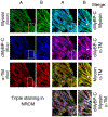
cMyBP-C connects thick and thin filaments. To determine the localization pattern of cMyBP-C along with thick and thin filament proteins, 3-day-old neonatal rat cardiomyocytes (NRCM) were used in chamber slides In vitro. The healthy NRCM were fixed and stained with the mouse monoclonal anti-α-myosin heavy chain (MF20, University of Iowa, Iowa), rabbit polyclonal anti-C0–C1 region of cMyBP-C [74] and sheep polyclonal anti-α-TM antibodies (Millipore, Billerica, MA). Secondary Alexa Fluor 488 goat anti-mouse IgG antibody for myosin (green color), Alexa Fluor Cy5 goat anti-rabbit IgG antibody for cMyBP-C (blue color) and Alexa Fluor 568 donkey anti-sheep IgG antibody for α-TM (red color) staining were used for immunofluorescence (Molecular Probes, Carlsbad, CA). Cardiomyocytes were analyzed with fluorescent microscopy (PCM-2000 scanner, Nikon, Melville, NY) and SimplePCI software (Compix Inc., Cranberry Township, PA). The rows of figures labeled B are enlarged from the inset in the rows of figures labeled A (60x magnification). Immunofluorescent staining of cMyBP-C (blue color) shows a typical doublet pattern of normal incorporation into the sarcomere. Co-staining with cMyBP-C with either myosin (green) or α-TM (yellow) show the co-localization of cMyBP-C with myosin (blue-green) and α-TM (pink). Co-localization of myosin and α-TM (yellow) confirm the presence of interaction between myosin and thin filament region. Triple staining shows that there is a defined merging of cMyBP-C, myosin and α-TM, but the presence of excess of pink (arrow) confirms the co-localization of cMyBP-C and α-TM. Nonoverlap regions are marked with arrows in respective panels. These data demonstrate that C0–C1 domains may directly interact with thin filament proteins via α-TM and thus connect thick and thin filaments.

Similar articles

Cited by

References

    1. Lloyd-Jones D, Adams R, Carnethon M, De Simone G, Ferguson TB, Flegal K, et al. Heart disease and stroke statistics–2009 update: a report from the American Heart Association Statistics Committee and Stroke Statistics Subcommittee. Circulation 2009;119:480–6. - PubMed
    1. Yano M, Ikeda Y, Matsuzaki M. Altered intracellular Ca2+ handling in heart failure. J Clin Invest 2005;115:556–64. - PMC - PubMed
    1. Messer AE, Jacques AM, Marston SB. Troponin phosphorylation and regulatory function in human heart muscle: dephosphorylation of Ser23/24 on troponin I could account for the contractile defect in end-stage heart failure. J Mol Cell Cardiol 2007;42:247–59. - PubMed
    1. Jacques AM, Copeland O, Messer AE, Gallon CE, King K, McKenna WJ, et al. Myosin binding protein-C phosphorylation in normal, hypertrophic and failing human heart muscle. J Mol Cell Cardiol 2008;45:209–16. - PubMed
    1. Hamdani N, Paulus WJ, van Heerebeek L, Borbely A, Boontje NM, Zuidwijk MJ, et al. Distinct myocardial effects of beta-blocker therapy in heart failure with normal and reduced left ventricular ejection fraction. Eur Heart J 2009;30: 1863–72. - PubMed

Publication types