. 2009 Dec 8;16(6):498-509.
doi: 10.1016/j.ccr.2009.10.014.

RETRACTED: miR-221&222 regulate TRAIL resistance and enhance tumorigenicity through PTEN and TIMP3 downregulation

Affiliations

RETRACTED: miR-221&222 regulate TRAIL resistance and enhance tumorigenicity through PTEN and TIMP3 downregulation

Michela Garofalo et al. Cancer Cell. .

Retraction in

Abstract

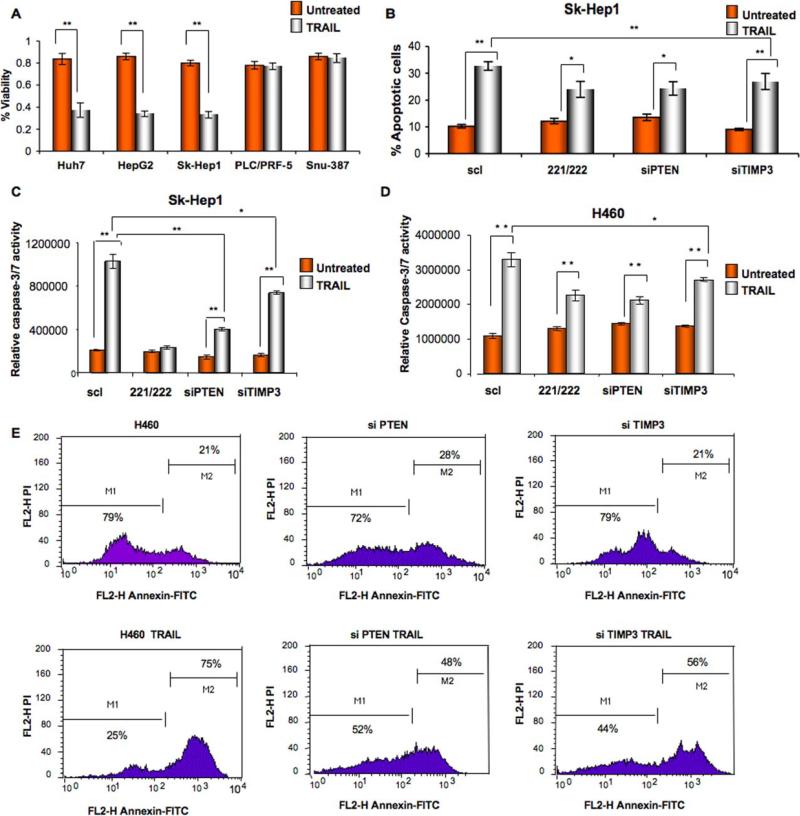
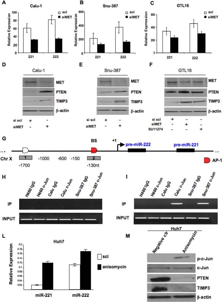
This article has been retracted: please see Elsevier Policy on Article Withdrawal (http://www.elsevier.com/locate/withdrawalpolicy). This article has been retracted at the request of the editors. This article was published on December 8, 2009. In November 2021, The Ohio State University notified the Cancer Cell editors that this article contains ten instances of plagiarized text; that Figures 1G, 5B, 5E, 7D, and 7F were falsified; and that part of Figure 1G in the article is the same as part of Figure 1B in another article that was later retracted (Garofalo et al., 2008, PLOS One 3, e4070, https://doi.org/10.1371/journal.pone.0004070). The university recommended the editors correct or retract the article. Given the extent and severity of these issues, the editors are retracting the article. S.-S. S. agrees with the retraction, and M.G., G.D.L., G.N., F.P., P.S., P.G., G.C., and C.M.C. disagree with the retraction; all other authors couldn't be reached or didn't respond.


Figures

Figure 1
Figure 1. PTEN and TIMP3 are targets of miR-221&222
(A) PTEN and TIMP3 3’UTRs contain one predicted miR-221 and miR-222 binding site. In the figure is shown the alignment of the seed regions of miR-221&222 with PTEN and TIMP3 3’UTRs. The sites of target mutagenesis are indicated in red. (B) qRT-PCR in MEG01 cells after enforced expression of miR-221 and miR-222. (C) PTEN and TIMP3 3’ UTRs are targets of miR-221&222. pGL3-PTEN and pGL3-TIMP3 luciferase constructs, containing a wild type (left side of the histograms) or mutated (right side of the histograms) PTEN and TIMP3 3’ UTRs, were transfected into MEG01 cells. Relative repression of firefly luciferase expression was standardized to a transfection control. The reporter assays were performed three times with essentially identical results. (D) qRT-PCR in H460 cells after enforced expression of miR-221 and miR-222. (E) MiR-221&222 enforced expression decreases endogenous levels of PTEN and TIMP3 proteins in H460 NSCLC. H460 cells were transfected with either scrambled or miR-221 or miR-222 for 72h. PTEN and TIMP3 expression was assessed by western blot. Loading control was obtained by using anti-β-actin antibody. (F) qRT-PCR showing miR-221&222 downmodulation in Calu-1 cells after anti-miRs transfection. (G) Western blot showing PTEN and TIMP3 expression after miR-221&222 downregulation by using anti-miR-221&222. Anti-miR-221 and -222 were able to increase PTEN and TIMP3 expression in Calu-1 cell line. (H) qRT-PCR of PTEN and TIMP3 mRNA after miR-221 and miR-222 forced expression in H460 cells. PTEN but not TIMP3 mRNA was downregulated by miR-221 and miR-222. Data are presented as ± SD.
Figure 2
Figure 2. PTEN and TIMP3 expression is inversely related to that of miR-221&222 in NSCLC and HCC
(A) MiR-221 and -222 expression levels was assessed by northern blot analysis using 15 μg of total RNA for NSCLC and 10 μg of total RNA for HCC cells. Western Blots anti-PTEN and TIMP3 were performed using total proteins extract (50 μg) isolated from the different NSCLC and HCC. (B) qRT-PCR of miR -221&222 and PTEN mRNA was performed by extracting RNA from the different NSCLC and HCC as described in the “Supplemental Experimental Procedures” section. MiR-221&222 were inversely related to PTEN mRNA expression in all the different NSCLC and HCC cells. Data are presented as ± SD.
Figure 3
Figure 3. PTEN and TIMP3 are direct targets of miR-221&222 in HCC in vitro and in vivo
(A) Western blot showing PTEN and TIMP3 expression in Sk-Hep1 and Snu-387 cells after miR-221&222 overexpression or downregulation. MiR-221 and miR-222 were able to downregulate PTEN and TIMP3 expression in Sk-Hep1; conversely, anti-miR-221&222 were able to increase PTEN and TIMP3 expression in Snu-387 cells. (B) qRT-PCR on 22 lung cancer patients and 10 normal lung tissues. The association between miR-221/222 and PTEN mRNA for the 10 subjects in the normal class and for the 22 subjects in the tumor class was calculated statistically by using the Pearson Correlation Coefficient (r) and the respective p-value, all significant at p 0.05. The Pearson correlation indicated an inverse relation between miR-221,-222 and PTEN mRNA in the normal and tumor samples. (C) IHC and ISH on hepatocarcinoma and normal liver tissues samples. MiR-221/222 (blue) and PTEN/TIMP3 (red) expression were inversely related in liver cancers and the adjacent normal/cirrhotic liver tissues. These tissues were analyzed for miR-221 and miR-222 expression by in situ hybridization, followed by immunohistochemistry for PTEN and TIMP3. Liver cancer cells abundantly expressed miR-221/222 and rarely expressed PTEN or TIMP3 (g,h,k,l) whereas the adjacent non-malignant liver abundantly expressed PTEN or TIMP3 and rarely had detectable miR-221/222 (a,b,e,f). In the cases of hepatocellular carcinoma where both miR-221/222 and TIMP3 expression were noted, the cancer cells expressing miR-221 (large arrow, panel k; TIMP3 is depicted by arrow in l) were distinct from those cells expressing TIMP3 (panel k, small arrow). c-i H&E; d-j miR-302, which is not express in liver, was used as negative control. 80 human HCC were analyzed. Scale bars indicate 25 μm.
Figure 4
Figure 4. MiR-221&222 induce TRAIL-resistance in NSCLC and HCC by targeting PTEN and TIMP3
(A) Proliferation assay on five different HCC. Cells were incubated with Super-Killer-TRAIL (400ng/ml) for 24h and viability evaluated as described in the supplemental methods. Huh7,HepG2 and Sk-Hep1 with low miR-221 and -222 expression, were more sensitive to TRAIL-induced apoptosis compared to PLC/PLF-5 and Snu-387, highly expressing miR-221/222. Mean ±SD of four independent experiments repeated in triplicate. (B) Cell death effects in Sk-Hep1 cells after miR-221/222 forced expression and PTEN or TIMP3 downregulation. Cells were transfected either with control miR or with pre-miR-221-222. 24h after transfection, cells were treated with Super-Killer TRAIL for 24 hours. Apoptosis was evaluated either with Annexin-FITC or (C) with caspase-Glo 3/7 kit. TRAIL resistance increased after miR-221/222 overexpression or PTEN and TIMP3 downmodulation. (D) Effects of miR-221/222 on cell death. H460 cells were transfected either with control siRNA or control miR or with 100 nmol of PTEN and TIMP3 siRNA. After 48 h from the transfection cells were treated with Super-Killer TRAIL for 24 hours. Apoptosis was evaluated by caspase 3/7 activity or (E) Annexin-V. Percentage of apoptotic cells decreased after PTEN and TIMP3 downregulation. Error bars indicate ±SD. *p < 0.05, **p < 0.001 by t test.
Figure 5
Figure 5. Anti-miR-221&222 override TRAIL-resistance in NSCLC and HCC through the inhibition of the AKT pathway
(A-C) Western Blots in H460 cells after miR-221/222 forced expression. MiR-221&222 forced expression induces the activation of the AKT/ERKs pathways and Metallopeptidases. (B) Western blots in Snu-387 cells after miR-221&222 knockdown by anti-miR-221/222. The inhibition of the AKT pathway is showed as result of miR-221&222 downregulation. (D-E) Western blots after PTEN or TIMP3 knockdown. Erks phosphorylation and PAK1 activation are both PTEN and TIMP3 dependent. The activation of the AKT pathway is PTEN-dependent, while TIMP3 silencing induces the expression of metallopeptidases.(F-G) Effects of anti-miRs and AKT pathway inhibition by API2/triciribine on cell death. Calu-1 and Snu-387 cells were transfected with anti-miR221/222 for 72h, or treated with API2/triciribine for 48h. MiR-221&222 downmodulation and/or the inhibition of the Akt pathway, induced an increase in apoptosis percentage in both Calu-1 and Snu-387 cell lines, as assessed by caspase 3/7 activity. Error bars indicate ±SD. **p < 0.001 by t test.
Figure 6
Figure 6. Ectopic expression of miR-221 and miR-222 affects the cell cycle distribution and migration/invasion capabilities of H460 cells
(A) Flow cytometric distributions of H460 cells transfected with pre-miR scrambled, miR-221&222, siRNA scrambled, siRNA PTEN. H460 transfected cells showed a decrease of G1 and a corresponding increase of the S and G2-M phases, as a consequence of PTEN downregulation. (B-C) miR-221&222 regulate cell migration ability in H460 cells. Migration Assay was performed as described in the “Experimental Procedures”. (D) miR-221&222 influences H460 and Sk-Hep1 cell invasion ability. Histogram reports the percentage of cells that invaded through Matrigel-coated membrane after transfection with negative control miRNA, miR-221, miR-222, siPTEN and siTIMP3. One-way analysis of variance (ANOVA) was performed to test the differences among means of invasion values. The Scheffe’ multiple-comparison method was used to test the differences between each pair of means. Significant differences were found between the scrambled vs miR-221&222, PTEN and TIMP3 H460 transfected cells (p-value <0.001). The same results were obtained using the Bonferroni and Sidak methods. Error bars indicate ±SD. *p < 0.001 by t test. Scale bar indicates 25 μm. The magnification is the same for all the panels.
Figure 7
Figure 7. MET oncogene regulates miR-221&222 activation
(A-B-C) Relative expression levels of miR-221&222 in Calu-1, Snu-387 and GTL16 after transfection with miR control and siRNA MET. MiR-221&222 expression decreased after MET knockdown. (D-E-F) Western blots after siRNA MET transfection in Calu-1, Snu-387 and GTL16 cells. MET knockdown decreased miR-221&222 expression levels, giving rise to PTEN and TIMP3 upregulation in all the different cell lines. GTL16 cells were moreover treated for 24h with 4μM of the MET inhibitor SU11274. MET inhibition increased miR-221&222 targets expression levels. (G-H-I) Identification of c-Jun (AP-1) interacting region by using 2 different amplicons across the miR-221/222 transcription start site. ChIP analysis was performed with chromatin from H460 c-Jun negative cells, Calu-1 and Snu-387 c-Jun positive cells. BS=binding site. (L) qRT-PCR of miR-221&222 in Huh7 cells after treatment with anisomycin (10 μM) for 30’. Anisomycin induced miR-221&222 upregulation. (M) Anisomycin induced c-Jun activation and PTEN and TIMP3 downregulation in Huh7 cells. Total lysate was analyzed by western blot using anti-PTEN and anti-TIMP3 antibody. Error bars indicate ±SD.
Figure 8
Figure 8
MET induces miR-221&222 activation through AP-1 (c-Jun) transcription factor. A model is reported in which growth factors determine c-Met activation which, in turn, through AP-1 and accordingly miR-221&222 upregulation, gives rise to PTEN and TIMP3 downregulation and subsequent apoptosis resistance, cellular migration and invasion.

Similar articles

Cited by

References

    1. Ahonen M, Baker AH, Kähäri VM. Adenovirus-mediated gene delivery of tissue inhibitor of metalloproteinases-3 inhibits invasion and induces apoptosis in melanoma cells. Cancer Res. 1998;58:2310–5. - PubMed
    1. Ambros V. The functions of animal microRNAs. Nature. 2004;431:350–5. - PubMed
    1. Bartel DP. MicroRNAs: genomics, biogenesis, mechanism, and function. Cell. 2004;116:281–97. - PubMed
    1. Baker AH, Zaltsman AB, George SJ, Newby AC. Divergent effects of tissue inhibitor of metalloproteinase-1, -2, or -3 overexpression on rat vascular smooth muscle cell invasion, proliferation, and death in vitro. TIMP3 promotes apoptosis. J. Clin. Invest. 1998;101:1478–87. - PMC - PubMed
    1. Bode W, Reinemer P, Huber R, Kleine T, Schnierer S, Tschesche H. The X-ray crystal structure of the catalytic domain of human neutrophil collagenase inhibited by a substrate analogue reveals the essentials for catalysis and specificity. EMBO J. 1994;13:1263–9. - PMC - PubMed

Publication types

MeSH terms

LinkOut - more resources