. 2009 Dec 8;16(6):510-20.
doi: 10.1016/j.ccr.2009.10.013.

Tissue-penetrating delivery of compounds and nanoparticles into tumors

Affiliations

Tissue-penetrating delivery of compounds and nanoparticles into tumors

Kazuki N Sugahara et al. Cancer Cell. .

Abstract

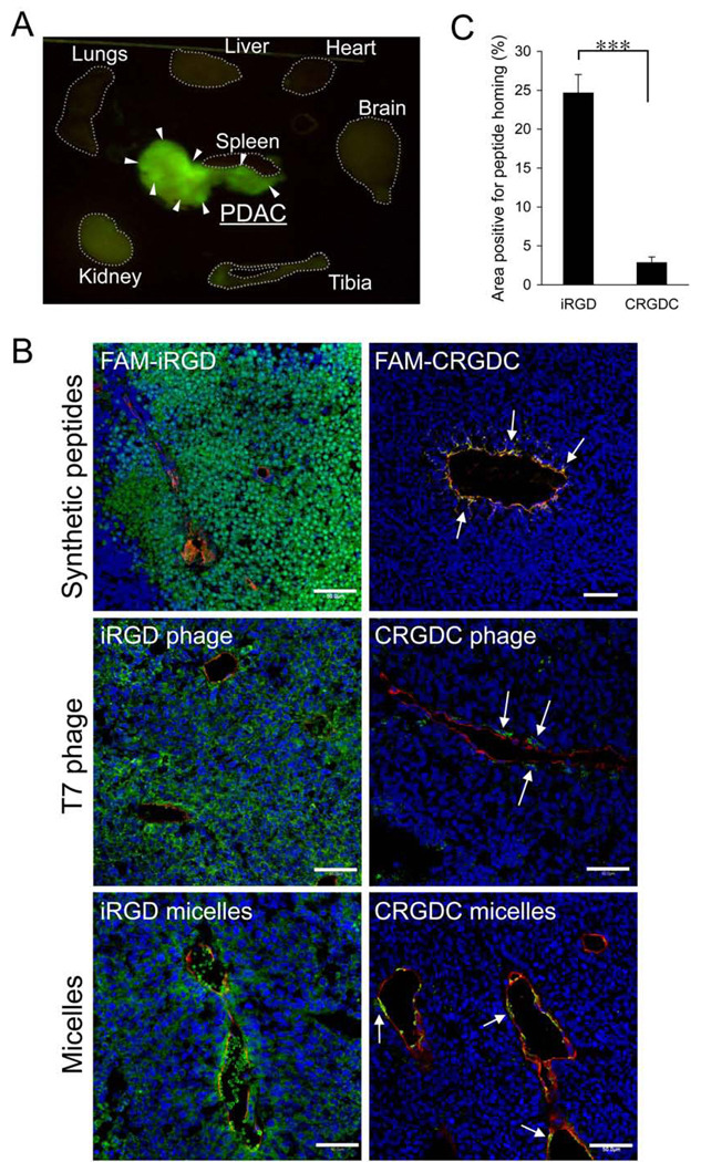
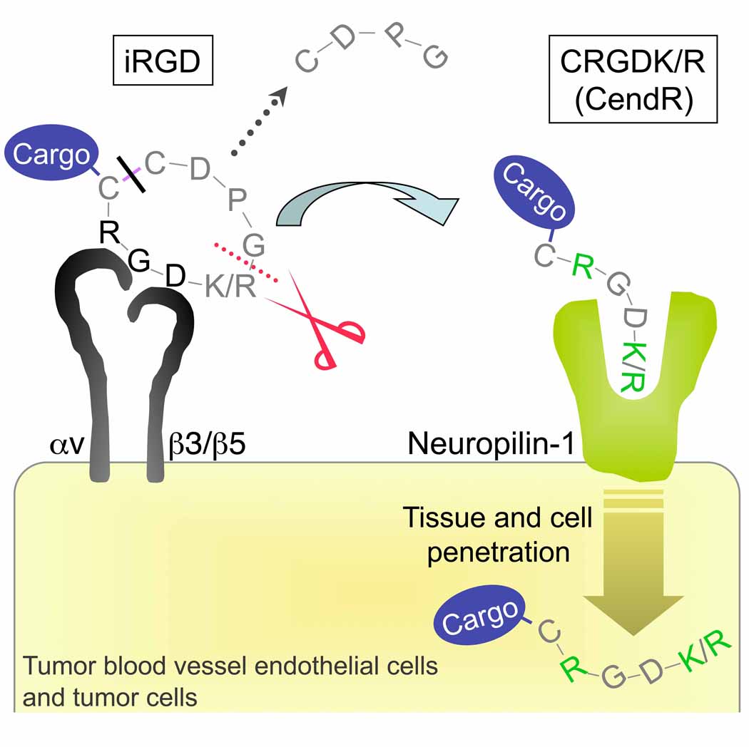
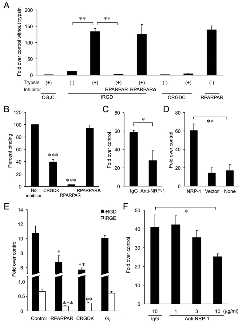
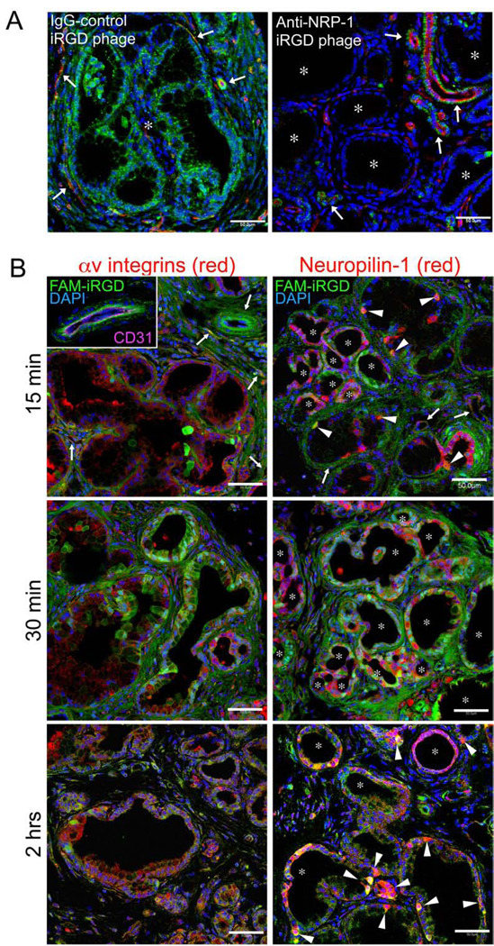
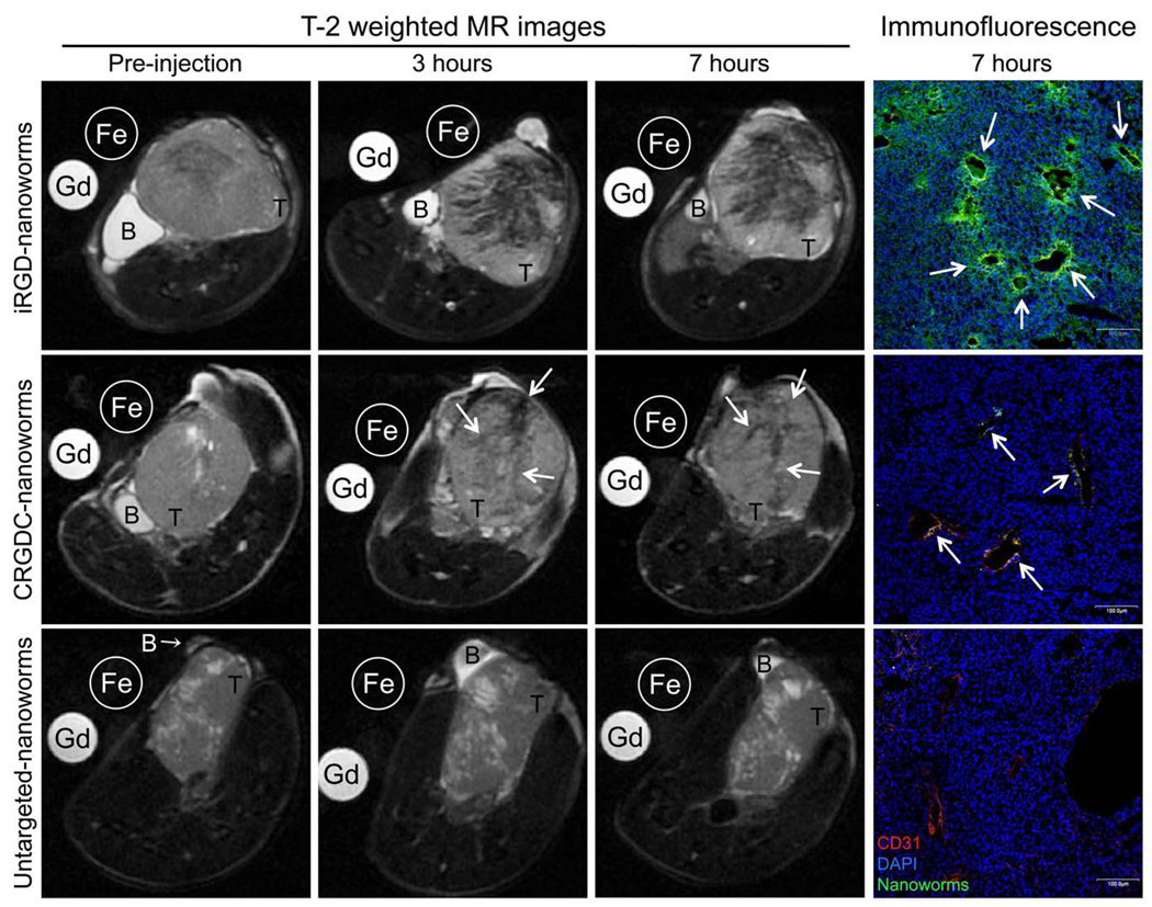
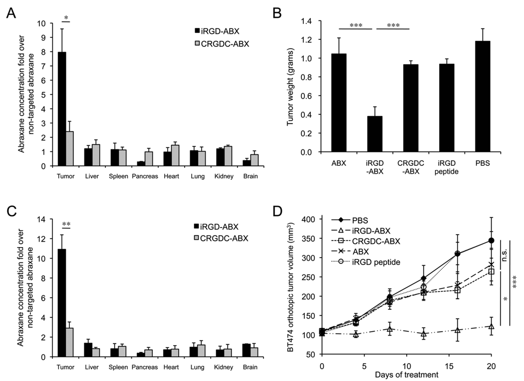
Poor penetration of drugs into tumors is a major obstacle in tumor treatment. We describe a strategy for peptide-mediated delivery of compounds deep into the tumor parenchyma that uses a tumor-homing peptide, iRGD (CRGDK/RGPD/EC). Intravenously injected compounds coupled to iRGD bound to tumor vessels and spread into the extravascular tumor parenchyma, whereas conventional RGD peptides only delivered the cargo to the blood vessels. iRGD homes to tumors through a three-step process: the RGD motif mediates binding to alphav integrins on tumor endothelium and a proteolytic cleavage then exposes a binding motif for neuropilin-1, which mediates penetration into tissue and cells. Conjugation to iRGD significantly improved the sensitivity of tumor-imaging agents and enhanced the activity of an antitumor drug.


Figures

Figure 1
Figure 1. Identification of iRGD peptide
(A) A representative example of the enrichment obtained in phage library screens on prostate cancer bone metastases. Three screens with similar results were performed. (B) RGD peptides selected in the screens. Approximately 50 individual clones were randomly picked for sequencing from phage pools recovered in the final round of ex vivo phage display. Clones that gave unsuccessful sequencing results were omitted during the analysis. The proportion of each RGD peptide is shown. (C) Binding and internalization to PPC1 human prostate cancer cells of phage expressing the CRGDKGPDC peptide. PPC1 cells were incubated with phage displaying CRGDKGPDC or a polyglycine control peptide CG7C for 1 hr at 4°C or 37°C. To assess internalization, phage bound at the cell surface was removed by washing the cells with an acid buffer. Note that the internalization of CRGDKGPDC phage occurs at 37°C but not at 4°C. Statistical analysis was performed with Student’s t-test. n = 3, error bars, s.e.m.; triple asterisk, p < 0.001.
Figure 2
Figure 2. In vivo tumor homing of iRGD peptide
(A) Approximately 200 µg of FAM-iRGD or control peptide in PBS was intravenously injected into KrasG12D, p48-Cre, Ink4a+/− mice bearing de novo pancreatic ductal adenocarcinoma. The peptides were allowed to circulate for 2 hrs and organs were collected and viewed under UV light. Arrowheads point to the tumors. Dotted lines show where the organs were placed. Representative images from four experiments are shown. (B) Confocal images of orthotopic 22Rv1 human prostate cancer xenografts from mice injected with the indicated peptides, phage, and micelles. iRGD was compared to a similar integrin-binding but non-penetrating peptide, CRDGC. The circulation time was 2 hrs for the free peptides, 15 min for the phage, and 3 hrs for the micelles. Red: CD31; green: peptides, phage, or micelles; blue: nuclei. Arrows point to CRGDC compounds in or just outside the vessel walls, illustrating its homing to the tumor vasculature. Representative fields from multiple sections of five tumors are shown. Scale bars = 50 µm. (C) Quantification of tumor homing area of iRGD and CRGDC peptides. Cryo-sections of 22Rv1 orthotopic tumors from mice injected with FAM-iRGD or FAM-CRGDC peptide were immunohistochemically stained with an anti-FITC antibody. The samples were subjected to image analysis with Scanscope CM-1 scanner for quantification of the FAM-positive areas. Statistical analysis was performed with Student’s t-test. n = 3; error bars, s.e.m.; triple asterisk, p < 0.001.
Figure 3
Figure 3. iRGD binds to αv integrins
(A) Quantification of the in vivo distribution of iRGD and control peptides. FAM-iRGD; a non-integrin-binding iRGD mutant, FAM-CRGEKGPDC (FAM-iRGE); and a FAM-labeled cyclic polyglycine control peptide, FAM-CG7C were injected into PDAC mice as described in the legend of Figure 2A. Fluorescence in each tissue was quantified with Image J. (B) Inhibition of the tumor homing of FAM-iRGD with non-labeled iRGD. A 10-fold excess of unlabeled iRGD peptide or iRGE peptide was injected 30 min before the injection of FAM-iRGD in PDAC mice. Fluorescence was quantified as above. FAM-iRGD homing without injection of unlabeled peptide was considered as 100%. (C) Dose-dependent inhibition of iRGD phage binding to PPC1 prostate cancer cells by synthetic iRGD peptide and iRGE. iRGD phage binding without inhibitors was considered as 100%. (D) Inhibition of iRGD phage binding to αv integrin expressing M21 cells by antibodies against integrins or control mouse IgG (left panel). Lack of iRGD phage binding to M21 cells selected for low level of αv integrin expression (M21L; right panel). Statistical analyses were performed with Student’s t-test in panels (A) and (B), and ANOVA in panels (C) and (D). n = 3; error bars, s.e.m.; single asterisk, p < 0.05; double asterisk, p < 0.01; triple asterisk, p < 0.001.
Figure 4
Figure 4. Multistep binding and penetration mechanism of iRGD
The iRGD peptide accumulates at the surface of αv integrin-expressing endothelial and other cells in tumors. The RGD motif mediates the integrin binding. The peptide is cleaved by a cell surface-associated protease(s) to expose the cryptic CendR element, RXXK/R, at the C-terminus (red dotted line). The CendR element then mediates binding to neuropilin-1, with resulting penetration of cells and tissues. The peptide can penetrate into tumor cells and tissues with a cargo, such as a simple chemical or a nanoparticle, provided that the cargo is attached to the N-terminus of the iRGD peptide because the disulfide bond apparently breaks before the peptide is internalized (black line).
Figure 5
Figure 5. CendR motif in iRGD penetration of tumor cells
(A) The penetration of trypsin-treated iRGD phage within PPC1 cells pre-treated or not with non-infectious RPARPAR or RPARPARA phage. (B) Inhibition of CRGDK phage binding to PPC1 by synthetic CRGDK, RPARPAR, and RPARPARA peptides. CRGDK phage binding without inhibitors was considered as 100%. (C) CRGDK phage binding to PPC1 cells treated with anti-neuropilin-1 blocking antibodies (anti-NRP-1) or control goat-IgG. (D) CRGDK phage binding to M21 cells transfected with neuropilin-1 cDNA to induce forced expression of neuropilin-1 (NRP-1), vector alone, or without transfection. (E) Inhibition of iRGD and iRGE phage penetration into PPC1 by non-infectious phage displaying the CendR peptides RPARPAR and CRGDK. (F) Dose-dependent inhibition of iRGD phage penetration of PPC1 cells by anti-neuropilin-1 antibodies (anti-NRP-1) to block neuropilin-1 function. Statistical analyses were performed with ANOVA in panels (A), (B), and (E), and Student’s t-test in panels (C), (D), and (F). n = 3; error bars, s.e.m.; single asterisk, p < 0.05; double asterisk, p < 0.01; triple asterisk, p < 0.001.
Figure 6
Figure 6. Penetration of iRGD within tumor tissue involves neuropilin-1
(A) Neuropilin-1-dependent spreading of iRGD phage within tumor tissue. Confocal images of PDAC tumors from transgenic mice pre-injected with a function-blocking anti-neuropilin-1 antibody (anti-NRP-1) or an IgG-control, followed by injection with the iRGD phage. Green, phage; red, CD31; blue, DAPI. Arrows and asterisks represent blood vessels and tumor ducts, respectively. Representative images from three independent sets of studies are shown. Scale bars = 50 µm. (B) Time-dependent homing of FAM-iRGD peptide (green) in relation to the expression of αv integrins (red, left panels) and neuropilin-1 (red, right panels) in PDACs. The blood vessels targeted by FAM-iRGD were positive for both αv integrins and neuropilin-1 (arrows). The inset shows CD31 staining (magenta) of a blood vessel targeted by FAM-iRGD. Nearly all tumor ducts examined were positive for αv integrins. Tumor cells (arrowheads) and tumor ducts (asterisks) also strongly positive for neuropilin-1 were particularly effective in internalizing and retaining FAM-iRGD. Representative images from three independently studied tumors at each time point are shown. Scale bars = 50 µm.
Figure 7
Figure 7. Tumor imaging with iRGD-coated iron oxide nanoworms
T2-weighted MR images of mice bearing orthotopic 22Rv1 human prostate tumors. The mice were injected intravenously with iron oxide nanoworms coated with iRGD, CRGDC, or no peptide (5 mg/kg of iron). Shown are axial images through the tumors acquired by repeated imaging before the nanoworm injection (Pre-injection) and 3 hrs or 7 hrs after the injection. The orientation of the tumors is slightly different between the time points because the mice were anesthetized for each scan and reintroduced in the MRI instrument. Gadolinium (Gd) was used as a reference for T1 and Feridex (Fe) for T2 imaging agents. Note the wide hypointensity areas indicating the spreading of iRGD-nanoworms in the tumor interstitium. CRGDC-nanoworms only decreased the intensity of tumor vessels, and the untargeted-nanoworms gave no signal in the tumor. Arrows point to the vasculature. T, tumor; B, urinary bladder. The right-most panels represent the nanoworm distribution in tumor tissue examined by confocal microscopy. Green, nanoworms; red, CD31; blue, DAPI. Scale bars = 100 µm. The images are representative of multiple tumor mice; iRGD-nanoworms, n = 5; CRGDC-nanoworms, n = 3; untargeted-nanoworms, n = 3.
Figure 8
Figure 8. Tumor treatment with iRGD-coated nanoparticles
(A and C) Abraxane quantification in orthotopic 22Rv1 (A) and BT474 (C) xenograft models. Abraxane was intravenously injected into tumor mice 3 hours earlier and captured from tumor extracts with a taxol antibody, followed by detection with a human albumin antibody. n = 3 for each group. (B and D) Long-term treatment of tumor mice with targeted abraxane conjugates. Mice bearing orthotopic 22Rv1 (B) or BT474 (D) xenografts were intravenously injected with peptide-coated abraxanes every other day at 3 mg paclitaxel/kg/injection. The treatment was continued for 14 days in (B) and 20 days in (D). Panel (B) shows one of three experiments, which all gave similar results. The total number of mice in (B) was: untargeted abraxane (ABX, n = 18), abraxane coated with CRGDC (CRGDC-ABX, n = 10) or iRGD (iRGD-ABX, n = 19), iRGD peptide alone as a control (iRGD peptide, n = 10), or PBS (n = 18). The number of mice per group was 8 in (D). Statistical analyses were performed with Student’s t-test in panels (A) and (C), and ANOVA in panels (B) and (D). Error bars, s.e.m.; n.s., not significant; single asterisk, p < 0.05; double asterisk, p < 0.01; triple asterisk, p < 0.001.

Similar articles

Cited by

References

    1. Acevedo LM, Barillas S, Weis SM, Göthert JR, Cheresh DA. Semaphorin 3A suppresses VEGF-mediated angiogenesis yet acts as a vascular permeability factor. Blood. 2008;111:2674–2680. - PMC - PubMed
    1. Arap W, Pasqualini R, Ruoslahti E. Cancer treatment by targeted drug delivery to tumor vasculature in a mouse model. Science. 1998;279:377–380. - PubMed
    1. Arbeit JM, Münger K, Howley PM, Hanahan D. Progressive squamous epithelial neoplasia in K14-human papillomavirus type 16 transgenic mice. J. Virol. 1994;68:4358–4368. - PMC - PubMed
    1. Arleth L, Ashok B, Onyuksel H, Thiyagarajan P, Jacob J, Hjelm RP. Detailed structure of hairy mixed micelles formed by phosphatidylcholine and PEGylated phospholipids in aqueous media. Langmuir. 2005;21:3279–3290. - PubMed
    1. Cheresh DA, Spiro RC. Biosynthetic and functional properties of an Arg-Gly-Asp-directed receptor involved in human melanoma cell attachment to vitronectin, fibrinogen, and von Willebrand factor. J. Biol. Chem. 1987;262:17703–17711. - PubMed

Publication types

LinkOut - more resources