. 2010 Feb 5;395(5):983-94.
doi: 10.1016/j.jmb.2009.11.062. Epub 2009 Dec 4.

Structural and biophysical studies of human PARP-1 in complex with damaged DNA

Affiliations

Structural and biophysical studies of human PARP-1 in complex with damaged DNA

Wayne Lilyestrom et al. J Mol Biol. .

Abstract

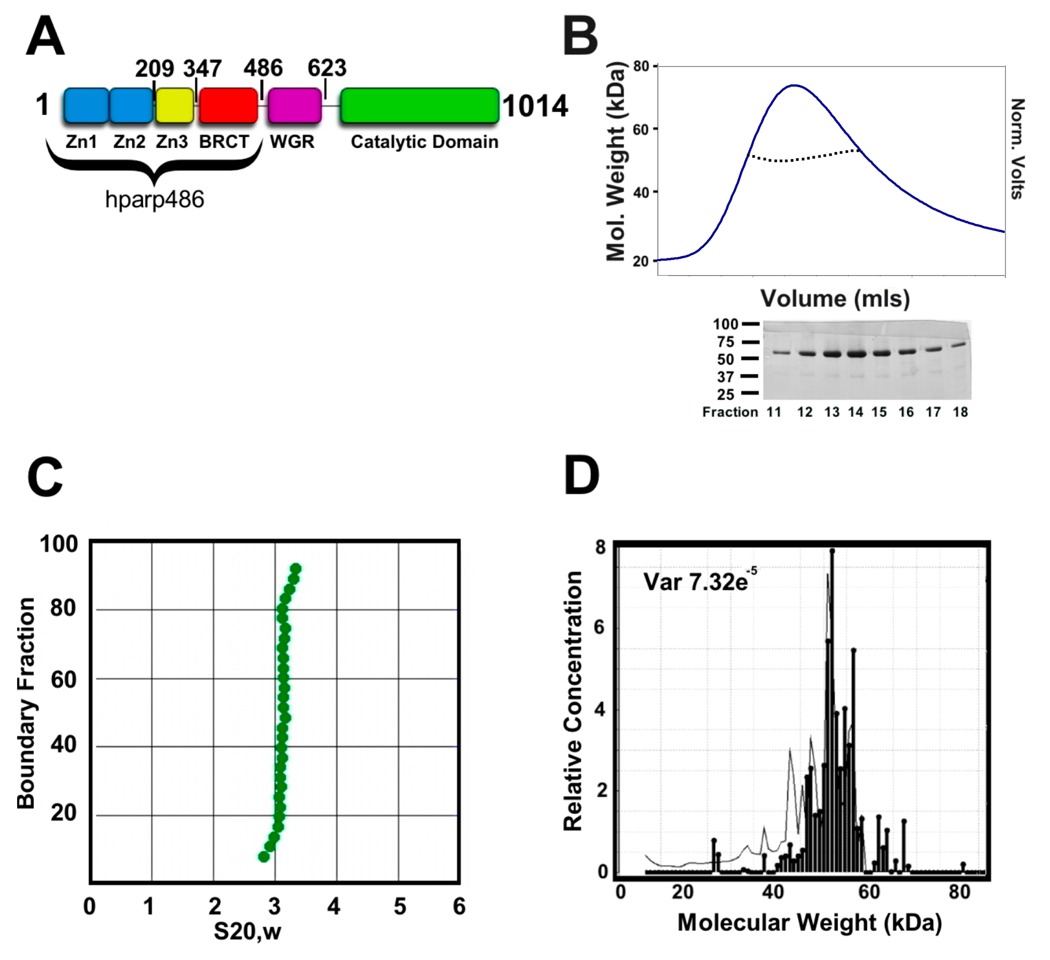
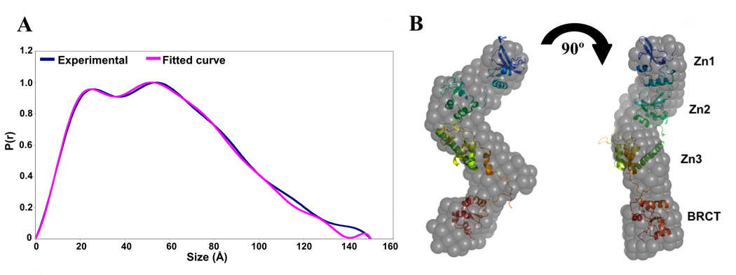
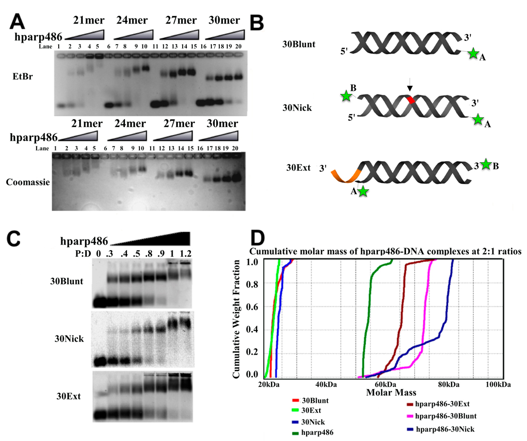
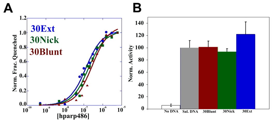
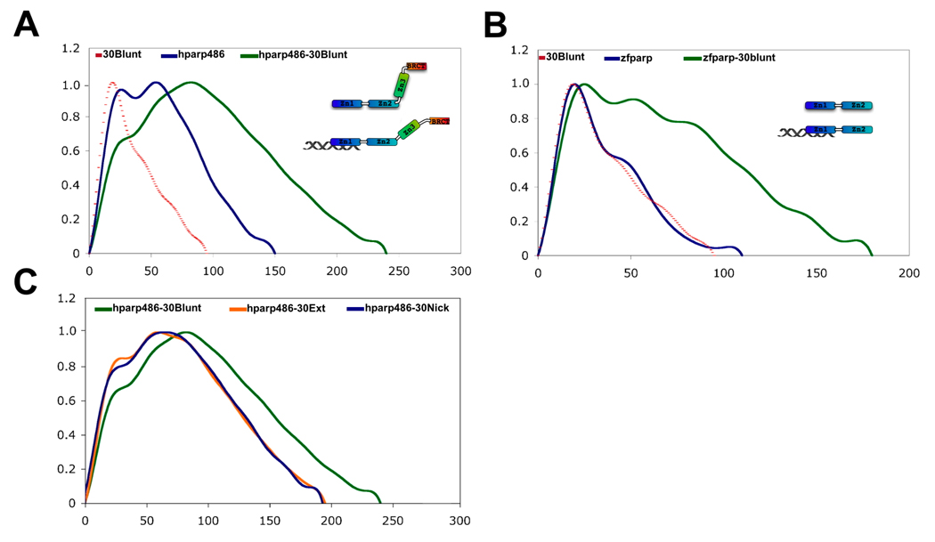
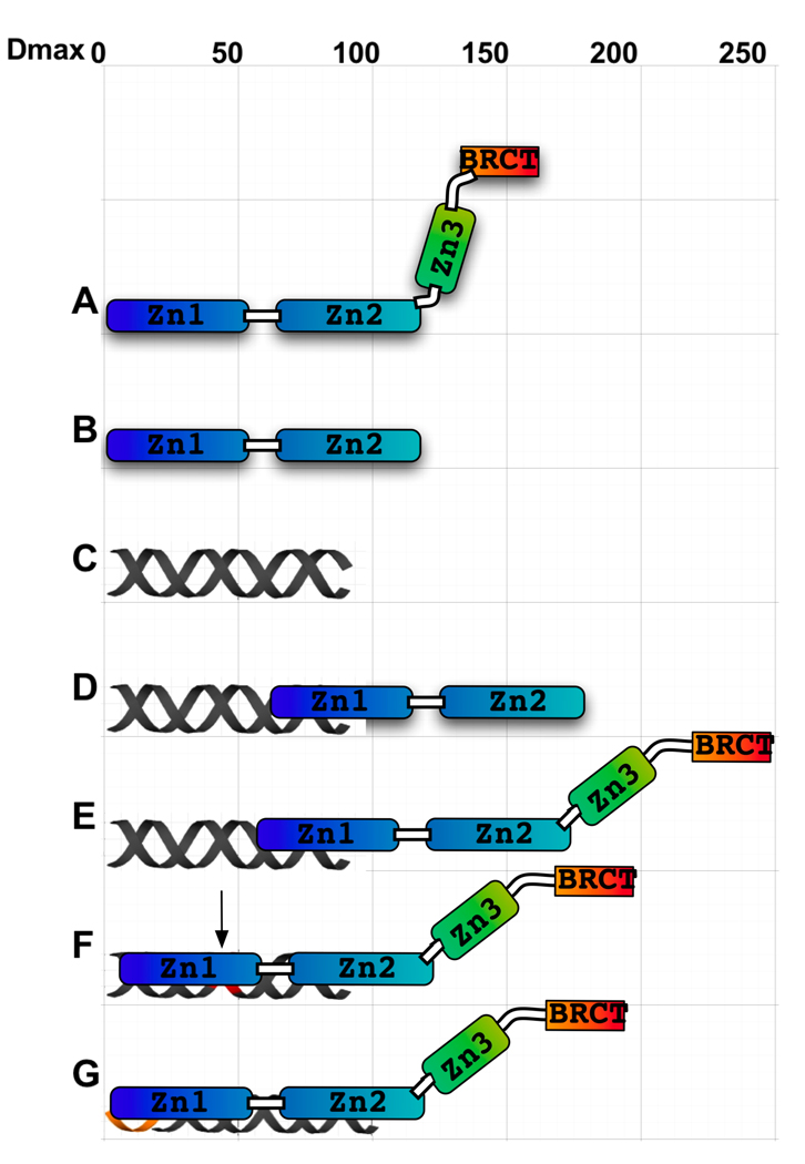
The enzyme poly(ADP-ribose) polymerase-1 (PARP-1) is a global monitor of chromatin structure and DNA damage repair. PARP-1 binds to nucleosomes and poly(ADP-ribosylates) histones and several chromatin-associated factors to expose specific DNA sequences to the cellular machinery involved in gene transcription and/or DNA damage repair. While these processes are critical to genomic stability, the molecular mechanisms of how DNA damage induces PARP-1 activation are poorly understood. We have used biochemical and thermodynamic measurements in conjunction with small-angle X-ray scattering to determine the stoichiometry, affinity, and overall structure of a human PARP-1 construct containing the entire DNA binding region, the zinc ribbon domain, and automodification domains (residues 1-486). The interaction of this PARP-1 protein construct with three different DNA damage models (DNA constructs containing a nick, a blunt end, or a 3' extension) was evaluated. Our data indicate that PARP-1 binds each DNA damage model as a monomer and with similar affinity, in all cases resulting in robust activation of the catalytic domain. Using small-angle X-ray scattering, we determined that the N-terminal half of PARP-1 behaves as an extended and flexible arrangement of individually folded domains in the absence of DNA. Upon binding DNA, PARP-1 undergoes a conformational change in the area surrounding the zinc ribbon domain. These data support a model in which PARP-1, upon binding DNA, undergoes a conformational change to become an active nuclear enzyme.


Figures

Figure 1
Figure 1. hparp486 is a monomer in the absence of DNA
A) hparp486 was designed to include the entire DNA binding region of the human PARP-1 protein. The first 486 residues encompass the three N-terminal Zn domains and the BRCT domain. The illustration indicates the C-terminal residue of each domain. B) Top: Size exclusion chromatography and inline static light scattering of hparp486. hparp486 elutes in a single peak from a KW803 size exclusion column. The static light scattering average molecular weight (~57kDa) for the central portion of the peak is illustrated by the black dotted line within the peak. Bottom: SDS-PAGE analysis of fractions from SEC purified hparp486. C) The sedimentation velocity profile of purified are presented as G(s) plots of the integral of S20,w against boundary fraction (%).van Holde - Weischet analysis was used to determine the f/f0 value of 1.8. D) Sedimentation equilibrium ultracentrifugation resolved that over 95% of the purified protein was monomeric at a concentration of 40µM.
Figure 2
Figure 2. The low resolution structure of hparp486 reveals a molecule with conformational flexibility
A) Superposed experimental and modeled distance distribution functions of hparp486. In blue, the SAXS distance distribution function P(r) for hparp486 is an asymmetric curve with two discrete maxima, representing two ordered regions connected by a flexible linker. The relatively shallow decent towards a maximum size of 150Å is reflective of an elongated, flexible molecule. Illustrated in violet, is the P(r) function resulting from the SASREF tertiary model shown in (B). B) Superposed particle reconstructions and tertiary structure models of hparp486. The DAMMIN particle reconstruction (grey) represents the filtered average of 30 models individually computed from a SAXS scattering curve and fits the SAXS scattering curve with a χ2 of 1.10. The SASREF tertiary structure model was computed by global rigid body modeling of the known domain structures of Zn1 (PDB id 2dmj, blue), Zn2 (PDB id 2cs2, green), Zn3 (PDB id 2jvn orange) and BRCT (PDB id 2cok, red) with steric restraints against the solution scattering data; this model has a χ2 of 1.21. In the orientation shown, the two models have a correlation coefficient of 0.56, while a 180° rotation of the polypeptide resulting in a switch in position of Zn1 and BRCT domains has a correlation coefficient of 0.54. The SASREF model illustrates the extended nature of the polypeptide and minimal inter-domain contacts.
Figure 3
Figure 3. hparp486 binds as a monomer to damaged DNA
A) EtBr and Coomassie stained 1% agarose gel EMSA assay of hparp486-DNA complexes. 1µM DNA was added to each lane. In this assay, all DNA constructs contain a 6-nucleotide 3’ extension, but vary in the length of the double stranded region (21–30 base pairs). hparp486 forms homogenous complexes as the DNA approaches 27–30 base pairs in length. B) A schematic representation of the three DNA damage models tested and the location of the fluorophores (green asterisk) used in this publication. Varying the fluorophore position (either in position A or B) allowed us to test the effect of its position on hparp486 association. The arrow and red region highlight the area of the single stranded break in the 30Nick DNA, while the orange area highlights the 6 nucleotide 3’ extension in 30Ext. C) EMSA of hparp486 association with 2µM fluorescently labeled 30Blunt, 30Nick and 30Ext DNA. hparp486 efficiently shifts the mobility of over 90% of each DNA at a 1:1 ratio, eliminating the possibility of cooperative dimerization under these conditions. D) Cumulative molar mass of DNA, hparp486 and hparp486-DNA complexes as determined by SEC-MALS. 2:1 molar ratios of hparp486:DNA were incubated for 30 minutes prior to subjecting the mixtures to SEC-MALS. The distribution of masses present in the major peaks eluting from the column for DNA, hparp486 or each hparp486-DNA complex is plotted as a fraction of cumulative molecular weight. hparp486 does not stably dimerize on any DNA fragment. Supplemental Figure 4 contains the elution profiles for each solution.
Figure 4
Figure 4. hparp486 has similar affinities for different forms of DNA damage; fulllength human PARP-1 is efficiently activated by DNA containing blunt ends, nicks or a 3’-extension
A) The affinity of hparp486 for fluorescently labeled 30 base pair DNA: 30Blunt (red), 30Nick (green), 30Ext(red) was determined by measuring the change in fluorescence of 2nM labeled DNA as a function of hparp486 concentration (0.5-6000nM) in solution. The affinity of hparp486 each DNA is shown in Table 3 and is in the range of ~70–300nM. The Hill coefficients calculated for each fluorescent titration were in the range of 0.92–0.99, demonstrating the lack of positive cooperativity in the individual interactions (Table 3). B) The ability of each DNA to activate full-length human PARP-1 was tested with a commercial assay that monitors the incorporation of biotinylated ADP-ribose onto immobilized histones. The activation induced by 1µM of 30Blunt, 30Nick or 30Ext DNA was normalized to the activity stimulated by sheared salmon sperm DNA. Regardless of the DNA damage model tested, PARP-1 was ~40 fold more active than in the absence of DNA.
Figure 5
Figure 5. The distance distribution functions of hparp486-DNA complexes portray a conformational change within hparp486 upon binding DNA
A) The P(r) functions of 30Blunt (dotted line), hparp486 (blue) and a 1:1 complex of hparp486-30Blunt complex (green). Upon binding 30Blunt, the profile of hparp486 in complex changes significantly when compared to the normalized profile of hparp486 in the absence of DNA. In particular, interacting with DNA changes the P(r) function from two discrete maximum of similar height to a single dominant maximum of 240Å, reflecting an increase in Dmax of 90Å over hparp486. We interpret this to represent a migration from a conformation containing two ordered regions connected by an internal flexible linker to an elongated, dynamic complex that does not have an internal highly flexible region. B) The P(r) function of zf-parp (blue), 30Blunt (dotted line) and a 1:1 complex of zf-parp-30Blunt. In contrast to hparp486, the P(r) function of zf-parp is characterized by a distinct single maxima, which reflects the lack of a high degree of internal flexibility. Upon binding 30Blunt DNA, no evidence of a conformational change is noted and with a maximum dimension of 180Å, the complex reflects an increase in size of only 65Å. C) The SAXS scattering profiles of solutions containing 1:1 ratios of hparp486 in complex with 30Blunt, 30Nick or 30Ext DNA were analyzed to determine the general shape and size of the complexes (green, blue, orange respectively). The P(r) functions of the complexes illustrate that hparp486 forms complexes of similar maximum size with 30Nick and 30Ext DNA (~200Å), but forms a much longer complex with 30Blunt DNA (~240Å maximum length).
Figure 6
Figure 6. A model for the interactions of hparp486 with damaged DNA
The overall shape and Dmax of hparp486 (A), zf-parp (B), 30Blunt DNA (C) and their complexes (D and E) have been experimentally determined by SAXS. The Dmax of 180Å (a 65Å increase) and asymmetric shape of the zf-parp-30Blunt complex is consistent with a single parp zinc finger interacting the blunt end (D). The hparp486-30Blunt complex is also elongated (E), but increases in size by 90Å (to 240Å). To account for both the change in profile of the P(r) function (Fig. 5 A, C) and greater Dmax,we model this with a conformational change that occurs in the Zn3-BRCT region of the protein. In contrast, when interacting with nicked DNA (F) or a 3’ extension region (G), hparp486 forms a more compact complex but a conformational change still needs to be taken into account to reach a Dmax of ~200Å. We propose that ultimately the conformational change that occurs during binding of DNA leads to activation of the PARP-1 enzymatic domain.
None

Similar articles

Cited by

References

    1. Ame JC, Spenlehauer C, de Murcia G. The PARP superfamily. Bioessays. 2004;26:882–893. - PubMed
    1. Bouchard VJ, Rouleau M, Poirier GG. PARP-1, a determinant of cell survival in response to DNA damage. Exp Hematol. 2003;31:446–454. - PubMed
    1. D'Amours D, Desnoyers S, D'Silva I, Poirier GG. Poly(ADP-ribosyl)ation reactions in the regulation of nuclear functions. Biochem J. 1999;342(Pt 2):249–268. - PMC - PubMed
    1. Jacobson EL, Antol KM, Juarez-Salinas H, Jacobson MK. Poly(ADP-ribose) metabolism in ultraviolet irradiated human fibroblasts. J Biol Chem. 1983;258:103–107. - PubMed
    1. Oleinick NL, Evans HH. Poly(ADP-ribose) and the response of cells to ionizing radiation. Radiat Res. 1985;101:29–46. - PubMed

Publication types

MeSH terms

LinkOut - more resources